 Research Article
Cecal Volvulus Caused by an Omphalo-Mesenteric Band: A Case Report of a Rare Complication of Meckel’s Diverticulum
Parmar Jitendra P1*, Patel Tapan C1, Desai Chirag J2, Shah Sandip R1 and Choksi Neel H1
1Department of Radiology, Apollo Hospitals International Limited, Ahmedabad, India
2Department of Gastrointestinal, Hepato-biliary and Liver transplant Surgery, Apollo Hospitals International Limited, Ahmedabad, India
Jitendra Parmar, Department of Radiology, Apollo Hospitals International Limited, Ahmedabad, India
Received Date: October 12, 2020; Published Date: November 03, 2020
Abstract
Introduction: Partial or complete developmental failure to close and absorb the omphalo-mesenteric duct results in a variety of congenital anomalies, Meckel’s Diverticulum (MD) with or without omphalo-mesenteric band being the most common. The Cecal volvulus (CV) caused by MD is extremely rare and only few case reports have been reported so far.
Case Summary: In this case report, a case of 40-years-old female patient with features of small bowel obstruction is reported. The patient subsequently diagnosed to have “Meckel’s Diverticulum” and “Loop Type of Cecal Volvulus” with ischemic changes involving cecum, Meckel’s diverticulum, and ascending colon on computed tomography of abdomen performed in emergency.
Conclusion: MD is often overlooked, and the patient usually presents with complications, appropriate knowledge of various pathophysiology of complications must be kept in mind for a prompt and effective management.
Key Points
i. The volvulus caused by Meckel’s Diverticulum is extremely rare but not unheard and the patient usually presents with the features of intestinal obstruction.
ii. As the diagnosis of Meckel’s Diverticulum is often overlooked and the patient usually presents with complications, appropriate knowledge of various pathophysiology of complications must be kept in mind for a prompt and effective management.
Keywords: Cecal Volvulus; Meckel’s Diverticulum; Intestinal Obstruction; Omphalo-Mesenteric Duct Anomalies
Abbreviations: MD-Meckel’s Diverticulum; CV- Cecal Volvulus; CT-Computed Tomography
Introduction
Meckel’s diverticulum (MD) is a true congenital diverticulum as it consists of all three layers of the bowel wall [1]. Partial or complete developmental failure to close and absorb the omphalomesenteric duct results in a variety of congenital anomalies, MD with or without omphalo-mesenteric band being the most common [2]. It is often asymptomatic, however, ~ 2-4% of patients may be symptomatic. Symptomatic MD is often descried by rule of 2s, that may clinically vary [3]. Children most commonly present with gastrointestinal bleeding, while adults present with features of small bowel obstruction and diverticulitis.
Cecal volvulus (CV) caused by MD is extremely rare and only few cases have been reported so far [4,5]. We present one of the rarest complications of MD, loop type of CV, causing large bowel obstruction in a middle-aged female patient. The diagnosis of CV with small bowel obstruction and changes of bowel ischemia of cecum and ascending colon caused by MD was made preoperatively on Computed Tomography Scan (CT scan), while omphalomesenteric band was found intra-operatively.
Case Presentation
History
A 40-yeard-old female patient presented in the emergency department with features suggestive of intestinal obstruction, including abdominal pain with abdominal distension and vomiting for 1 day. The patient had no previous operative history.

Procedures and Investigations
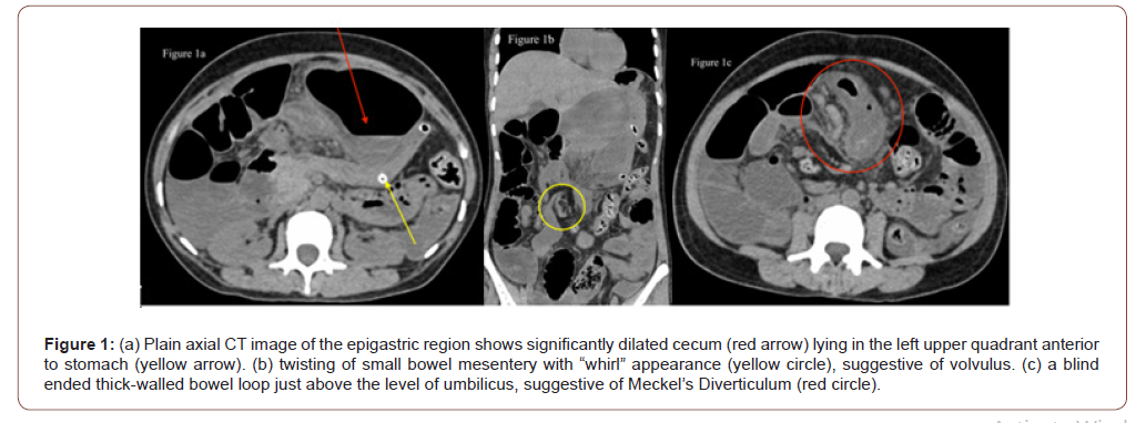
Nasogastric tube was inserted in emergency department and urgent CT scan of abdomen was performed (Figure 1). The CT scan of abdomen revealed significantly dilated caecum (red arrow), lying in the left upper quadrant, anterior to the collapsed stomach (yellow arrow). It was associated with twisted small bowel mesentery just above the level of umbilicus, suggestive of “Whirl” sign (Yellow circle), resulting in significant dilatation with air-fluid levels in small bowel loops. The findings were suggestive of “Loop Type of Cecal Volvulus” with changes of closed loop obstruction and ischemic wall thickening of cecum and part of ascending colon. The transverse colon and descending colon were collapsed. In addition, a thick walled blind ended bowel loop with specks of intraluminal air was also noted adjacent to the dilated cecum, appearing to be communicating with collapsed distal ileum, which probably represents inflamed and ischemic MD (Red circle). However, no obvious fibrous band could be appreciated on CT scan.
The laboratory investigations were within normal limits and unremarkable.

Therapeutic Intervention
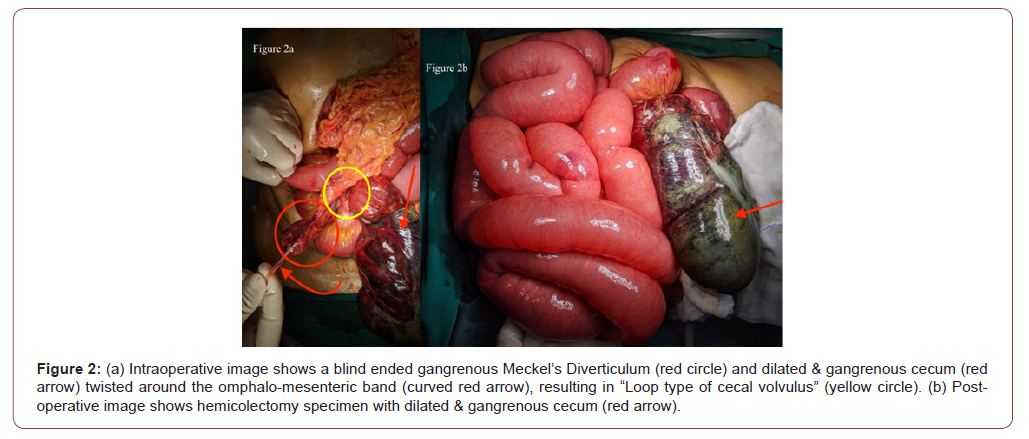
The patient underwent emergency exploratory laparotomy (Figure 2), which revealed over-distended and gangrenous cecum lying in the left hypochondrium. It was twisted around an omphalomesenteric band on a broad-based inflamed MD, resulting in closed loop obstruction. The diverticulum, cecum and ascending colon were resected.
Final Diagnosis
Loop type of Cecal Volvulus caused by Meckel’s diverticulum with an Omphalo-Mesenteric Band, resulting in acute intestinal obstruction and ischemic changes involving cecum, ascending colon, and Meckel’s Diverticulum.
Discussion
MD with or without an omphalomesenteric ligament or fibrous band is the most common omphalomesenteric duct anomaly and most common congenital anomaly of the gastrointestinal tract. It is caused by partial or complete developmental failure to close and absorb the omphalomesenteric duct [2,3,6,7]. Symptomatic MD is often traditionally descried by rule of 2s, that may clinically vary: seen in ~ 2% of the population; often detected 2 feet proximal to the ileocecal valve on the antimesenteric border of terminal ileum; about 2 inches long; approximately two-thirds of cases have ectopic mucosa; two types of mucosa in bleeding diverticulum (native intestinal and ectopic gastric); two types of ectopic mucosa (gastric and pancreatic); two times as likely to be symptomatic in male patients as in female patients; develop complications in 2% of patients; and usually present before the age of 2 years [3].
MD is often asymptomatic, however, ~ 2-4% of patients may be symptomatic and often present with complications including bleeding, torsion, small bowel obstruction, inflammation, and neoplastic changes. Intestinal obstruction is the most common presenting feature of MD in adults, which may be caused by inverted diverticulum, intussusception, volvulus, internal or external herniation, neoplasm, and, rarely, an enterolith [3,8,9]. CV can typically be classified into two types of twists [10]. In the first type, the cecum twists in the axial plane around its long axis, and the cecum lies in the right lower quadrant [11]. In the second type, also known as the “loop type” of cecal volvulus, cecum both twists and inverts, typically lying in the left upper abdominal quadrant [11]. In certain rare entity, known as a “cecal bascule”, the cecum dilates and folds anteriorly, and lies in mid abdomen without any twisting [10].
Although CT has low sensitivity for detection of uncomplicated MD, it may appear as a fluid-filled or air-filled blind-ending pouch that arises from the antimesenteric side of the distal ileum. The CT appearance of MD often varies, depending on the complications. Contrast-enhanced CT of the abdomen is the imaging modality of choice for intestinal obstruction and ileocolic intussusception is the most common cause for obstruction related to MD. Cecal volvulus often has a characteristic appearance at conventional radiography, which may be further confirmed by a contrast enema study or CT [10]. A dilated gas-filled bowel, often located ectopically in the left upper quadrant or mid abdomen, is a classic imaging feature of cecal volvulus, which may be associated with “whirl” sign that is an area of swirling of the bowel and its mesentery [12].
Symptomatic cases should usually be managed by open or laparoscopic diverticulectomy or segmental resection of the small bowel, while incidentally detected asymptomatic MD should be untouched [13]. In conclusion, although MD is the most prevalent congenital anomaly of the gastrointestinal tract, volvulus caused by MD is extremely rare but not unheard, and the patient usually presents with the features of intestinal obstruction. As the diagnosis of MD is often overlooked and the patient usually presents with complications, appropriate knowledge of various pathophysiology of complications must be kept in mind for a prompt and effective management.
Acknowledgements
We appreciate the help of Dr. Drashti, Dr. Niyati, Dr. Akshay, and Dr. Uvaish for their unconditional help while preparing the manuscript.
Conflict of Interest
No conflict of interest.
References
- Turgeon DK, Barnett JL (1990) Meckel's diverticulum. Am J Gastroenterol 85(7): 777-781.
- Levy AD, Hobbs CM (2004) From the archives of the AFIP. Meckel diverticulum: radiologic features with pathologic Correlation. Radiographics 24(2): 565-587.
- Dumper J, Mackenzie S, Mitchell P, Sutherland F, Quan ML, et al. (2006) Complications of Meckel's diverticula in adults. Can J Surg 49(5): 353-357.
- Neidlinger NA, Madan AK, Wright MJ (2001) Meckel's diverticulum causing cecal volvulus. Am Surg 67(1): 41-43.
- Altaf A, Aref H (2014) A case report: Cecal volvulus caused by Meckel's diverticulum. Int J Surg Case Rep 5(12): 1200-1202.
- Elsayes KM, Menias CO, Harvin HJ, Francis IR (2007) Imaging manifestations of Meckel’s diverticulum. AJR Am J Roentgenol 189(1): 81-88.
- Thurley PD, Halliday KE, Somers JM, Al Daraji WI, Ilyas M, et al. (2009) Radiological features of Meckel's diverticulum and its complications. Clin Radiol 64(2): 109-118.
- Lee NK, Kim S, Jeon TY, Kim HS, Kim DH, et al. (2010) Complications of congenital and developmental abnormalities of the gastrointestinal tract in adolescents and adults: evaluation with multimodality imaging. Radiographics 30(6): 1489-1507.
- Chatterjee A, Harmath C, Vendrami CL, Hammond NA, Mittal P, et al. (2017) Reminiscing on Remnants: Imaging of Meckel Diverticulum and Its Complications in Adults. AJR Am J Roentgenol 209(5): W287-W296.
- Peterson CM, Anderson JS, Hara AK, Carenza JW, Menias CO (2009) Volvulus of the gastrointestinal tract: appearances at multimodality imaging. Radiographics 29(5): 1281-1293.
- Moore CJ, Corl FM, Fishman EK (2001) CT of cecal volvulus: unraveling the image. AJR Am J Roentgenol 177(1): 95-98.
- Frank AJ, Goffner LB, Fruauff AA, Losada RA (1993) Cecal volvulus: the CT whirl sign. Abdom Imaging 18(3): 288-289.
- Zani A, Eaton S, Rees CM, Pierro A (2008) Incidentally detected Meckel diverticulum: to resect or not to resect? Ann Surg 247(2): 276-281.
-
Parmar Jitendra P, Patel Tapan C, Desai Chirag J, Shah Sandip R. Cecal Volvulus Caused by an Omphalo-Mesenteric Band: A Case Report of a Rare Complication of Meckel’s Diverticulum. Anaest & Sur Open Access J. 2(3): 2020. ASOAJ.MS.ID.000537.
-
Cecal Volvulus, Meckel’s Diverticulum, Intestinal Obstruction, Omphalo-Mesenteric Duct Anomalies, Abdominal pain, Congenital anomalies, Patient, diagnosis
-

This work is licensed under a Creative Commons Attribution-NonCommercial 4.0 International License.


