


 Research Article
Role of Neutrophil-Lymphocyte Ratio and Platelet-Lymphocyte Ratio in Differential Diagnosis Between Crystal Induced Arthritis and Septic Arthritis
Received Date:March 18, 2024; Published Date:March 26, 2024
Introduction
Globally, the frequency of ED and inpatient visits for crystal induced arthritis including, gout have increased substantially [1- 3]. However, the clinical presentations of gout and pseudogout are sufficiently similar to that of septic arthritis that history and examination cannot reliably differentiate the two conditions [4]. Similarly, peripheral blood tests including white cell count (WCC), C- reactive protein (CRP) and erythrocyte sedimentation rate (ESR) are elevated in both conditions [5] and the serum uric acid level may be normal during an acute gout flare [6]. Microscopy, WCC and culture of aspirated synovial fluid is currently required to fully differentiate between septic arthritis and gout [7,8]. With the frequent delay in obtaining joint culture results and not all joints being easily accessible, synovial fluid results are not always easily achievable.
To further complicate matters, management for crystal induced arthritis and septic arthritis may be mutually exclusive at times due to the need for immunosuppression in severe gout and pseudogout which can exacerbate infection [9]. Delayed treatment of patients with septic arthritis risks at least, joint damage and at worst in severe morbidity or mortality Thus the recommendation that patients receive treatment for suspected septic arthritis pending investigation results [10].
Shifts in platelet, neutrophil and lymphocyte counts are recognized disease severity indicators in rheumatic disease, especially if combined with acute phase reactants [11]. Increased peripheral blood neutrophils and platelets occur as part of the acute phase response to inflammation and infection [12,13], along with and lymphocyte apoptosis [14]. The neutrophil lymphocyte ratio (NLR) is an established marker of some types of systemic inflammation [15]. It has been shown to be significantly elevated in a number of inflammatory conditions including systemic lupus erythematosus (SLE) especially with nephritis [16,17], juvenile arthritis [18], rheumatoid arthritis [19] and sarcoidosis [20]. Likewise, the platelet lymphocyte ratio (PLR) has similarly be a marker of inflammation in SLE [21], rheumatoid [22] and psoriatic arthritis [23]. Both NLR and PLR have previously been shown to be both diagnostic and prognostic indicators of sepsis [24, 25]. No previous data has been used in crystal induced arthritis
A common problem is the difficulty of clinically differentiating septic from crystal induced arthritis. This study set out to investigate whether NLR and PLR derived from peripheral blood sample at presentation were able to differentiate the 2 conditions In addition, machine learning algorithms were utilized to evaluate their diagnostic accuracy against conventional acute phase reactants.
Methods
Study design
The study was performed on data derived from the Austin Hospital data warehouse records from patient visits between January 1st 2011 and June 31st 2019 in Melbourne Australia. The records included all patient characteristics (age, gender), major clinical information (diagnosis, medication) and all investigation results per hospital visit. All investigation results presented are of first set of samples per admission. Patients are included in this study if they have a primary diagnosis of one of septic arthritis, gout or pseudogout according to the International Classification of Diseases, Tenth Revision, Australian Modification (ICD-10-AM) and had an intraarticular joint aspirate confirming the diagnosis. Patients who also had a diagnosis which included as part of that an inflammatory arthritis were excluded. For the purpose of this study, we included both Emergency Department (ED) visits as well as inpatient admissions.
Data for this study was collected retrospectively on visits of patients over the age of 18 and who had consented their information be available for research purposes (i.e. electronic files were not suppressed). All visits recorded in this study contains a primary diagnosis based on ICD-10-AM of septic arthritis, gout, or pseudogout which was confirmed on findings from an intraarticular joint aspirate. Patients with a secondary diagnosis with either any immunodeficiency disease nor another other type of inflammatory arthritis were excluded.
Information on patient age, gender and, results of full blood examination (FBE), inflammatory markers including C-reactive protein (CRP) and erythrocyte sedimentation rate (ESR) were recorded as were the results of any intraarticular aspiration.
Ethics approval was granted by the local ethics committee.
Statistical analysis
Statistical analyses were performed using R Studio version 1.1.442 on Mac OS using packages Tidyverse, caret, random Forest and Optimal Cut points. Descriptive statistics were performed on stratified data based on primary diagnosis. PLR was calculated dividing platelet count by lymphocyte count and NLR dividing neutrophil count by lymphocyte count. Performance characteristics of CRP, ESR, neutrophil count, platelet count, NLR and PLR were analyzed using receiver operating curves (ROC) and respective Youden’s index was calculated. Two individual data sets were created for the purpose of machine learning. The core data set consisting age, gender, platelet count, lymphocyte count, neutrophil count and CRP, the ratio data set which includes NLR and PLR in addition to the core data set. Prior to machine learning both data sets were pre-processed, by scaling and centered to achieve standardized inputs with mean of zero and standard deviation of 1 for all pathology data. Data was randomized and split into training and testing sets using a ratio of 80% training and 20% testing using the R function create Data Partition. The 80% training data with confirmed diagnosis retained is used to train two separate predictive models using random forest algorithm with 10-fold cross validation. Partitioning is identical for both models such that identical participants are used for the two models’ training and testing processes individually.
Each individual model was used to predict the diagnosis of patients on the testing set which has the diagnosis removed. The predicted diagnoses were then compared to the known diagnosis to evaluate the model accuracy which is reported in the form of a confusion matrix.
Results
Descriptive statistics
A total of 446 patients were included in this study, from 455 episodes of admission. The median age of included patients was 76 (IQR 65, 85) and 66% were male. Patient demographics characteristics and biochemical data are outlined in Table 1. Of the 455 episodes, 160 (35.2%) were septic arthritis, 275 (60.4%) were gout and 20 (4.4%) were pseudogout A diagnosis of septic arthritis concurrent with the presence of crystals on joint aspirate occurred in 26 episodes and, these patients were categorised as having septic arthritis Results were available on all patients for joint aspirate culture and microscopy, a CRP and FBE. An ESR result was performed on only 94 patients and a synovial fluid cell analysis 164 samples, the most common reason for a lack of these results latter results being clotting of the synovial fluid.
Table 1:Basic participant clinical information.
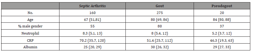
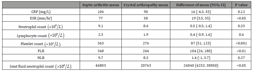
Table 2 outlines compares the means of serum inflammatory markers, joint aspirate neutrophil count and calculated NLR/ PLR between septic arthritis patients and crystal arthropathy. Using a t-test, a significant difference was observed in the mean platelet count, that of the septic arthritis patients being 363 compared with 276 for those with crystal arthropathy (p < 0.001). Similarly, there was a significant difference between the mean PLR of septic group (348) compared with crystal arthropathy (244) (p <0.01). In contrast, no significant differences were observed between the two groups in NLR, CRP and serum lymphocyte nor neutrophil counts Within the synovial fluid, septic arthritis patients did have significantly higher synovial neutrophils (P < 0.05) but not total white cell count thank non septic patients. No correlation was found between white cell level within the synovial fluid and serum.
Table 2:Comparison of t-tests between septic and crystal induced arthritis.
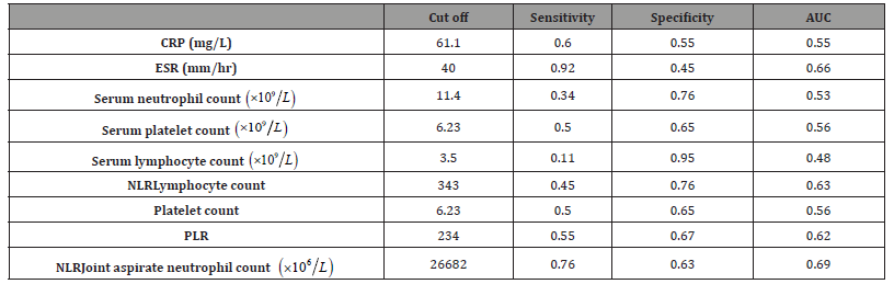
Youden’s index is computed for the serum inflammatory markers and joint aspirate neutrophil count individually. Their respective cut offs, sensitivity, specificity and area under the receiver operating characteristic curve (AUC) are shown in Table 3 Using the jJoint aspirate neutrophil count appeared to have the best overall performance with an AUC of 0.69. Serum neutrophil and platelet counts returned the highest specificity whereas serum ESR returned the highest sensitivity at 0.92. PLR achieved an AUC of 0.62 with sensitivity of 0.55 and specificity of 0.67 while NLR had an AUC of 0.63, sensitivity of 0.45 and specificity of 0.765.
Table 3:Predictive values of serum inflammatory markers and joint aspirate neutrophil count.
Predictive Modelling
The machine learning model created from core data produced a model with prediction accuracy of 66.6%, sensitivity of 34.4% and specificity of 84.5%. The top contributors to the core data model in descending order is platelets count, CRP, neutrophil count and then, lymphocyte count. Model trained from the ratios data set produced a superior predictive model with a prediction accuracy of 75.5% with, sensitivity and specificity of 62.5% and 82.8% respectively. The independent variables which contributed to the model in descending order were platelet count, PLR, NLR and CRP. The addition of NLR and PLR into the decision-making progress improved accuracy by 8.9%, majority of the improvement was in the form of a 28.1% improvement in the sensitivity.
Discussion
Single joint crystal induced arthritis has near identical presentations to septic arthritis and similar investigation findings, however, it is crucial to differentiate the two due to different managements. In this study, we established two novel markers-NLR and PLR, for the differentiating septic arthritis from crystal induced arthritis who presents with mono arthritis.
Significant differences were observed between crystal arthropathy and septic arthritis in traditional inflammatory markers such as serum ESR, serum platelet count, as well as joint fluid neutrophil count in contrast to previous studies, CRP and serum white cells could not be used as a distinguishing factor between crystal induced arthritis and septic arthritis [26,27]. We were unable to apply the diagnostic approaches established from previous studies to safely crystal arthritis from septic arthritis in our cohort [28,29]. Hariharan et all’s finding of ESR >10mm/h and CRP >20mg/L would individually return sensitivity >90% for septic arthritis was not replicated in this current study [30]. We found that serum platelet count is a significant predictor both within and outside the machine learning algorithms related to its role in response to sepsis, while thrombocytosis was previously documented, platelets are not commonly used clinically to identify sepsis [31,32]. These differences are likely secondary to the selection of a more homogenous cohort based on clinical need for intraarticular joint aspirates which is suggestive of the diagnostic dilemma, in contrast to previous studies in which patient selections are purely based on retrospective diagnosis.
In our cohort of patients, all serum markers contributed to the final diagnostic algorithm in distinction between septic arthritis and crystal induced septic arthritis. Elevated NLR and PLR individually are more suggestive than elevation in their respective ratio compositions. Interestingly, NLR and PLR improved diagnostic accuracy and sensitivity in the context of their derived components also included into the prediction algorithm, suggesting that while small, their improvements are additive to the measured serum markers.
The combination model including both NLR and PLR outperformed the standard model by 9%, both ratios were also included within the top 3 predictors.
Given the presence of inflammation in these conditions, traditional acute phase reactants would therefore be unreliable in separating the two diagnoses. The differences in inflammatory drivers lies with IL-6 in sepsis, which is a potent platelet production driver as well as a driver of neutrophil apoptosis [33-36] This may demonstrate a more amplified response in both NLR and PLR in those with septic arthritis than those without and justifies PLR as a more sensitive measure.
The use of random forest a supervised tree-based machine learning algorithm is both novel and an efficient tool in medical research and risk modelling [37,38]. Compared to conventional covariate analysis, random forest is less likely to produce false positives [39]. To our knowledge, this is the first study which utilises machine learning algorithms to establish predictive models in the separation of crystal induced arthritis from septic arthritis patients. Likewise, this is also the first study which evaluates and ranks the contribution of multiple investigation results. By the inclusion of only intraarticular proven disease we sampled the population of patients whom poses the specific clinical dilemma that we sought to differentiate, and thus likely explains the differences in our findings to previous studies.
This study potentially has some limitations. Firstly, the diagnosis accuracy and ranking of the patients relied on accurate transcription by hospital administrators of the synovial fluid pathology findings into the correct ICD-10 coding While we did test the model on remainder 20% of patient data which were involved in model development, future studies prospectively evaluating the performance is pertinent to establish its utility. Lastly, being a tertiary centre, not every participant had serum ESR taken, we did not record information on antimicrobial use or causative organism, but it is likely that most patients would have received treatment prior to joint aspiration.
Conclusion
Our findings suggest that both PLR and NLR, when adapted into a random forest model or used independently, can aid in distinguishing between septic arthritis and crystal induced arthritis. Given the availability of both PLR and NLR, both of which act as acute phase reactants, these findings suggest their elevation favor’s a diagnosis of septic arthritis more so than crystal induced arthritis. Septic arthritis is a major differential diagnosis from crystal induced arthritis, if these markers can be further validated in a larger prospective trial, we may be able to reduce patient morbidity by early diagnosis.
Acknowledgement
None.
Conflict of Interest
No conflict of interest.
References
- Fisher MC, Pillinger MH, Keenan RT (2014) Inpatient gout: a review. Curr Rheumatol Rep 16(11): 458.
- Jinno S, Hasegawa K, Neogi T, Goto T, Dubreuil M (2016) Trends in Emergency Department Visits and Charges for Gout in the United States between 2006 and 2012. J Rheumatol 43(8): 1589-1592.
- Rai SK, Avina Zubieta JA, McCormick N, De Vera MA, Lacaille D, et al. (2017) Trends in Gout and Rheumatoid Arthritis Hospitalizations in Canada from 2000 to 2011. Arthritis Care Res (Hoboken) 69(5): 758-762.
- Visser S, Tupper J (2009) Septic until proven otherwise: approach to and treatment of the septic joint in adult patients. Can Fam Physician 55(4): 374-375.
- Roseff R, Wohlgethan JR, Sipe JD, Canoso JJ (1987) The acute phase response in gout. J Rheumatol 14(5): 974-977.
- Urano W, Yamanaka H, Tsutani H, Nakajima H, Matsuda Y, et al. (2002) The inflammatory process in the mechanism of decreased serum uric acid concentrations during acute gouty arthritis. J Rheumatol 29(9): 1950-1953.
- Margaretten ME, Kohlwes J, Moore D, Bent S (2007) Does this adult patient have septic arthritis? JAMA 297(13):1478-1488.
- Ma L, Cranney A, Holroyd-Leduc JM (2009) Acute monoarthritis: what is the cause of my patient's painful swollen joint? CMAJ 180(1): 59-65.
- Burns CM, Wortmann RL (2012) Latest evidence on gout management: what the clinician needs to know. Ther Adv Chronic Dis 3(6): 271-286.
- Suresh E (2005) Diagnosis and management of gout: a rational approach. Postgrad Med J 81(959): 572-579.
- Gasparyan AY, Ayvazyan L, Mukanova U, Yessirkepov M, Kitas GD (2019) The Platelet-to-Lymphocyte Ratio as an Inflammatory Marker in Rheumatic Diseases. Ann Lab Med 39(4): 345-357.
- Lerman YV, Kim M (2015) Neutrophil migration under normal and sepsis conditions. Cardiovasc Hematol Disord Drug Targets 15(1): 19-28.
- Nurden AT (2011) Platelets, inflammation and tissue regeneration. Thromb Haemost Suppl 1: S13-33.
- Wesche DE, Lomas Neira JL, Perl M, Chung CS, Ayala A (2005) Leukocyte apoptosis and its significance in sepsis and shock. J Leukoc Biol 78(2): 325-337.
- Imtiaz F, Shafique K, Mirza SS, Ayoob Z, Vart P, et al. (2012) Neutrophil lymphocyte ratio as a measure of systemic inflammation in prevalent chronic diseases in Asian population. Int Arch Med 5(1): 2.
- Li L, Xia Y, Chen C, Cheng P, Peng C (2015) Neutrophil-lymphocyte ratio in systemic lupus erythematosus disease: a retrospective study. Int J Clin Exp Med 8(7): 11026-11031.
- Oehadian A, Suryadinata H, Dewi S, Pramudyo R, Alisjahbana B (2013) The role of neutrophyl lymphocyte count ratio as an inflammatory marker in systemic lupus erythematosus. Acta Med Indones 45(3): 170-174.
- Gunes A, Ece A, Sen V, Uluca U, Aktar F, et al. (2015) Correlation of mean platelet volume, neutrophil-to-lymphocyte ratio, and disease activity in children with juvenile idiopathic arthritis. Int J Clin Exp Med 8(7): 11337-11341.
- Fu H, Qin B, Hu Z, Ma N, Yang M, et al. (2015) Neutrophil- and platelet-to-lymphocyte ratios are correlated with disease activity in rheumatoid arthritis. Clin Lab 61(3-4): 269-273.
- Gungor S, Akturk UA, Yalcinsoy M, Kocak ND, Goksenoglu NC, et al. (2016) What is the neutrophil/lymphocyte ratio in sarcoidosis? Bratisl Lek Listy 117(3): 152-155.
- Qin B, Ma N, Tang Q, Wei T, Yang M, et al. (2016) Neutrophil to lymphocyte ratio (NLR) and platelet to lymphocyte ratio (PLR) were useful markers in assessment of inflammatory response and disease activity in SLE patients. Mod Rheumatol 26(3): 372-376.
- Peng YF, Cao L, Zeng YH, Zhang ZX, Chen D, et al. (2015) Platelet to lymphocyte ratio and neutrophil to lymphocyte ratio in patients with rheumatoid arthritis. Open Med (Wars) 10(1): 249-253.
- Kim DS, Shin D, Lee MS, Kim HJ, Kim DY, et al. (2016) Assessments of neutrophil to lymphocyte ratio and platelet to lymphocyte ratio in Korean patients with psoriasis vulgaris and psoriatic arthritis. J Dermatol 43(3): 305-310.
- Kaushik R, Gupta M, Sharma M, Jash D, Jain N, et al. (2018) Diagnostic and Prognostic Role of Neutrophil-to-Lymphocyte Ratio in Early and Late Phase of Sepsis. Indian J Crit Care Med 22(9): 660-663.
- Shen Y, Huang X, Zhang W (2019) Platelet-to-lymphocyte ratio as a prognostic predictor of mortality for sepsis: interaction effect with disease severity-a retrospective study. BMJ Open 9(1): e022896.
- Soderquist B, Jones I, Fredlund H, Vikerfors T (1998) Bacterial or crystal-associated arthritis? Discriminating ability of serum inflammatory markers. Scand J Infect Dis 30(6): 591-596.
- Tagoe CE, Raza Y (2013) Differences in Acute Phase Reactants between Gout and Pseudogout. International Journal of Clinical Medicine 4(12): 13-19.
- Genes N, Chisolm-Straker M (2012) Monoarticular arthritis update: Current evidence for diagnosis and treatment in the emergency department. Emerg Med Pract 14(5): 1-19; quiz -20.
- Janssens HJ, Fransen J, Van de Lisdonk EH, Van Riel PL, Van Weel C, et al. (2010) A diagnostic rule for acute gouty arthritis in primary care without joint fluid analysis. Arch Intern Med 170(13): 1120-1126.
- Hariharan P, Kabrhel C (2011) Sensitivity of erythrocyte sedimentation rate and C-reactive protein for the exclusion of septic arthritis in emergency department patients. J Emerg Med 40(4): 428-431.
- Guclu E, Durmaz Y, Karabay O (1994) Effect of severe sepsis on platelet count and their indices. Afr Health Sci 13(2): 333-338.
- Yohannan MD, Higgy KE, Al-Mashhadani SA, Santhosh-Kumar CR (1994) Thrombocytosis. Etiologic analysis of 663 patients. Clin Pediatr (Phila) 33(6): 340-343.
- Busso N, Ea HK (2012) The mechanisms of inflammation in gout and pseudogout (CPP-induced arthritis). Reumatismo 63(4): 230-237.
- Tissi L, Puliti M, Barluzzi R, Orefici G, Von Hunolstein C, et al. (1999) Role of tumor necrosis factor alpha, interleukin-1beta, and interleukin-6 in a mouse model of group B streptococcal arthritis. Infect Immun 67(9): 4545-4550.
- Ishibashi T, Kimura H, Shikama Y, Uchida T, Kariyone S, et al. (1989) Interleukin-6 is a potent thrombopoietic factor in vivo in mice. Blood 74(4): 1241-1244.
- Asensi V, Valle E, Meana A, Fierer J, Celada A, et al. (2004) In vivo interleukin-6 protects neutrophils from apoptosis in osteomyelitis. Infect Immun 72(7): 3823-3828.
- Hashizume M, Higuchi Y, Uchiyama Y, Mihara M (2011) IL-6 plays an essential role in neutrophilia under inflammation. Cytokine 54(1): 92-99.
- Breiman L (2001) Random Forests. Machine Learning 45(1): 5-32.
- Weissler EH, Naumann T, Andersson T, Ranganath R, Elemento O, et al. (2021) Correction to: The role of machine learning in) clinical research: transforming the future of evidence generation. Trials 22(1): 593.
- Rigdon J, Baiocchi M, Basu S (2018) Preventing false discovery of heterogeneous treatment effect subgroups in randomized trials. Trials 19(1): 382.
-

This work is licensed under a Creative Commons Attribution-NonCommercial 4.0 International License.