

 Research Article
Muscles and their Osteoarthritis Pathogenic Linkages 2024
Received Date:July 29, 2024; Published Date:August 05, 2024
Abstract
Osteoarthritis, the most prevalent musculoskeletal disease remains relatively impervious to a desired reversal of its progressive impact on mobility and life quality. Moreover, even if available, standard treatments may not be indicated, or efficacious, or safe and commonly fail to impact the disease directly. A disease with multiple progressively degenerating physical manifestations including joint tissue damage, inflammation, and muscle pathology, more needs to be done to alleviate its prevalence and public health burden. Building on prior reviews capturing almost all data on this topic prior to 2020, here we present some newer data on this topic published since then that furthers the view that interventions harnessed to mediate, moderate, or even prevent muscle dysfunction may have a bearing on osteoarthritis joint damage and life quality and health costs. Its vast protective and interactive attributes with joint and brain tissues show sufficient potential in the context of osteoarthritis pain and inflammation, as well as joint structure and cartilage physiology that may be of paramount importance, but commonly overlooked, with dire consequences.
Keywords:Aging; Disability; Joint; Intervention; Muscle; Osteoarthritis; Pain; Prevention
Introduction
It is apparent that osteoarthritis, the most common joint disease, and one which disables many older adults in all countries has been well researched for more than a century, but to no conclusive avail as to its origins and to the application of precision medicine to any meaningful degree. Often only diagnosed in its later disease stages, it is accepted that the condition commonly induces considerable pain, stiffness, perpetual discomfort, a declining capacity for self-care and functions of daily living, and while some form of medical management or medical therapies may help to mitigate the condition to some degree, osteoarthritis is generally deemed irreversible and progressive and is observed to raise the risk for all-cause mortality in the older population [1]. Most commonly attributed to age and injury associated linkages there is a limited or accepted universal understanding of the complex disease mechanisms of osteoarthritis and their temporal trajectories, and why some interventions fail, while others appear somewhat efficacious, such as exercise [2,3].
The problem here may however stem from the limited conceptual model of the disease and its common characterization as a disease of articular cartilage origins, plus problems in extrapolating laboratory studies that employ invasive or inflicted joint pathology in selected animal models to simulate those that emerge without any visible cause in the clinical realm. Additionally, in clinical studies, failing to ensure control subjects are indeed ‘disease free’ with comparable health status, and gender distributions prevents the acceptance of either observed differences or group similarities. Additionally, the use of multiple subjective measures, rather than singular objective muscle and kinetic oriented measures with well-established measurement properties renders disease determinant data and its interpretation highly challenging at best. Moreover, even though more recent acceptance of osteoarthritis as a disease encompassing all joint tissues, such as the joint capsule, joint ligaments, tendons, and synovial membrane lining, and that includes extensive focal as well as systemic mobility changes [4], including those associated with muscles, most articles still refer to the articular cartilage lining of freely moving joints such as that at the knee as the key tissue that fails over time and is impervious to intervention because once diseased the tissue inevitably degenerates. At the same time, several studies that focus on the molecular changes in osteoarthritis and/ or possible disease modifying drugs to counter cartilage pathology [5] do not appear to embrace any clear role for any form of muscular antecedent or mediating factor in the disease cycle.
Since the body of research directed towards the search for a viable form of osteoarthritis mitigation is currently limited in this regard, we propose it may be useful to examine the idea that osteoarthritis may stem in some cases from the prolonged interaction of subnormal reflexive interactions between muscles that may be infiltrated by fat or other non-contractile components [6] and their reduced strength and adverse joint based tissue mechanical and physical stresses and strains. This idea is not new, but an approach that accepts, rather than ignores, the ability of the human motor and skeletal systems to adapt to biological conditions both negative and positive, such as deficient, subnormal or excessive joint load and/or associated muscle or soft tissue impacts or impairments that can induce a state of limb disuse and a possible host of progressive negative alterations in bone and articular cartilage tissues [7,8]. It was hence felt an updated review and one that acknowledges the close links between form and function, injury, and mechanical loading effects might lend some needed insight into the disease origins and its variations.
Muscle was the focus of this discourse because i) it is possible the focal distribution of osteoarthritis may be mediated by muscle mechanisms, ii) muscle starts to undergo structural age associated attritional changes quite uniformly, iii) treating muscle appears to be one aspect of osteoarthritis that appears respond to its practice and is a low-cost factor that can generally be safely examined, measured, intervened upon, and improved.
Aims
To extend what is known in this regard, this current report aimed to specifically examine if the idea that some forms of osteoarthritis may represent an adverse adaptive response of one or more joint tissues to one or more insults, in the face of deficient or unrelenting subnormal reactive and protective muscle mechanisms this report aimed to summarize what trends exist in this regard. Indirectly, it strove to provide support for the idea that carefully tailored interventions designed to improve one or more aspects of muscle function may set the stage for joint healing or disease retardation and with this more favorable rather than adverse outcomes.
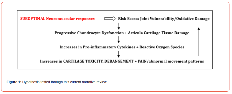
Tested in this respect is the idea that osteoarthritis and its numerous molecular and biochemically deranged compositional and metabolic features may have emerged as a structural-functional adaptation or a temporal response to a variety of cascading extrinsic as well as intrinsic injurious stimuli that may manifest more readily in an older person than a younger person and that may induce perpetual and increasing cortical, sub-cortical, spinal segmental, muscle, and joint level atypical functional and structural impacts as well as articular surface damage and motor control strategies (see Figure 1)
Methods
To achieve these abovementioned review aims, we elected to build on an extensive review of available documents housed in PUBMED, PubMed Central, and Google Scholar from 1990 up until 2019 using the key terms Osteoarthritis/Muscle/Muscle Dysfunction/Muscle Impairment/Muscle Pathology. More specifically, an updated overview of related research published between January 1 2019-July 25 2024 was sought.
To this end, all available articles on these websites were scanned for relevance, and salient research articles or reviews that addressed some aspect of the current topic of interest were then reviewed in more depth without regard to research design. An attempt was made to include all modes of experimentation, but the focus was on clinically derived data. No systematic review was conducted, and while it is acknowledged the body of data may not be exhaustive-it does arguably highlight some telling lines of research and tentative conclusions using three reputable data bases, taken to presumably house and capture the state of the art and gold standard papers on this topic sufficient for arriving at a reasoned opinion through a narrative lens. Readers can learn more by exploring the current cited reports as well as some prior reports, for example Chen et al. [9], De Ceuninck et al. [10], Suter et al. [11] and Shiavi et al. [12].
Results
General observations
Among the literature currently reviewed, it is evident many studies published in recent years have discussed the epidemiology and costs of osteoarthritis to society. Most however, fail to offer any definitive explanations as to why osteoarthritis is increasing in prevalence and why few efficacious approaches for mitigating this widespread disease to any population have been demonstrated to date. No longer considered a disease of articular cartilage alone, osteoarthritis, which induces or is induced by abnormal bone remodeling processes and some degree of joint swelling and joint inflammation [13] its link to muscle as a causative agent is commonly less well accepted. This is surprising because several early studies link osteoarthritis to induced injury related muscle mechanisms [e.g. Herzog], while sarcopenia, described as a pathological state of muscle weakness associated with altered muscle mass, strength, and function is also a prevalent factor in the older adult with osteoarthritis that may be fostering its progression.
In addition, increasing evidence strongly implies that in addition to declines in lower limb muscle strength associated with knee or hip osteoarthritis, pain, altered joint stability, maladapted postures, excess force production, and defective neuromuscular transmission and reception processes might play a role, as observed by Herzog [14]. In addition, it appears cellular and molecular level changes in cartilage chondrocytes as well as muscle myoblasts share a common set of pathological targets and pathways that could explain their impact on osteoarthritis risk [10-15].
In particular, the presence of chronic problems within the neuromuscular system may provoke cartilage, bone, synovial membrane and capsular changes, plus the production of inflammatory cytokines [16] subnormal limb kinetics and kinematics [17] and various muscle structural alterations that are hard to reverse [18]. In other cases, alterations in one or more of the highly complex networks of sensory nerves that normally serve to coordinate joint mobility and stability appear to have the potential to undermine joint biomechanics, limb posture, stability, and mobility [19,20], as do muscle inflammation, muscle fiber degeneration, muscle blood flow alterations and joint effusion [21,22].
Mansfield et al. [23] found individuals with osteoarthritis of the knee to not only have motor deficits associated with decreased neural activation and central nervous system sensitization, but decreases in quadriceps muscle spindle responsiveness, along with increased calf muscle activity. Ling et al. [24] implicate muscle activation changes at the motor unit level in the context of symptomatic knee osteoarthritis that is mediated by radiological disease severity. Recently Schaefer et al. [25] reported strength deficits of the hamstrings in patients with knee osteoarthritis wherein the more affected side seemed to show further specific impairments regarding neuromuscular control. This research is fairly novel and as indicated recently by Lopes et al. [26] should not be ignored as there may be more to the pathology of knee osteoarthritis and its muscular associations than is widely acknowledged, such as possible deficits in muscle rate of force development [27].
Other observations
In addition to the aforementioned muscle associations studied in the context of the osteoarthritis disease cycle – it appears muscle weakness or wasting or both found in osteoarthritis may also serve as possible disease causative or progression factors [11,28]. Moreover, a role for reflex muscle inhibition and poor muscle activation patterns consequent to joint inflammation as well as joint destruction can be considered as probable and serious osteoarthritis pathogenic correlates, regardless of whether these are reactive or causal [24]. Pain and related immobility responses that promote muscle disuse atrophy and weakness, poor muscle quality, muscle fat infiltration, and increased muscle stiffness are additional muscle related pathogenic mediators or moderators [29] as are alterations in muscle architecture and morphology plus muscle volume losses [30-32] and muscle inflammation [33].
Other reports imply muscle fatigue/poor muscle endurance may be expected to foster rather than retard damaging bouts of joint loading and load distribution and strain [34]. Similarly, a general or selective loss of muscle-based enzyme production and functional losses [35] may have an independent or cumulative bearing on joint loading deficiencies and aberrations. In particular, decreases in timely as well as appropriately modulated voluntary activation efficiencies, may hasten its progression quite markedly and significantly [34,36,37] as may muscle weakness, inefficient bouts of muscle force production, alterations in muscle growth differentiation factor, abnormal movement synergies, muscle atrophy and disuse, and a proportionate change in non-contractile muscle tissue presence [35,38-44]. Other recent examples of muscle joint interactions that may play a pivotal role in cartilage damage and irreversible alterations of articular cartilage biochemistry plus variable responses to joint damage and surgery are: muscle-based circuits associated with protein degradation and fibro-adipogenic cell gene expression, muscle inflammation, and altered gene muscle dysfunction [45]. In addition, differential muscle activation patterns, and excess or suboptimal joint stiffness may mediate osteoarthritis progression and severity [32,46-50]. Other factors that can play a unique or additive role in osteoarthritis progression are various degrees or combinations of muscle atrophy, muscle pathology, and muscle stiffness [51-56].
In short, even if negative studies prevail, as outlined above and generally consistently for more than 30 years, it appears joint status at any age may be degraded progressively in some cases as a result of preexisting and/or emergent muscle deficiencies or abnormalities that impact the efficiency of static or dynamic joint loading efforts. These include, but are not limited to a) Those residing in the central, spinal, and peripheral nervous systems; b) Those due to alterations in muscle contractile properties, muscle quality, muscle fat content, muscle transcription factors, and selective muscle fiber losses, rearrangement, derangement, or mal distribution; c) Those due to persistent abnormalities in muscle reflex activity and force production, muscle agonist/antagonist imbalances and contractures, uncoordinated and muscle force timing and directional changes; d) Those due to muscle inhibition, inflammation, impaired muscle metabolism, and microcirculation; e) Those due to muscle pain, muscle injury, and subnormal muscle afferent and efferent transmission and reception deficits; f) Those due to muscle disuse, abnormal motor unit activation, muscle fatigue, muscle co contraction, and uncoordinated movement patterns, and their impact on joint, bone, and surrounding supportive tissues; g) Those due to unresolved or poorly treated cardiovascular conditions and obesity; h) Those due to poor nutrition and vitamin D intake or exposure.
On the other hand, and accepting this work is not all inclusive, or a critically oriented examination of the papers currently reviewed, the majority that represent the key peer reviewed studies and comments published in English in 2024, point to the likelihood of one or more muscular abnormalities having a clinically significant role in osteoarthritis manifestations among older adult populations as per Figure 1 and regardless of research approach or issue examined. Indeed, while many other factors may be involved, when viewed collectively, most current authors as well as those that have led this line of research study indicate this muscle is a highly promising osteoarthritis topic to pursue as well a possible insightful one and hence advocate for more careful well controlled longitudinal studies in this regard in the future. Even if the idea is eventually refuted, a well-designed series of comparative pre-clinical and clinical studies, can undoubtedly help reveal how osteoarthritis emerges in some and not others rather than jumping to well-trodden and repeated conclusions and can thus potentially help in framing more targeted efforts to mitigate the disease prevalence and/or its severity in the older adult population more readily and optimally than not. As such, the number of current calls for more insights into the present role of muscle as revealed is clearly not simply an academic one, but is urged because it will likely have the potential to reduce suffering and the immense costly osteoarthritis burden over time and is reaping some clinically beneficial applications and insights even at this time of any solid conclusions.
Discussion
Despite over a century of study, osteoarthritis, the most common form of arthritis, and an immense disabler of older adults continues to elude clinicians as well as scientists when considering its origins, why it is frequently progressive, and what remediation or mitigation endeavors are likely to prove most beneficial as well as cost effective and safe. We thus asked ourselves whether this is because osteoarthritis is inevitable or is it a predictable response occurring over a protracted period as a consequence of a limited ability to reduce its cellular responses to repetitive loads that exceed the joint tissues protective threshold and the ability to withstand these or due to differences in the way osteoarthritis is studied. Since most current standard remedies fail to reverse or mitigate the condition and many approaches do not commonly consider the structural features found at the affected joint other than cartilage, or their sensitivity to subnormal joint loads and stresses, can a role for neuromuscular factors be more firmly established as an explanatory as well as a significant osteoarthritis mediating determinant that can be applied to enhance more favorable longterm outcomes for many? The rationale for this aforementioned line of thought, while not conclusive, has been and is currently supported to a modest degree in our view by varied evidence that has emerged of multiple reflexive oriented and other underlying or emergent adaptations of the motor system that appear linked to the disease progression or its persistence, and severity. However, since the force generated by muscle is attributable to muscle cross sectional areas and level of motor unit activation, if muscle abnormalities remain unaddressed or overlooked in the clinical realm as well as the research realm, it may be challenging to fully comprehend the nature of osteoarthritis pathology and its required treatment and especially optimal treatment.
In particular, if there is an influential osteoarthritis-muscle linkage, and this is overlooked, possible adverse compensatory muscle mechanisms may arise that may collectively advance joint destruction rather than joint protection. At the same time, the presence of undetected and untreated muscle weakness, including sarcopenia and/or excessive muscle fat infiltration processes a perpetual state of joint vulnerability and dysfunction may well ensue [6,41,57]. Associated preexisting or progressive sensory input impairments and strength may further explain the cycle of joint instability, and kinetics often reported in osteoarthritis cases [38,58-60].
According to Li et al. [61], until more is known in this realm, researchers and clinicians should be encouraged to keep an open mind as to the potential significance of one or more motor alterations that could have a bearing on osteoarthritis degradation processes and its seeming inability to regress or stabilize readily, but that are not commonly examined or identified in the clinic or research realms. Others imply that a thoughtful combination of muscle related variables assessed routinely and with more contemporary tools, might enable a more profound understanding of the disease in the individual and how to foster more normal biomechanical, energy saving and osteoarthritis kinetic and functional responses that do not provoke inflammation, distress, and pain during or following joint loading. Establishing the relative importance of muscle fiber pathology or loss, muscle fat mass, muscle inflammation [44,62], effusion and deficient muscle circulation on muscle growth differentiation and functional performance and weakness [43] could also probably help in explaining the considerable variance in osteoarthritis disease manifestations and that could have multiple clinically important disease associated treatment and mitigation implications [63-67]. In this regard it appears paramount to consider a role for muscle composition [46] and prevailing muscle stiffness [42] in fostering subnormal movement and stability functions that may well contribute inadvertently to the osteoarthritis pain cycle, and its progressive failure to attenuate excess or day to day joint impacts if ignored [68-70]. Since muscle is mutable, and can adapt to its cumulative sensorimotor inputs, allied efforts to establish if treating one or more muscle abnormalities where present, can yield favorable muscle, plus joint structural and functional adaptations, and possibly thereby impact cartilage maintenance or regeneration plus pain favorably is advocated as well.
In the interim, while the important factors contributing to osteoarthritis clearly require more extensive insightful study, and description, many older adults may currently be suffering intently from osteoarthritis muscle correlates that can actually be remediated. Indeed, a high number of treatable cases may be resorting to unsafe remedies or narcotics to relieve their pain and avoid exercise, and may hence die prematurely or encounter worse functional outcomes than not if their treatment personnel fail to measure, identify and intervene upon one or more muscle factors that may be reversible or mutable. Moreover, it is possible multiple muscle alterations predate or appear to prevail in the context of osteoarthritis and that influence functional performance and if unaddressed or not addressed in a timely manner may explain a) the challenges in reducing this disease burden as time progresses, b) why not all older adults are affected by this disease, and c) why one area of the body and not others is commonly affected. As well, it may explain the spread of the disease to other joints over time, and if possibly already present before surgery, may yet remain persistently inefficient or possibly worsened after surgery.
Conclusion
Despite disagreements over the actual initiating factors underpinning osteoarthritis, it is evident that a role for neuromuscular factors in the osteoarthritis disease cycle cannot be readily dismissed.
It is further concluded it is vital to examine muscle attributes alongside functional, biochemical, and radiologic attributes in order to arrive at sound disease associated understandings concerning its diverse manifestations and treatment needs.
On the other hand, a failure to consider the importance of remediable factors such as muscle imbalances, muscle contractile delays, as well as muscle weakness, and poor endurance, among other muscular related problems common in aging and osteoarthritis, one can anticipate multiple adverse structural adaptations of the bone and joint tissues as well functional performance declines.
In particular, since the synthetic and degradation processes of normal articular cartilage are maintained, in part, by intermittent loading imparted to the articular cartilage chondrocyte by muscle, and impact loads are best absorbed by muscles that are not fatigued and that work efficiently every effort to enhance optimal muscle function and its properties and biochemistry in the affected older adult whether or not they have been diagnosed as having osteoarthritis or not is strongly advocated.
Finally, since muscle function and structure decline quite significantly with age, concerted efforts to keep aging adults active and educated about joint safety and protection, and the dangers of exercise adoption without expert consultation, especially in the sarcopenic older adult appears imperative.
In the meantime, to render current clinical decisions that can
help avert the immense burden suffered by many older adults with
osteoarthritis, current practitioners who take the time to reflect
on the following evidence-based items regarding what we know
of muscle contributes to structural and functional joint changes
and direct their skills accordingly are likely to do immense ‘good’
rather than not on behalf of their clients. Patients who understand
why they need to be active treatment partners as well as active
may experience a higher rather than lower quality of life, as well
as lower health needs and costs. Researchers can help immensely
in this regard by validating and expanding upon the degree to
which the issues evidenced to date and listed below are of clinical
relevance to the abatement of osteoarthritis disability.
• Defective muscle transcription and functional problems
• Muscle fiber and muscle architectural damage
• Muscle stiffness and decreased shock absorption
• Muscle ischemia, quality changes and fat mass functions
• Muscle spasm, contractures, inflammation and volume
changes
• Subnormal muscle spindle responses, endurance capacity,
and muscle weakness
• Sarcopenia and muscle injury
• Muscle asymmetry and imbalances
It may also be that abnormal inputs from muscle might combine with abnormal joint inputs referring pain to other sites. This can perhaps explain the presence of widespread discomfort and a generally observed unwillingness on the part of the osteoarthritis patient to exercise consistently, and which might provoke further cartilage destruction and pain. In addition, careful early intervention is strongly advocated because abnormal or awkward compensatory movement the patient may use to avert pain on one joint may place loads on others that cause further pain.
In essence, we conclude much can be done to advance our understandings and capacity to reduce osteoarthritis suffering wherever it exists, and clinicians and researchers who examine the role of motor systems in osteoarthritis realms will likely enable many older adults to age ‘in place’, and are hence encouraged to do so sooner rather than later.
Acknowledgement
None.
Conflict of Interest
No conflict of Interest.
References
- Palazzo C, Nguyen C, Lefevre-Colau MM, Rannou F, Poiraudeau S (2016) Risk factors and burden of osteoarthritis. Ann Phys Rehabil Med 59(3): 134-138.
- Dantas GAF, Sacco ICN, Ferrari AV, Matias AB, Watari R, et al. (2023) Effects of a foot-ankle muscle strengthening program on pain and function in individuals with knee osteoarthritis: a randomized controlled trial. Braz J Phys Ther 27(4): 100531.
- Karimi S, Derakhshan M, Tondro A (2024) The effect of aquatherapy on pain intensity and performance in patients with osteoarthritis. J Family Med Prim Care 13(5): 1793-1796.
- Abramoff B, Caldera FE (2020) Osteoarthritis: pathology, diagnosis, and treatment options. Med Clin North Am 104(2): 293-311.
- Xia B, Di Chen, Zhang J, Hu S, Jin H, et al. (2014) Osteoarthritis pathogenesis: a review of molecular mechanisms. Calcif Tissue Int 95(6): 495-505.
- Rasch A, Byström AH, Dalen N, Berg HE (2007) Reduced muscle radiological density, cross-sectional area, and strength of major hip and knee muscles in 22 patients with hip osteoarthritis. Acta Orthop 78(4): 505-510.
- Cigercioglu NB, Bazancir-Apaydin Z, Apaydin H, Baltaci G, Guney-Deniz H (2024) Differences in ankle and knee muscle architecture and plantar pressure distribution among women with knee osteoarthritis. J Foot Ankle Res 17(2): e12028.
- Ma X, Zhang K, Ma C, Zhang Y, Ma J (2024) Physical activity and the osteoarthritis of the knee: A Mendelian randomization study. Medicine (Baltimore) 103(26): e38650.
- Chen CT, Burton-Wurster N, Lust G, Bank RA, Tekoppele JM (1999) Compositional and metabolic changes in damaged cartilage are peak-stress, stress-rate, and loading-duration dependent. J Orthop Res 17(6): 870-879.
- De Ceuninck F, Fradin A, Pastoureau P (2014) Bearing arms against osteoarthritis and sarcopenia: when cartilage and skeletal muscle find common interest in talking together. Drug Discov Today 19(3): 305-311.
- Shiavi R, Zhang LQ, Limbird T, Edmondstone MA (1992) Pattern analysis of electromyographic linear envelopes exhibited by subjects with uninjured and injured knees during free and fast speed walking. J Orthop Res 10(2): 226-236.
- Suter E, Herzog W, Leonard TR, Nguyen H (1998) One-year changes in hind limb kinematics, ground reaction forces and knee stability in an experimental model of osteoarthritis. J Biomech 31(6): 511-517.
- Gong Z, Li S, Cao P, Ruan G, Zhang Y, et al. (2023) The association between quadriceps strength and synovitis in knee osteoarthritis: an exploratory study from the Osteoarthritis Initiative. J Rheumatol 50(4): 548-555.
- Herzog W, Longino D, Clark A (2003) The role of muscles in joint adaptation and degeneration. Langenbecks Arch Surg 388(5): 305-315.
- Jeong YG, Jeong YJ, Koo JW (2016) Effect of chronic knee osteoarthritis on flexion-relaxation phenomenon of the erector spinae in elderly females. J Phys Ther Sci 28(7): 1964-1967.
- Mathiessen A, Conaghan PG (2017) Synovitis in osteoarthritis: current understanding with therapeutic implications. Arthritis Res Ther 19(1): 18.
- Carvalho C, De Oliveira MPB, Pisani GK, Marolde IB, Serrão PRMDS (2022) Biomechanical characteristics and muscle function in individuals with patellofemoral osteoarthritis: a systematic review of cross-sectional studies. Clin Biomech (Bristol, Avon) 98: 105721.
- Siqueira MS, Souto LR, Martinez AF, Serrão FV, De Noronha M (2022) Muscle activation, strength, and volume in people with patellofemoral osteoarthritis: a systematic review and meta-analysis. Osteoarthritis Cartilage 30(7): 935-944.
- Cardoneanu A, Macovei LA, Burlui AM, Mihai IR, Bratoiu I, et al. (2022) Temporomandibular joint osteoarthritis: pathogenic mechanisms involving the cartilage and subchondral bone, and potential therapeutic strategies for joint regeneration. Int J Mol Sci 24(1): 171.
- Li W, Gong Y, Liu J, Guo Y, Tang H, et al. (2021) Peripheral and central pathological mechanisms of chronic low back pain: a narrative review. J Pain Res 14: 1483-1494.
- Bandak E, Boesen M, Bliddal H, Riis RG, Gudbergsen H, et al. (2015) Associations between muscle perfusion and symptoms in knee osteoarthritis: a cross-sectional study. Osteoarthritis Cartilage 23(10): 1721-1727.
- Kim JR, Pham THN, Kim WU, Kim HA (2023) A causative role for periarticular skeletal muscle weakness in the progression of joint damage and pain in OA. Sci Rep 13(1): 21349.
- Mansfield CJ, Culiver A, Briggs M, Schmitt LC, Grooms DR, et al. (2022) The effects of knee osteoarthritis on neural activity during a motor task: a scoping systematic review. Gait Posture 96: 221-235.
- Ling SM, Conwit RA, Talbot L, Shermack M, Wood JE, et al. (2007) Electromyographic patterns suggest changes in motor unit physiology associated with early osteoarthritis of the knee. Osteoarthritis Cartilage 15(10): 1134-1140.
- Schaefer LV, Dech S, Carnarius F, Rönnert F, Bittmann FN, et al. (2024) Adaptive force of hamstring muscles is reduced in patients with knee osteoarthritis compared to asymptomatic controls. BMC Musculoskelet Disord 25(1): 34.
- Lopes HS, Waiteman MC, Priore LB, Glaviano NR, Bazett-Jones DM, et al. (2024) There is more to the knee joint than just the quadriceps: A systematic review with meta-analysis and evidence gap map of hamstring strength, flexibility, and morphology in individuals with gradual-onset knee disorders. J Sport Health Sci 13(4): 521-536.
- Suzuki Y, Iijima H, Nakamura M, Aoyama T (2022) Rate of force development in the quadriceps of individuals with severe knee osteoarthritis: A preliminary cross-sectional study. PLoS One 17(1): e0262508.
- Shorter E, Sannicandro AJ, Poulet B, Goljanek-Whysall K (2019) Skeletal muscle wasting and its relationship with osteoarthritis: a mini-review of mechanisms and current interventions. Curr Rheumatol Rep 21(8): 40.
- Chopp-Hurley JN, Wiebenga EG, Bulbrook BD, Keir PJ, Maly MR (2020) Evaluating the relationship between quadriceps muscle quality captured using ultrasound with clinical severity in women with knee osteoarthritis. Clin Biomech (Bristol, Avon) 80: 105165.
- Karapınar M, Ayyıldız VA, Unal M, Fırat T (2024) Effect of intramuscular fat in the thigh muscles on muscle architecture and physical performance in the middle-aged women with knee osteoarthritis. J Orthop Sci 29(1): 194-199.
- Mohajer B, Moradi K, Guermazi A, Dolatshahi M, Zikria B, et al. (2023) Diabetes-associated thigh muscle degeneration mediates knee osteoarthritis-related outcomes: results from a longitudinal cohort study. Eur Radiol 33(1): 595-605.
- Li Z, Leung KL, Huang C, Huang X, Su S, et al. (2024) Associations amongst dynamic knee stiffness during gait, quadriceps stiffness, and the incidence of knee osteoarthritis over 24 months: a cohort study with a mediation analysis. BMC Musculoskelet Disord 25(1): 511.
- Sanchez-Ramirez DC, Van der Leeden M, Van der Esch M, Gerritsen M, Roorda LD, et al. (2013) Association of serum C-reactive protein and erythrocyte sedimentation rate with muscle strength in patients with knee osteoarthritis. Rheumatology (Oxford) 52(4): 727-732.
- Burgess LC, Taylor P, Wainwright TW, Swain ID (2022) Strength and endurance deficits in adults with moderate-to-severe hip osteoarthritis, compared to healthy, older adults. Disabil Rehabil 44(19): 5563-5570.
- Tallon MJ, Harris RC, Maffulli N, Tarnopolsky MA (2007) Carnosine, taurine and enzyme activities of human skeletal muscle fibres from elderly subjects with osteoarthritis and young moderately active subjects. Biogerontology 8(2): 129-137.
- Tayfur B, Charuphongsa C, Morrissey D, Miller SC (2023) Neuromuscular joint function in knee osteoarthritis: A systematic review and meta-analysis. Ann Phys Rehabil Med 66(2): 101662.
- Arnold JB, Halstead J, Grainger AJ, Keenan AM, Hill CL, et al. (2021) Foot and leg muscle weakness in people with midfoot osteoarthritis. Arthritis Care Res 73(6): 772-780.
- Ghazwan A, Wilson C, Holt CA, Whatling GM (2022) Knee osteoarthritis alters peri-articular knee muscle strategies during gait. PLoS One 17(1): e0262798.
- Chen C, Zhou H, Yin Y, Hu H, Jiang B, et al. (2023) Rotator cuff muscle degeneration in a mouse model of glenohumeral osteoarthritis induced by monoiodoacetic acid. J Shoulder Elbow Surg 32(3): 500-511.
- Akagawa M, Saito H, Takahashi Y, Iwamoto Y, Iida J, et al. (2024) The impact of reduced skeletal muscle mass on patients with knee osteoarthritis. Acta Med Okayama 78(3): 245-250.
- Lawrenson PR, Crossley KM, Vicenzino BT, Hodges PW, James G, et al. (2019) Muscle size and composition in people with articular hip pathology: a systematic review with meta-analysis. Osteoarthritis Cartilage 27(2): 181-195.
- Van den Noort JC, Vander Leeden M, Stapper G, Wirth W, Maas M, et al. (2022) Muscle weakness is associated with non-contractile muscle tissue of the vastus medialis muscle in knee osteoarthritis. BMC Musculoskelet Disord 23(1): 91.
- Brightwell CR, Latham CM, Keeble AR, Thomas NT, Owen AM, et al. (2023) GDF8 inhibition enhances musculoskeletal recovery and mitigates posttraumatic osteoarthritis following joint injury. Sci Adv 9(48): eadi9134.
- Drummer DJ, McAdam JS, Seay R, Aban I, Lavin KM, et al. (2022) Perioperative assessment of muscle inflammation susceptibility in patients with end-stage osteoarthritis. J Appl Physiol 132(4): 984-994.
- Drummer DJ, Lavin KM, Graham ZA, Obryan SM, McAdam JS, et al. (2022) Muscle transcriptomic circuits linked to periarticular physiology in end-stage osteoarthritis. Physiol Genomics 54(12): 501-513.
- Ericsson YB, Roos EM, Owman H, Dahlberg LE (2019) Association between thigh muscle strength four years after partial meniscectomy and radiographic features of osteoarthritis 11 years later. BMC Musculoskelet Disord 20(1): 512.
- Hatfield GL, Costello KE, Astephen Wilson JL, Stanish WD, Hubley-Kozey CL (2021) Association between knee joint muscle activation and knee joint moment patterns during walking in moderate medial compartment knee osteoarthritis: implications for secondary prevention. Arch Phys Med Rehabil 102(10): 1910-1917.
- Lee JY, Han K, Park YG, Park SH (2019) Association of leg muscle symmetry with knee osteoarthritis. Clin Rheumatol 38(12): 3549-3556.
- Wang HY, Ho CY, Pan MC (2024) Evaluation of lumbar and hip movement characterization and muscle activities during gait in patients with knee osteoarthritis. Gait Posture 108: 1-8.
- Yagi M, Taniguchi M, Tateuchi H, Hirono T, Yamagata M, et al. (2022) Relationship between individual forces of each quadriceps head during low-load knee extension and cartilage thickness and knee pain in women with knee osteoarthritis. Clin Biomech 91:105546.
- Lee B, Lee SE, Kim YH, Park JH, Lee KH, et al. (2021) Severe atrophy of the ipsilateral psoas muscle associated with hip osteoarthritis and spinal stenosis-a case report. Medicina (Kaunas) 57(1): 73.
- Wu Q, Xu Z, Ma X, Li J, Du J, et al. (2024) Association of low muscle mass index and sarcopenic obesity with knee osteoarthritis: a systematic review and meta-analysis. J Int Soc Sports Nutr 21(1): 2352393.
- Wu Y, Boer CG, Hofman A, Schiphof D, Van Middelkoop M, et al. (2024) Weight-bearing physical activity, lower-limb muscle mass, and risk of knee osteoarthritis. JAMA Netw Open 7(4): e248968.
- Chen W, Li C, Wang Y, Shen X, Wu Z, et al. (2023) Comparison of the asymmetries in muscle mass, biomechanical property and muscle activation asymmetry of quadriceps femoris between patients with unilateral and bilateral knee osteoarthritis. Front Physiol 14: 1126116.
- Guven AE, Schönnagel L, Camino-Willhuber G, Chiapparelli E, Amoroso K, et al. (2024) Relationship between facet joint osteoarthritis and lumbar paraspinal muscle atrophy: a cross-sectional study. J Neurosurg Spine 14:1-9.
- Pan F, Tian J, Scott D, Cicuttini F, Jones G (2022) Muscle function, quality, and relative mass are associated with knee pain trajectory over 10.7 years. Pain 163(3): 518-525.
- Taniguchi M, Umehara J, Yamagata M, Yagi M, Motomura Y, et al. (2024) Understanding muscle coordination during gait based on muscle synergy and its association with symptoms in patients with knee osteoarthritis. Clin Rheumatol 43(2): 743-752.
- Metcalfe A, Stewart C, Postans N, Barlow D, Dodds A, et al. (2013) Abnormal loading of the major joints in knee osteoarthritis and the response to knee replacement. Gait Posture 37(1):32-36.
- Tuna S, Balcı N, Özçakar L (2016) The relationship between femoral cartilage thickness and muscle strength in knee osteoarthritis. Clin Rheumatol 35(8): 2073-2077.
- Macías-Hernández SI, Miranda-Duarte A, Ramírez-Mora I, Cortés-González S, Morones-Alba JD (2016) Knee muscle strength correlates with joint cartilage T2 relaxation time in young participants with risk factors for osteoarthritis. Clin Rheumatol 35(8): 2087-2092.
- Li HZ, Zhang JL, Yuan DL, Xie WQ, Ladel CH, et al. (2024) Role of signaling pathways in age-related orthopedic diseases: focus on the fibroblast growth factor family. Mil Med Res 11(1): 40.
- Bamman MM, Ferrando AA, Evans RP, Stec MJ, Kelly NA, et al. (2015) Muscle inflammation susceptibility: a prognostic index of recovery potential after hip arthroplasty? Am J Physiol Endocrinol Metab 308(8): E670-E679.
- Peng P, Wu J, Fang W, Tian J, He M, et al. (2024) Association between sarcopenia and osteoarthritis among the US adults: a cross-sectional study. Sci Rep 14(1): 296.
- Yesiltepe S, Kılcı G, Gök M (2022) Evaluation of the lateral pterygoid muscle area, attachment type, signal intensity and presence of arthrosis, effusion in the TMJ according to the position of the articular disc. J Stomatol Oral Maxillofac Surg 123(6): e973-e980.
- Tossini NB, Lessi GC, Simões Zacharias AL, Corrêa E Silva GR, Seraphim Abrantes LS, et al. (2021) Impairment of electrical activation of wrist flexor and extensor muscles during gripping and functional activities in the early stage of hand osteoarthritis: a cross-sectional study. J Hand Ther 34(1): 109-115.
- Tossini NB, Zacharias ALS, Abrantes LSS, Da Silva Serrão PRM (2022) Initial stages of hand osteoarthritis do not affect the extrinsic muscles of the hand: a cross-sectional study. Sci Rep 11(1): 5381.
- Tibrewala R, Pedoia V, Lee J, Kinnunen C, Popovic T, et al. (2021) Automatic hip abductor muscle fat fraction estimation and association with early OA cartilage degeneration biomarkers. J Orthop Res 39(11): 2376-2387.
- Taniguchi M, Fukumoto Y, Kobayashi M, Kawasaki T, Maegawa S, et al. (2015) Quantity and quality of the lower extremity muscles in women with knee osteoarthritis. Ultrasound Med Biol 41(10): 2567-2574.
- Kameda M, Tanimae H, Kihara A, Matsumoto F (2020) Does low back pain or leg pain in gluteus medius syndrome contribute to lumbar degenerative disease and hip osteoarthritis and vice versa? A literature review. J Phys Ther Sci 32(2): 173-191.
- Takiyama K, Kubota K, Yokoyama H, Kanemura N (2024) Speed-dependent modulations of muscle modules in the gait of people with radiographical and asymptomatic knee osteoarthritis and elderly controls: case-control pilot study. J Biomech 171: 112194.
-

This work is licensed under a Creative Commons Attribution-NonCommercial 4.0 International License.