




 Research article
The Spinal Cord and Pyramidal Tract of The Aardvark (Orycteropus Afer)
Received Date:June 27, 2025; Published Date:July 09, 2025
Abstract
Keywords:Spinal cord; orycteropus afer; häggqvist technique; pyramidal tract
Introduction
Orycteropus afer (Figure 1), better known by its colloquial name Aardvark or Erdvark, is the representative of the Order of the Tubulidentata, with only one Family Orycteropodidae and only one living species. Simpson [1] considered the most probable ancestry the Condylarthra (early primitive hoofed animals) based on Colbert’s research [2]. Paleosystematics of the Orycteropodidae has been studied by Lehmann [3] describing several extinct species that belonged to this family. Aardvark’s own phylogenetic position has been studied by its myoglobin and the aardvark lineage is one of the most ancient among the Eutheria [4]. Relations with Erinaceidae (hedgehog), Chiroptera (bats) and Probiscidae (elephants) are supported by this study. α-Crystallin studies favor genealogical relations to elephants, hyraxes (dassies) and to sea cows [5]. Mitochondrial genome data confirms the strong relation with elephants [6]. Modern research therefore undermines the statement that the relation is mainly with anteaters (Edentata, originally based on teeth and termite nutrition, see also remark in 7), and Aardvark’s “primitive” encephalization should now be considered separately (see 8).
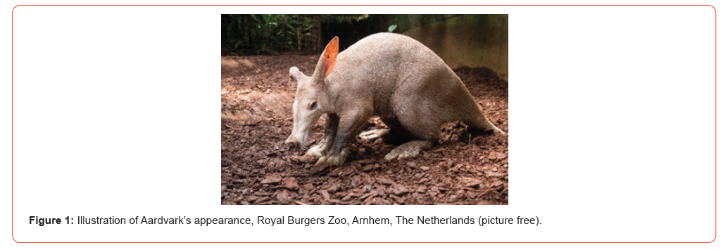
Aardvark’s habitat is below the Sahara in whole Africa except the Congo delta. Nutrition is nearly solely by ants and termites [9]. Known for its digging capacity and knocking the termite mounds, its forepaw (4 toes) and hind paw (5 toes) have been studied with CT images by Watanabe et al. [10]. Different functions are attributed to forepaw medial two digits compared to the lateral third and fourth digit: respectively for crushing and for sweeping soil. In fact, grouped digits can be moved rather independently due to their different functions but skilled movements are absent: “Skilled movements are likely to have been derived from, and elaborated through, food-handling behavior. In addition, it is now thought that the role played by the lateral and medial descending pathways of the spinal cord in the execution of skilled forelimb movements could be synergistic, rather than the exclusive responsibility of an individual pathway” [11]. Detailed reports on the architecture of the cerebral hemispheres and cerebellum with emphasis on the enormously developed olfactory system were published by Sonntag and Woollard [12].
An earlier report on Edentata, including Aardvark, was published by Elliot-Smith [13]. Brainstem studies were performed by Ziehen [14] and Verhaart [15], but a spinal cord description is lacking. Brainstem descriptions have been based on Weigert- Pal-stained material and includes the localization of red nucleus, mamillary and cerebral peduncles, fasciculus retroflexes and Cajal’s interstitial nucleus in the mesencephalon [15], while among others the corpora geniculate medial and laterale with its commissura, the commissural posterior, lemniscus medialis and substantia nigra are indicated by Ziehen [14]. At the pes pontis level the brachium conjunctivum, cerebral peduncle, lateral and medial lemniscus, rubrospinal tract, ventral spinocerebellar tract and Wallenberg’s dorsal trigeminal tract [15], has been completed at a lower level by [14] with the pyramidal tract, trigeminal entrance, corpus restiforme, cerebellar peduncles.
Nevertheless, not earlier than at a rather high medullary level of Orycteropus, Verhaart [15] indicates the pyramidal tract, together with the medial lemniscus that is crossed by the trapezoid body, the genu of the facial nerve, nucleus abducens, oliva superior and dorsal acoustic stria, and at mid-olivary level together with the descending trigeminus, inferior olive, nucleus hypoglossus, rubro-spinal tract and ventral spinocerebellar tract. These medulla oblongata levels are more extensively described by Ziehen [14]. However, his lower medulla oblongata is damaged at the ventral side, disrupting the pyramidal tract. In general, a nearly complete overview of structures presents in the lower brainstem, pes pontis and lamina quadrigemina with mesencephalon are given by these two studies. Confirmation of the course of the pyramidal tract at lower levels is missing. This study contains descriptions of the spinal cord and the course of the pyramidal tract using the Häggqvist technique [16].
Material and Methods
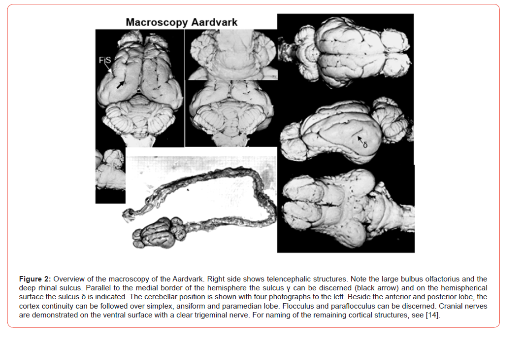
The specimen under consideration was an adult, apparently healthy male. Its weight was about 55 kg. The animal was killed by an overdose of Ketalar® (ketamine) and exsanguinated. Skull and vertebral column were opened by lateral incisions in the vault and vertebral arcs to expose dorsal brain and spinal cord. Consequently, spinal cord with cut off spinal nerves together with the entire brain and cut off cranial nerves were removed (Figure 2). The dura was opened, spinal cord and brain rinsed with water and transferred to Baker’s 10% formalin. Available for the present study were sections of the mesencephalon (rostral and caudal), pons, medulla oblongata as well as parts of the spinal cord at cervical, thoracic, lumbar and sacral levels. The blocks were processed for the Häggqvist technique on 6 μm paraffin sections [16]. The Häggqvist technique for paraffin sections (17) is a modification of Alzheimer-Mann’s staining in short: formalin fixed blocks were rinsed and mordanted in potassium bichromate and embedded in paraffin.
Deparaffinized sections were rinsed and mordanted in a saturated solution of phosphomolybdic acid. Sections were stained in Alzheimer-Mann’s solution, a mixture of methyl blue and of eosin in distilled water (staining introduced by Verhaart [18] and see, Sie Pek Giok [19] for the first descriptions respectively in Macaca ira and Man, see [16] for modifications). In Häggqvist-stained sections, axons are colored blue and myelin light red. Cell nuclei, cytoplasm, dendrites and glia are stained in different shades of blue. This method was chosen on account of its superior ability to distinguish individual fibers and fiber patterns of various tracts in normal material, but can also detect degenerated systems (see 20,14). In order to obtain a better picture of the cells additional slides were stained with the Klűver-Barrera method see [16]. The original Weigert-Pal results of the Aardvark (series H6397) studied by W.J.C. Verhaart were available.
Results
Macroscopy of Brain and Spinal Cord
The length of the brain (telencephalon with olfactory bulb) was estimated 7 cm, maximal breadth of telencephalon 4.5 cm, while the spinal cord reached 58.8 cm, from the first cervical roots till the last emerging coccygeal rootlets, and was oval-shaped with a breadth of 1.2 cm. In this specimen the telencephalic sulci and fissures are shallow compared to the Ziehen’s [14] results (Figure 2). The sulcus rhinalis is clearly present. The large olfactory bulbs easily catch the eyes. On the telencephalic surface the fissure of Sylvius (FiS in Figure 2) is absent and represented by a cortical dell, the sulcus γ (sulcus lateralis) and δ (suprasylvian gyrus) are discernable. The sulcus β (gyrus praesylvia) is clearly present as a deep groove restricted to the rostral frontal lobe (Figure 2; see 22; for naming of gyri, see 2). Cerebellar topography as described by Voogd [24] for the cortex continuity is recognized (Figure 2).
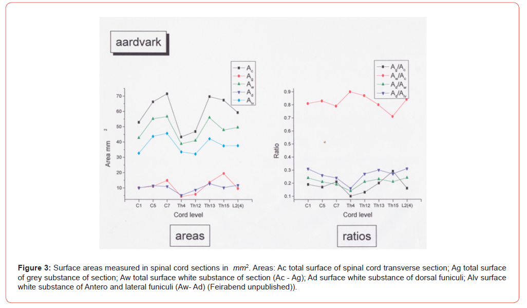
The flocculus (small) and paraflocculus (relatively large) are present at the dorso-lateral surface of the brainstem. Taking into account the vertebral formula of this animal the following roots were counted: cervical 8, thoracic 13, lumbar 8, sacral 5. (Th 14 and 15 are presumably L1 and L2 in Figure 2). The exact number of the thin coccygeal roots is unsure. Since only one specimen is studied that arrived in formalin fixed blocks gross anatomy of this spinal cord was difficult to perform, although cervical and lumbosacral enlargements were noted after opening of the dura. Alternatively, surface measuring of transverse sections (Figure 3) showed that two intumescences are present with increase in their grey substances; while at the thoracic level surface areas are comparatively reduced. Ratios showed the grey substance at intumescences to increase despite the increase of the whole transverse section. Moreover, the ratio white matter surface to total transverse cord surface at the thoracic level did not differ from intumescences ratios (Figure 3).
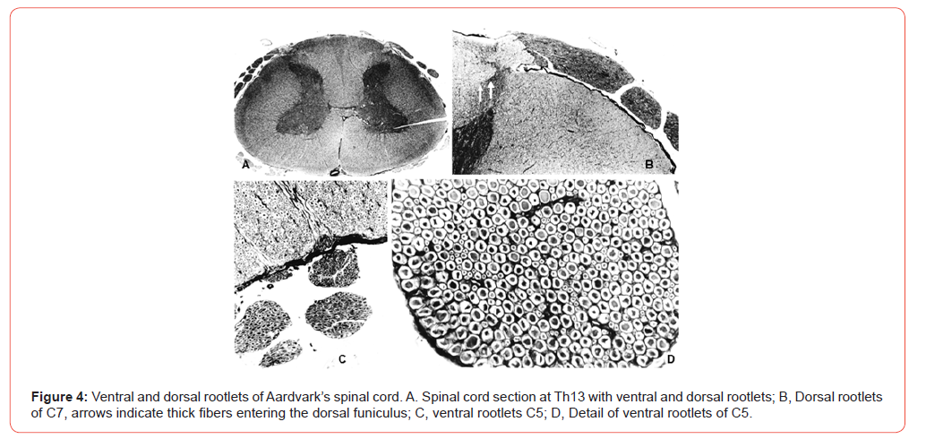
Spinal Cord Ganglion, Dorsal and Ventral Rootlets (Figure 4)
The spinal cord ganglia are mainly constituted by large and small cells (Figure 7B) and intermediate cell sizes are hardly encountered.
Spinal cord C5 ventral root was studied for its diameter and area of myelinated fibers. A clear bimodal distribution was found by Feirabend (unpublished):
a. max. area 50μm2 which is a mean of 8 μm diameter of
small myelinated fibers
b. max. area 160 μm2 which is a mean of 14 μm diameter
of large myelinated fibers. Areas are recalculated into circle
diameters.
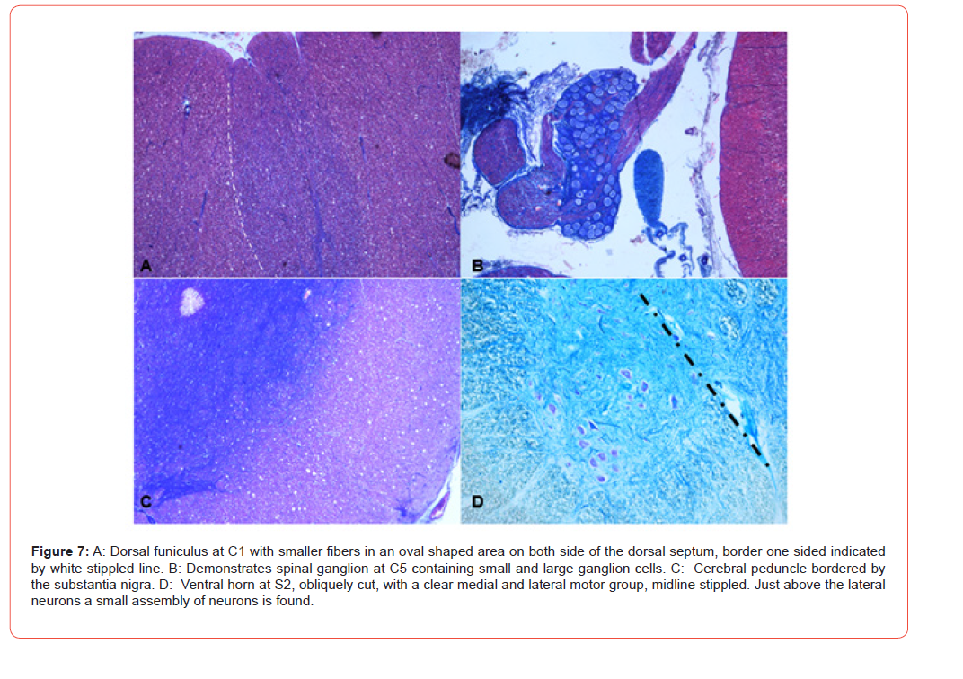
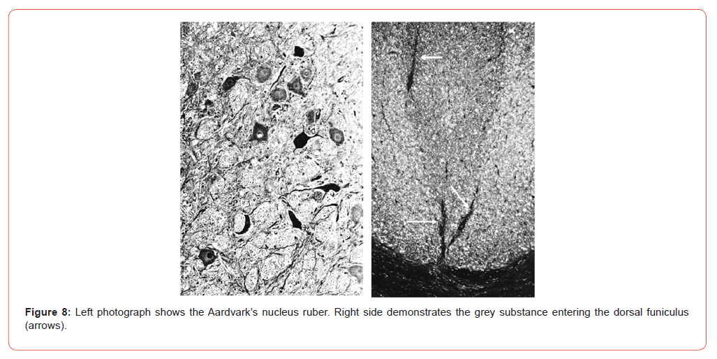
characteristic fiber pattern around the column (so they stand out) is noted. Lateral and ventral motor cell groups are present in the enlargements, whereas medial cell-groups (Rexed’s lamina VIII) are not numerous, but are clearly distinguished in the lumbosacral enlargements. At the sacral level a clear lateral group of cells in the ventral grey matter appears (Figure 7D). The grey commissure is short and thin, not widening at the intumescences. Motor rootlets left the ventral horns in a hooked manner. A cervical nucleus could not be identified. At the first cervical segment small longitudinal areas emerged from the medial part of the dorsal grey matter. It contained numerous round cells of medium size. A small strand of grey matter continued into the dorsal funiculus, containing few neurons (Figure 8).
Spinal Cord Fiber Systems
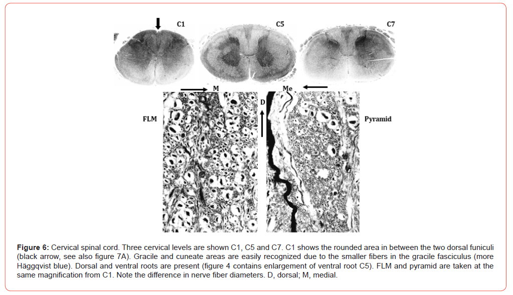
Pyramidal tract fibers were found in a small group along the dorsal part of the ventral median fissure only in the first cervical segment (Figure 6). Densely packed large fibers, identified as dorsal spinocerebellar fibers, were localized along the dorsolateral border of the lateral funiculus at cervical and thoracical levels. These fibers cannot be distinguished from the more ventrally situated ventral spinocerebellar fibers. Medial to the dorsal spinocerebellar fibers, less densely packed, probably rubrospinal fibers were seen in the dorsal one third to one quarter of the dorsolateral funiculus. These fibers are larger than those of the dorsal spinocerebellar tract.
Coarse fibers, localized to the most dorsolateral corner of the lateral funiculus are not observed caudal to the lower thoracic segments. In the ventral funiculus the fasciculus longitudinal is medialis, characterized by its large number of thick fibers could be identified at cervical (Figure 6) and thoracic levels. More caudally its borders are extra blurred due to the adjacent thick fibers, mainly in the central regions. Thin fibers of uncertain identity lined the ventral border of the ventral funiculus at cervical levels. In the dorsal funiculus a medial and medial dorsal area with relatively small medium-sized fibers is sharply demarcated from the adjacent more lateral areas in the cervical (Figure 6&7A), and to a much lesser extent in the remaining segments.
The Pyramidal Tract
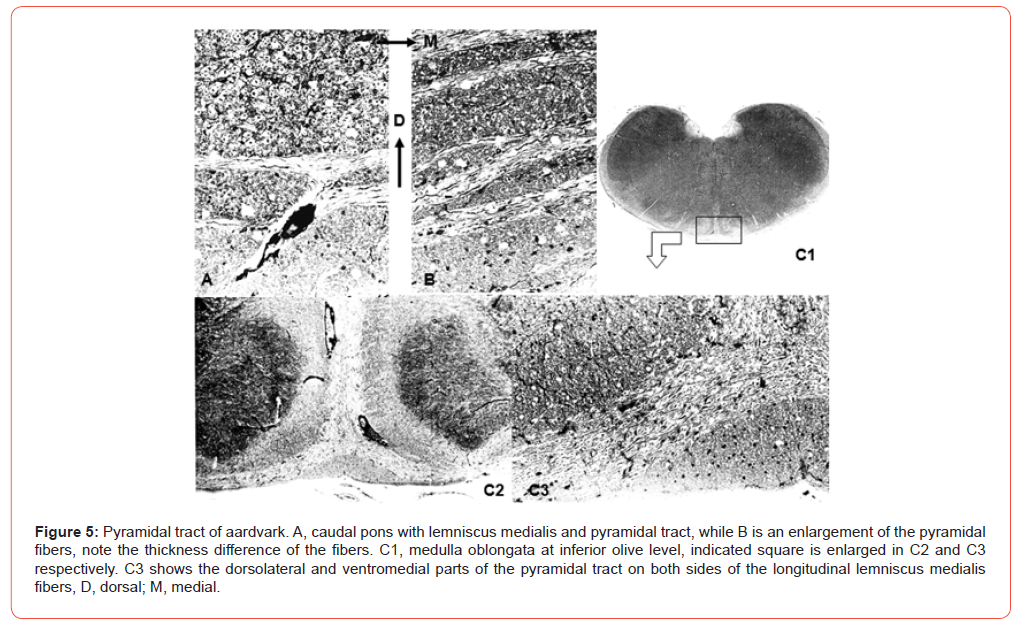
At mesencephalic levels the reticular part of the substantia nigra forms the dorsal border of the peduncle (Figure 7C). There is no sharp demarcation between these two systems. Whorls of peduncular fibers hampered the distinction of various parts. Nevertheless, the dorsomedial area contains more relatively thick fibers than its surrounding parts (Figure 7C). The medial part of the peduncle contains only very small fibers. Bagley’s bundles [25] were not seen in this material. Cranial pontine slides revealed a compact, somewhat oval area of peduncular fibers, its long axis in these transverse sections is headed in an oblique medio-lateral direction. This area clearly exceeds the region of pyramidal fibers at the Pontomedullary junction. Dorsally adjacent to the peduncular fibers the medial lemniscus can be delimited. Pontine nuclei surround the remaining parts of the peduncle. Fibers from a rather densely packed lateral group of these nuclei traverse the pedunculi to join the other pontocerebellar fibers in the pons. A sharp demarcation of different areas within the peduncle was feasible: a dorsomedial area could be identified by its larger content of thicker fibers than in its surrounding territory. At caudal pontine levels (Figures 5A&5B) a similar division by external arcuate fibers separated the tract into a triangular medio-ventral and a dorso-lateral part, the latter being split into several groups by coarse fibers.
The fiber patterns between the two groups appeared to be slightly different. The medial lemniscus characterized by its very different pattern of myelinated axons, lined the pyramidal system dorsally. The caudal medullary levels showed the pyramidal tract divided into two parts by coarse external arcuate fibers: a medioventral triangular area and a strand of fibers along the medioventral olivary complex (Figure 5C1-3). There was a suggestion that this triangular region contained more fibers with a relatively larger diameter than the lateral bands. At the transition cord-medulla pyramidal fibers were recognized in a small area on both sides of the posterior part of the ventral medial fissure (Figure 6). These fibers were continuous with the medio-ventral group of pyramidal fibers at caudal medullary levels. This tiny group of fibers, with its own characteristically fiber pattern, could not be traced further than the caudal part of the first cervical segment (Figure 6). The nucleus rubber is well developed and is pictured in Figure 8.
Discussion
“The scientific name of the aardvark is Orycteropus afer. The genus name Orycteropus is a concatenation of the Greek word όρνκτήρ — meaning miner — and ποΰς, the genitive form of ποδος — meaning foot. The epithet on afer is the Latin word for African, describing the origin of the aardvark” [26].
Spinal Cord
Heavy and powerful fore- and hind legs are among the distinctive features of the Aardvark (Figure 1) [10]. It is therefore not surprising that the two spinal cord enlargements are well developed. Gross anatomy of Aardvark’s spinal cord does not differ of the general cord patterns, except for the higher number of spinal segments. The large number of thick fibers in the rootlets of the ventral (14 μm, Figure 4) and dorsal horns is noteworthy in this respect. The absence of intermediate fiber calibers in the ventral rootlets is supported by the bimodal fiber distribution. Due to the small size of the neurons of the nucleus thoracalis the fibers of the dorsal spinocerebellar tract are moderately large and smaller than those of the fasciculus longitudinal is medialis. The mutual relationship between these fiber systems has been described in various mammalian orders by Verhaart [15], who did not include the Aardvark in this respect.
The oval shaped area of small to medium sized fibers in the dorsal funiculi, but most conspicuously in the upper cervical segments is not unique, most likely this ‘gracile’ area comprises many primary afferents from lumbosacral segments. Rearrangement of fibers is well-known to occur in long tracts, which may explain variations in shape of such areas at different levels. The absence of coarse fibers in the dorso-lateral corner of the lateral funiculus, caudal to the lower thoracic segments is noteworthy, because such fibers are well-recognized in the cat and in the spider monkey. These spinothalamic fibers are related to tail function (27) and Aardvark’s tail has pronounced features and is sometimes compared to the Kangaroo’s tail [26]. The course of the coccygeal fibers in the Aardvark is unknown. A small strand of grey matter with very few neurons was seen in the dorsal funiculus of the first cervical segment. Its significance is unknown.
Pyramidal tract
An overview of animal pyramidal tracts has been published by Voogd in the chapter Mammals in the “Central nervous system of vertebrates” [28], together with the human pyramidal tract [29]. The pyramidal tract of the Aardvark was studied by Ziehen [14] and Verhaart [15] using Weigert-Pal sections of which Ziehen’s series were ventrally damaged at the lower medulla oblongata. The typical distribution of the fibers as seen in the Häggqvist coloration is absent in the Weigert-Pal staining. Verhaart indicated the cerebral peduncle from the mesencephalon on and points to the pyramid also in the mid-olivary level. At this level he distinguished two small bands, separated by external arcuate fibers. The cerebral peduncle at pontine levels was localized dorsal to the pontine nuclei. Ziehen indicates the pyramidal tract caudal from the facialis nucleus on to the pes pedunculi. Both authors did not decide on the presence of the pyramidal tract at lower spinal cord levels [18].
This Häggqvist study showed that the pyramidal tract does not enter the spinal cord, presumably only reaches the upper cervical level, and a clear-cut pyramidal crossing was not found. In several species like cow, deer, hedgehog, rabbit the pyramidal tract ends in the upper cervical levels. Best known for early termination of the pyramidal tract is the goat, where the longest pyramidal fibers end in the lower cervical segments (see 30, 28). The present investigation supports Verhaart’s report [15] of the pyramidal system and refutes Ziehen’s conclusion that the pyramid in the medulla oblongata is large, despite the general knowledge that the number of pyramidal fibers can vary widely in different specimens (see 31). The Häggqvist stain permitted to discern a ventro-medial and a dorso-lateral part by its pattern of myelinated fibers. These two parts were accentuated by the traversing coarse external arcuate fibers. The very dorsal position of the peduncle at rostral pontine levels, already observed by Ziehen and Verhaart is noteworthy, but not a unique feature.
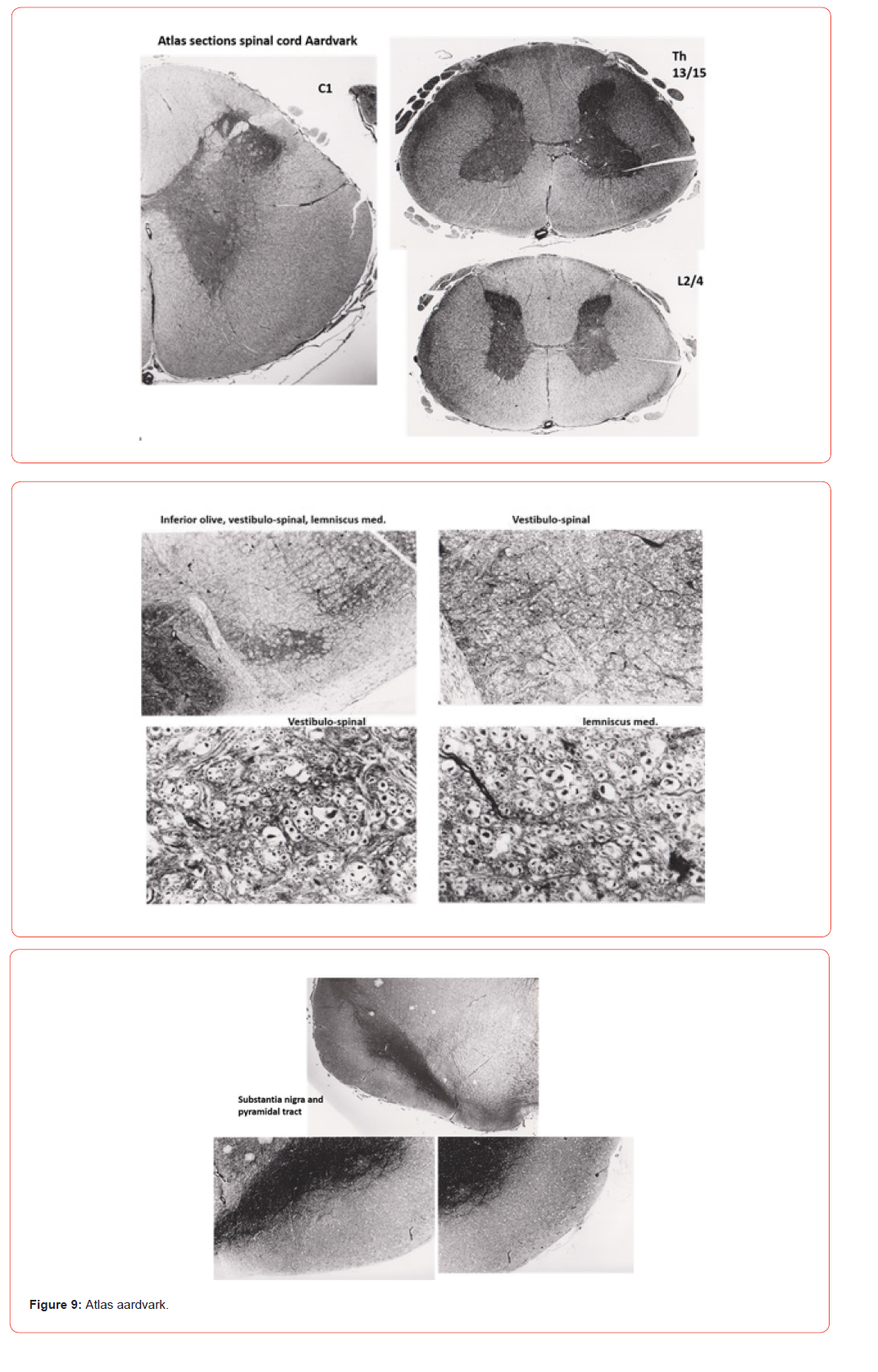
Both Lawrence and Kuypers [32,33] and Heffner and Masterton [30] indicated that direct pyramidal connections onto the motorneurons are needed to perform independent finger movements. Independent digit motions are not found in the Aardvark’s study [10]. However, such a coincidence hardly can be the explanation for the missing pyramidal tract endings in the Aardvark’s spinal cord as shown by the next examples: The pyramidal decussation at the transition brainstem to spinal cord is absent in European hedgehog, the mole and the klipdassie, but the pyramidal tracts in these animals descend into the spinal cord, except for the hedgehog. The rabbit has a crossed pyramidal tract that ends at C3, the cow contains a crossed and uncrossed pyramidal tract reaching C2-3, the goat possesses a crossed and uncrossed pyramidal tract that stops at C5 and the elephant’s crossed pyramidal tract ends at C5, while the uncrossed pyramidal tract reaches T6 [34,15,28]. A general rule for the absence or presence of the crossed and/or uncrossed pyramidal tract in the spinal cord is lacking in view of the enormous variation of pyramidal tract constructions present among various species (Figure 9).
Epilogue
The central nervous system of this Aardvark was obtained by Andries de Graaf † (Leiden thesis) [35] at the end of the 1980’s, because the South African farmers wanted the specimen to be killed due to the damage caused to their crops. In January 1991 Andries contacted his old group of the Academic Hospital of the Leiden University now Leiden University Medical Centre. The presented data were a combined effort of A.S. de Graaf † (also by his unfinished manuscript) and of the members of the Neuro regulation group: H.K.P. Feirabend, J.M. Guldemond, H. Choufoer, E. Marani, but has not been published, except for a web draft.
- Simpson GG (1961) Principles of animal taxonomy. Columbia University Press, NY, London.
- Colbert EH (1941) A study of Orycteropus gaudryi from the Island of Samos. Bull Amer Mus Nat Hist 78: 305-351.
- Lehmann T (2009) Phylogeny and systematics of the Orycteropodidae (Mammalia, Tubulidentata). Zool J Linnean Soc 155(3): 649-702.
- Dene H, Goodman M, Walz DA, Romero-Herrera AJ (1983) The phylogenetic position of Aardvark (Orycteropus afer) as suggested by its myoglobin. Hoppe Seylers Z Physiol Chem 364(11): 1585-1595.
- De Jong WW, Zweers A, Goodman M (1981) Relationship of aardvark to elephants, hyraxes and sea cows from α-crystallin sequences. Nature 292(5823): 538-540.
- Cao Y, Fujiwara M, Nikaido M, Okada N, Hasegawa M (2000) Interordinal relationships and timescale of eutherian evolution as inferred from mitochondrial genome data. Gene 259(1-2):149-158.
- Endo H, Watanabe K, Hayashi Y, Matsuzaki M, Sakai T, et al. (2003) Three-dimensional CT image analysis of the digging system in the Aardvark. Ann Anat 185(4): 367-372.
- Iwaniuk AN, Whishaw IQ (2000) On the origin of skilled forelimb movements. Trends Neurosci 23(8): 372-376.
- Sonntag CF, Woollard HH (1925) A monograph of Orycteropus afer: II Nervous system, Sense-organs and hairs. In Republished in J. Zoology 95(3): 1185-1235.
- Pirlot P, Kamiya T (1983) Quantitative brain organization in Anteaters (Edentata-Tubilidentata). J Hirnforsch 24(6): 677-689.
- Elliot Smith G, Sydney MD (1899) The brain of the Edentata. Trans Linn Soc Lond, 2nd ser Zool 7(7): 277-394.
- Ziehen Th (1909) Zur Kenntnis des Faserverlaufs im Gehirn von Orycteropus. Jenaische Denkschriften XV: 477(106)-510(138).
- Verhaart WJC (1970) Comparative anatomical aspects of the mammalian brain stem and the cord. Vol I and II, Studies in Neuroanatomy 9. Van Gorcum, Assen, The Netherlands 2(9): 1-312.
- Marani E, Schoen JHR (2005) A reappraisal of the ascending systems in Man, with emphasis on the medial lemniscus. Adv Anat Embryol Cell Biol 179: 1-76.
- Häggqvist G (1936) Analyse der Faserverteilung in einem Rückenmarkquerschnitt (Th3). Zeitschr f mikrosc-anat Forschung 39: 1-4.
- Verhaart WJC (1954) Fiber tracts and fiber patterns in the anterior and lateral funiculus of the cord in Macaca ira. Acta Anat 20(4): 330-373.
- Sie Pek Giok (1956) Localization of fiber systems within the white matter of the medulla oblongata and the cervical cord in Man. Thesis University Leiden.
- Voogd J, Feirabend HKP (1981) Classic methods in neuroanatomy. In: Lahue R (ed) Methods in Neurobiology. Vol 2 Plenum Press NY, PP: 207-234.
- Verhaart WJC (1963) The brain stem of the anteater, Myrmecophaga jubata L. J Hirnforsch 6: 205-221.
- Bagley CH (1922) Cortical motor mechanism of the sheep brain. Arch Neurol Psychiat 7(4): 417-453.
- Taylor WA, Lindsey PA, Skinner JD (2002) The feeding ecology of the Aardvark Orycteropus afer. J Arid Environm 50(1): 135-152.
- Knőthig J (2005) Biology of the Aardvark (Orycteropus afer). Diplom Arbeit, Fakultät Biowissenschaften, Ruprecht-Karls-Universität, Heidelberg.
- Chang HT, Ruch TC (1947) Topographical distribution of spinothalamic fibers in the thalamus of the spider monkey. J Anat 81(Pt 2):150-164.
- Voogd J, Glickstein M (1998) The anatomy of the cerebellum. Trends Neurosci 21(9): 370-375.
- Voogd J, Nieuwenhuys R, van Dongen PAM, ten Donkelaar HJ (1998) Mammals. Chap 22 in: eds. The central nervous system of vertebrates. PP: 1637-2098, Springer, Berlin.
- Nieuwenhuys R, Ten Donkelaar HJ, Nicholson C (1998) The central nervous system of vertebrates. Vol 3. Springer, Berlin.
- Schoen JH (1969) The corticofugal projection on the brain stem and spinal cord in Man. Psychiatr Neurol Neurochir 72(1): 121-128.
- Lawrence DG, Kuypers HG (1968a) The functional organization of the motor system in the monkey. I The effects of bilateral pyramidal lesions. Brain 91(1): 1-14.
- Lawrence DG, Kuypers HG (1968b) The functional organization of the motor system in the monkey. II Effects of lesions of the descending brain-stem pathways. Brain 91(1): 15-36.
- Heffner R, Masterton B (1975) Variation in form of the pyramidal tract and its relationship to digital dexterity. Brain Behav Evol 12(3): 161-200.
- Benoit J, Crumpton N, Mérigeaud S, Tabuce R (2013) A memory already like an Elephant’s. The advanced brain morphology of the last common ancestor of Afrotheria (Mammalia). Brain Behav Evol 81(3): 154-169.
- Verhaart WJC (1967) The non-crossing of the pyramidal tract in Procavia capensis (Storr) and other instances of absence of the pyramidal crossing. J Comp Neurol 131: 387-392.
- De Graaf AS (1967) Anatomical aspects of the cetacean brain stem. Thesis Leiden. Van Gorcum, Assen.
- Noorduyn NJA (1959) Quantitative aspects of the pyramidal tract. Thesis Leiden University.
- Usunoff KG, Marani E, Schoen JHR (1997) The trigeminal system in Man. Adv Anat Embryol Cell Biol 136: 1-126.
-

This work is licensed under a Creative Commons Attribution-NonCommercial 4.0 International License.




