 Research Article
The Role of Hands-on Ultrasound Sessions in Medical School Teaching of Gross Anatomy
Ahmed Mahgoub MD*1, Joseph Asemota MD, MPH1,2, Deepak Sharma MD1, Andre Granger MD1,3, Danny Burns MD1,
1Department of Anatomical Sciences, St. George’s University, West Indies
2Department of Internal Medicine, Howard University Hospital, USA
3NYU Langone Hospital, Brooklyn, New York, USA
Ahmed Mahgoub, Department of Anatomical Sciences, St. George’s University School of Medicine, Grenada, West Indies.
Received Date: May 10, 2021; Published Date: June 02, 2021
Abstract
Purpose: Imaging concepts and skills are increasingly integrated into pre-clinical medical education, often with the aim of consolidating gross anatomy principles. This study investigates students’ attitudes towards ultrasound education, perceived effects understanding anatomy, and perceived confidence in identifying commonly encountered structures.
Method: At the end of the gross anatomy course, students completed a questionnaire consisting of 25 items on a 5-point Likert scale. Several items assessed students’ perceptions of the role and effectiveness of ultrasound teaching in gross anatomy education. The responses were analyzed, and significant findings synthesized.
Results: 197 students completed the survey. Fewer than 10% of respondents had previous ultrasound experience. Most students agreed that ultrasound sessions stimulated their interest in learning gross anatomy (67%), aided their understanding of human anatomy (81%), or helped them to identify structures on other imaging modalities such as CT and MRI (56%). Most students expressed confidence in identification of the mitral and aortic valves (55%), the hepatorenal space (58%), the abdominal aorta and its major branches (53%).
Conclusion: Students’ perception of the role of ultrasound is important for its future in medical education. Our survey shows that students’ perceptions towards ultrasound are generally positive by supplementing their understanding of gross anatomy.
Keywords:Ultrasound; Teaching; Anatomy; Medical Education
Introduction
The existing body of knowledge on human gross anatomy, as well as dissection, prosection and surface anatomy content delivery has remained relatively unchanged for decades [1]. Over the last decade, however, gross anatomy courses have incorporated and integrated imaging content with the aim to supplement the gross anatomy courses or modules to better prepare medical students for their transition to the clinical setting [2-6].
With new developments and technological advances, the use of ultrasound has expanded in the last two decades. Ultrasonography is a portable, low-cost, non-invasive imaging modality with an excellent safety record. Its B-mode (brightness-mode) displays two-dimensional “slices” composed of varying gray-scale value pixels, representing ultrasound echoes in real-time [7, 8]. This real-time imaging provides dynamic anatomical and functional information, such as arterial pulsations and cardiac motion thus allowing for visualization and evaluation of anatomical structures as well as for guided interventional diagnostic and therapeutic procedures [9]. Its utility in the quick evaluation of hypotension, shock, thoracic and abdominal injuries make it an integral tool in emergency and critical care settings and as such is increasingly being used as an extension of physical examination [2,10-12]. Given the widespread utilization of ultrasound examinations, it is fast becoming a necessary skill for future physicians across a wide range of specialties and heightened emphasis on ultrasound competencies is emerging in postgraduate training and specialty board examinations. Consequently, some degree of knowledge and skill in ultrasonography will soon be an essential part of the competencies that every medical student should acquire prior to graduation [12,13]. In fact, while radiographs, CT and MRI scans have traditionally been the primary modalities used for teaching imaging in medical students’ curriculum, ultrasound imaging has recently been added to the essential imaging modalities that every student should be exposed to.
Although the integration of ultrasound imaging into the medical school curriculum has steadily increased over the past several years, there still remains significant variability in ultrasound training across institutions. In many instances, medical student training is limited to reviewing only a few ultrasound images during anatomy instruction while in a small number of medical schools there has been a vertical integration into their existing learning modules. Surprisingly, despite the significant variation in medical student ultrasound training, not very many studies have investigated these variations in ultrasound education and the effects that specific teaching methods have on students’ educational satisfaction and perceived level of competence. Therefore, the authors of this paper sought to identify students’ perceptions and level of satisfaction regarding the effectiveness of hands-on ultrasound sessions during the human gross and developmental anatomy course at St. George’s University in Grenada. This study also highlights many of the benefits of sonography in pre-clinical studies and would serve as a vital resource for developing effective standardized teaching methods.
Material and Methods
This study was approved by St. George’s University Institutional Review Board (number 14080).
Informed consent was obtained verbally by asking participants to use their own interactive clicker devices.
During the human gross and developmental anatomy course, students receive hands-on ultrasound scan training under the guidance of physician facilitators in groups of 4 students. These sessions are held for 30 minutes, once weekly for a duration of 12 weeks. Sonographic anatomy is demonstrated on live participants selected from a pool of paid simulated patients. In order to standardize the learning experience for each student group, ultrasound training is provided to the physician facilitators prior to each session and areas of emphasis reiterated. The facilitators train and evaluate the students in identifying anatomical structures in each of the 12 sessions.
Over a 12-week period, students acquire knowledge and skills necessary to identify a list of structures in the neck, upper limb, thorax, abdomen, and lower limb using B-mode ultrasound. Sessions build on one another, with each session including basic knobology, probe selection, image optimization techniques, and recognition of tissues, organs, and structures. Tissues that are emphasized throughout the course are: fat, fascia, ligaments, muscles, bones, tendons, arteries, veins, and nerves. Each session is organized around an aspect of regional anatomy. Ultrasound labs during the first half of the course focus on identifying: the radial nerve from the mid-humerus to its terminal divisions, brachial plexus in the scalene interval, cords of the brachial plexus in the axilla, pleural lines/pleural sliding, contents of the cubital fossa and carpal tunnel. The second half of the course includes: ultrasound of the heart, the abdomen and pelvis, the knee, the ankle and the neck. The final ultrasound session introduces the students to FAST (focused assessment with sonography for trauma) exam, which is a combination of most of the topics previously taught. At the conclusion of the 2015 spring course, participants completed a questionnaire consisting of 25 items to be rated on a 5-point Likertscale. Specific items on the questionnaire required respondents to rate their degree of agreement/disagreement with statements about the effects of ultrasound training on interest in learning anatomy, understanding anatomy, understanding other imaging modalities as well as their overall satisfaction with the training group size, duration of teaching sessions and effectiveness of group facilitators. Respondents also rated their confidence in the ability to consistently identify commonly encountered tissues such as bones, muscles, blood vessels, tendons and nerves. In addition, respondents rated their confidence in identifying selected specific structures/groups of structures, which had been emphasized in one or more of the ultrasound sessions. These responses were analyzed and significant findings were synthesized.
Data Sources
Participants for this study were recruited on a voluntary basis from first year medical students enrolled in the spring term of the human gross and developmental anatomy course in 2015. This course had hands-on ultrasound education incorporated into it and the data for this project was obtained using clicker response technology by Turning Technologies. 25 questions were projected using PowerPoint on screens at the start of last anatomy small group session. 24 questions were analyzed for this study.
Results
There was a total of 197 respondents. 9.6% of the respondents had prior ultrasound experience. The majority of students agreed that ultrasound teaching stimulated their interest in learning gross anatomy (67%), helped their understanding of human anatomy (81%), and helped them to identify structures in other imaging modalities such as CT and MR (56%). (Figure 1)

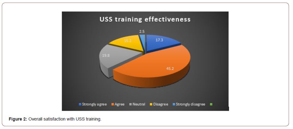
Students reported satisfaction with group size for training sessions (66%) and the effectiveness of group facilitators (81%). However, most students (67%) did not agree that the duration of the sessions was sufficient. Figure 1. Among our respondents, there was a 63% overall satisfaction with the ultrasound training. (Figure 2).

Regarding the sonographic appearance of commonly encountered tissues, a majority of participants reported confidence in their ability to consistently recognize muscles (67%), bones (62%) and blood vessels (56%), while only a minority of participants reported confidence in their ability to recognize tendons (32%) and nerves (38%) (Table 1).
Table 1: Basic Tissues.

With respect to selected structures encountered in thoracic ultrasound sessions, a majority of students reported confidence in their ability to identify the aortic and mitral valves (55%) and the pleural line/pleural sliding (55%), while a minority reported confidence in their ability to identify heart chambers and pericardium (41%) (Table 2).
Table 2: Thoracic Ultrasound.

As regards selected structures encountered in abdominal ultrasound sessions, most participants reported confidence in their ability to identify the liver, right kidney, hepatorenal space (58%), the abdominal aorta and its major branches (53%), the spleen, left kidney and splenorenal space (50%) (Table 3).
Table 3: Abdominal Ultrasound.

When asked about other selected groups of structures encountered in one or more ultrasound sessions, the majority of students reported confidence in their ability to identify several structures at the base of the neck including thyroid gland, common carotid artery, internal jugular vein and interscalene components of the brachial plexus (63%), as well as major structures of the carpal tunnel (57%) and popliteal fossa (52%). Slightly fewer than half of participants reported confidence in their ability to identify major contents of the tarsal tunnel (47%) (Table 4).
Table 4: Ultrasound of the Neck, Carpal Tunnel, Popliteal fossa and Tarsal Tunnel.

Discussion
In 2011, the department of Anatomical Sciences at St. George’s University in Grenada introduced hands-on ultrasound sessions during the human gross and developmental anatomy course and implemented ultrasound stations for the objective structured clinical examination (OSCE). This provides an opportunity for students to learn applied ultrasound earlier in their career with significant benefits accruing well beyond their preclinical training and even into their clinical years. A study by Brown et al., reported that incorporating ultrasound training into the anatomy curriculum could increase students’ confidence in performing invasive procedures during their residency. In another study on a similar approach employed at Loma Linda University, a 22-point ultrasound OSCE (US-OSCE) evaluation showed a significant difference in ultrasound skills of trained students compared to ultrasound-naïve students. Overall, our study findings are similar to other studies where students reported that cross-sectional anatomy teaching along with exposure to ultrasound examination improves their confidence in identifying anatomical structures. In our study, most students (77.3%) expressed confidence in their ability to identify the structures and tissues from the core objectives of ultrasound teaching sessions. Students generally reported high confidence in identifying structures that received substantial attention in other aspects of the course such as lectures and small group discussions, and which were emphasized and revisited in ultrasound sessions. This finding buttresses the utility of ultrasound teaching as a tool in reinforcing anatomy teaching. It also underpins the need for a plurality of resources in the effective teaching of anatomy.
In the present study, most respondents (81.8%) felt that ultrasound training stimulated their interest in learning anatomy and made anatomy and other imaging modalities easier to understand. This finding is again consistent with results reported in previous studies. According to a study by Hammoudi et al., ultrasound examination of the heart during the anatomy course improved the understanding of gross anatomy and at the same time enhanced the motivation to learn cardiac anatomy and physiology. While a majority of participants were confident in their ability to identify the median nerve and flexor tendons in the carpal tunnel, only 30% of participants were confident in their general ability to identify tendons and other nerves. Students also expressed that they were able to identify clinically relevant structures more easily. These findings suggest the importance of utilizing pertinent clinical correlations in the teaching of anatomy and is in keeping with the evolution of anatomy education from traditional, large quantities of detailed anatomy to a more clinically oriented method. With regards to identifying the mitral and aortic valves, 55% students reported confidence in their ability while approximately 21% of students were not confident in their ability to identify these structures. This is probably due to the fact that small adjustments in the orientation of the probe while scanning the heart will produce a new view that may be unfamiliar to students. This however raises the question, “How much do you rely on pattern recognition versus understanding of spatial orientation and 3-dimensional relationships?”. It is also unclear whether notions about students desired future residency training requirements affected their immediate interest in the ultrasound training sessions ultimately affecting the effectiveness of the training sessions. This raises yet another question: “Are students who are entering careers which are not heavily dependent on imaging interested in learning or understanding various modalities of imaging?”
Finally, even though participants expressed overall satisfaction with the ultrasound sessions and specifically with the group size and effectiveness of group physician facilitators, more than half expressed a desire for more time in ultrasound sessions. Considering that ultrasound requires hands-on practice, 30-minute sessions for groups of 4-5 students may not permit sufficient practice for each student. Thus, increasing the allotted time for hands-on ultrasound sessions and creating opportunities to access ultrasound machines after scheduled sessions could significantly assist student learning. Additionally, an ultrasound manual detailing scheduled session can be provided beforehand so that students may pre-read to gain an understanding of material.
Limitations
This study has some important limitations and thus the results should be interpreted cautiously. Since clicker responses were obtained only from students present for the last session of the anatomy course, this opens up the possibility of selection bias. Also, the relatively small sample size, and the fact that our study population was obtained from a single institution may impose limitations on the generalizability of the study findings. Because of the small sample size limitations, stratification of students into desired specialization subgroups for further analysis was restricted. Furthermore, student ability was self-reported and not objectively assessed. Despite these limitations, this study highlights the benefits of a hands-on teaching approach to ultrasound imaging and assesses the perceived effect this method has on student satisfaction. In light of the limitations, more prospective studies are needed to confirm the results of this study.
Conclusion
This study highlights many of the benefits of sonography in pre-clinical studies. Students’ perception of the role of ultrasound is important in measuring its success and determining its future in medical education. Ultrasound teaching supplements students understanding of Gross Anatomy and boosts their confidence in identifying commonly encountered structures. We, however, recommend that future studies implement an ultrasound manual and increase session length as well as objectively measure students’ ability to identify structures instead of focusing solely on students’ perceptions about their abilities. It would also be interesting and greatly enlightening for future studies to tease out the nuances between students’ desired future residency training aspirations and their immediate interest in ultrasound training.
Acknowledgement
None
Conflict of Interest
None.
References
-
Ahmed Mahgoub MD, Joseph Asemota MD, MPH, Deepak Sharma MD, Andre Granger MD, Danny Burns MD. The Role of Hands-on Ultrasound Sessions in Medical School Teaching of Gross Anatomy. Anat & Physiol Open Access J. 1(1): 2021. APOAJ.MS.ID.000504.
-
Anatomy principles, Abdominal aorta, Clinical setting, Ultrasound, Teaching, Anatomy, Medical education, Ultrasonography, Brachial plexus, Carpal tunnel, Cubital fossa, Tendons, Arteries, Veins, Terminal divisions, Knobology, Probe Selection, Image optimization techniques, Organs, Structures
-

This work is licensed under a Creative Commons Attribution-NonCommercial 4.0 International License.


