




 Case Report
Smoke Without Fire - When is a Cardiac Biomarker Not a Cardiac Biomarker ?
Received Date:October 21, 2025; Published Date:November 04, 2025
Abstract
Keywords:Troponin T; Troponin I; SLE systemic lupus erythematosus; connective tissue disease; myocarditis
Abbreviations:TnT: Troponin T; TnI: Troponin I; SLE: Systemic Lupus Erythematosus
Case Description
A 40-year-old male was referred to rheumatology with arthralgia and myalgia. He had juvenile onset Systemic Lupus Erythematosus (SLE) with previous class IV lupus nephritis, and had been in remission for over 10 years. He had subsequent chronic kidney disease (stage G1 A2) and well-treated hypertension. On examination, he had tight skin in his proximal phalanges and a malar rash, but showed normal muscle strength and no overt synovitis. Cardiorespiratory examination was unremarkable, and he was normotensive. Baseline investigations assessed SLE activity including full blood count, renal profile, liver function tests, Creatine Kinase (CK), C-reactive protein, Erythrocyte Sedimentation Rate (ESR), DsDNA, C3 and C4. Results indicated a flare of his SLE (see Table 1). Subsequent investigations were requested including NT-proBNP and troponin T (TnT), echocardiogram, Computed Tomography (CT) thorax to evaluate cardiorespiratory involvement and Magnetic Resonance Image (MRI) of the thighs.
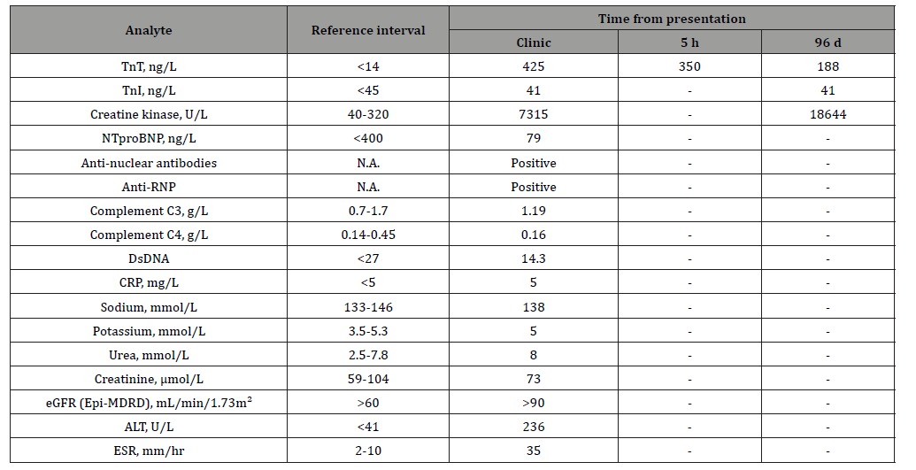
Blood results were largely unremarkable apart from an extremely high TnT result of 425 ng/L (Roche, Lewisham, UK, reference interval <14 ng/L). CK, ESR and alanine transaminase were also increased (Table 1). His ECG and echocardiogram were normal, but cardiac MRI confirmed subtle high signal change in the anterior septum consistent with myocardial oedema, inflammation and preserved left ventricular function MRI of the thighs revealed widespread fasciitis, myositis and synovitis of the knee. The duty biochemist and requesting clinician discussed the TnT result as it was not consistent with clinical history (i.e., no evidence of acute cardiac event) and arranged for the measurement of troponin I (TnI, Siemens Atellica, Manchester, UK) at a local laboratory, which was within the reference range at 41 ng/L (reference interval <45 ng/L).
Follow-Up
The TnI result was communicated to the acute medical and rheumatology teams, highlighting the differences in the TnT and TnI results. It was concluded that the patient had myocarditis, not associated with a cardiac failure or an acute cardiac event. He was treated with intravenous methylprednisolone and then a weaning course of prednisolone alongside rituximab and methotrexate for a severe, multiorgan flare of SLE including myocarditis. The patient was closely observed in light of the grossly abnormal TnT result. An echocardiogram revealed a normal ejection fraction >55% (reference value >50%). Muscle biopsy confirmed ongoing skeletal myositis. A further cardiac MRI confirmed improvement and resolution of previous areas of inflammation seen on the first cardiac MRI. He improved over the next few months. Following a short exercise session, he reported worsening myalgia and on prompting mentioned some retrosternal burning. His ECG was normal, but similar to the previous instance, TnT and CK were elevated and TnI was within the reference range (Table 1). He was managed with a short increase in oral steroids, clinically and biochemically resolving his myositis.
Table 1: Blood results obtained from patient during the two presentations. TnT assay was performed on Roche Cobas e801, TnI assay was performed on Siemens Attelica IM. Sodium and potassium were measured using Roche 8000 ISE module. Urea, creatinine, CK and ALT were measured using Roche Cobas c701 module. NTproBNP was measured using Roche Cobas e801 module. eGFR was calculated using the 2009 CKD-EPI equation.
Questions to Consider
1. Why are patients with connective tissue disease at
increased risk of myocarditis?
2. What non-cardiac physiological factors can cause elevated
TnT?
3. How do TnT and TnI differ?
4. Is TnI more suitable than TnT to assess myocardial
infarction in patients with connective tissue disease?
5. Why does the interference appear only in the TnT assay?
Discussion
Why are patients with connective tissue disease at increased risk of myocarditis?
SLE is a multisystemic, autoimmune, connective tissue disease which can cause inflammation in a wide range of tissues and organs throughout the body. The common presentation is associated with the skin and musculoskeletal system, though attacks on different organs can alter the clinical presentation [1]. The heart is particularly vulnerable to this inflammation, with cardiac involvement being reported in over 50% of patients with SLE [2]. This can manifest as pericarditis, inflammation in the lining of the heart or, more dangerously as lupus myocarditis if the inflammation occurs in the cardiac muscle. Lupus myocarditis occurs in 5-10% of patients with SLE and has associated morbidity in approximately half of these patients [3].
The increased risk of these patients developing myocarditis highlights the utility of cardiac biomarkers and the importance of understanding their associated pitfalls. Typically, patients with myocarditis may have a normal TnI, highlighting the need for a high index of suspicion and subsequent specialised cardiac imaging including cardiac MRI. Patients with SLE are also at increased risk of ischaemic heart disease so it is imperative that this is diagnosed quickly because of diverging acute clinical management [4].
What non-cardiac physiological factors can cause elevated TnT?
The diagnostic dilemma in this case was an elevated cardiac biomarker, which did not accurately reflect an acute cardiac event. TnT is an intramuscular biomarker, which is released into circulation when cardiac muscle is damaged. Several factors such as strenuous exercise and pulmonary embolism can contribute to its elevation through the strain they place on the heart. Additionally, chronic kidney disease can decrease renal clearance to the point that the resultant inflammation and uremic toxins contribute to an elevated TnT. These factors, while notable, would not be expected to cause the scale of TnT elevation observed in the case of this patient. In this case, the TnT was grossly elevated, but on further investigation, no cardiac damage was found.
This raises the question, how did this amount of TnT enter the bloodstream if not via damaged cardiac muscle? While TnT has assumed cardiac specificity in adults, during fetal development, an isoform of TnT which bears structural similarity to mature cardiac TnT is expressed in skeletal muscle. This becomes less prominent throughout maturation, culminating in its absence from the skeletal muscle of a healthy, mature population [5]. It is theorised that in connective tissue diseases such as SLE, this fetal isoform is re-expressed in the diseased skeletal muscle and subsequently released into circulation through the associated tissue damage [6,7]. This provides an explanation for the non-cardiac elevation of this biomarker. Fetal TnI is not expressed in skeletal muscle, preventing an equivalent effect on its cardiac specificity (Table 1).
How are TnT and TnI different?
TnT and TnI are two subunits of the troponin complex within cardiac muscle cells. They are released into circulation by necrotic cardiomyocytes and are removed from circulation by the liver and kidneys, demonstrating similar kinetics [8]. While, they are broadly treated as interchangeable biomarkers of myocardial infarction, they do display distinct characteristics which gives each an individual utility. TnI is a more specific marker of myocardial injury, it reaches higher concentrations and is cleared more quickly, owing to its lower molecular weight. As demonstrated by this case, TnT is less specific as the elevation observed was a consequence of diseased skeletal muscle and not damaged cardiac muscle. For a more in-depth review comparing TnT and TnI see Espinosa et al [8]
Is TnI more suitable than TnT to assess myocardial infarction in connective tissue disease?
The suspected interference from fetal TnT in the Roche TnT assay has drawn into question the utility of TnT as a biomarker of myocardial infarction in cases of connective tissue disorders such as SLE. In situations such as these, the validity of the TnT results cannot be solely relied upon to indicate cardiac damage, and in this case caused a diagnostic dilemma leading to the patient being recalled as the increase in TnT was so marked. The TnI assay does not experience the same kind of interference as there is not an equivalent structural analogue of TnI released from diseased skeletal muscle. As a result, TnI has been established as a more specific marker of myocardial injury than TnT [9].
Why does the interference appear only in the TnT assay?
The latest generation of the Roche TnT assay is based on a sandwich immunoassay utilising electrochemiluminescent detection [10]. It has been reported that these techniques are subject to interference by heterophilic antibodies, which are able to replace the TnT in the ‘sandwich’ arrangement and result in a false positive. The Siemens Atellica TnI method similarly utilises the sandwich immunoassay arrangement, but utilises three antigen binding sites whereas the Roche immunoassay utilises two antigen binding sites [10]. While the Siemen’s assay should theoretically have an advantage in specificity granted by its additional binding site, it should also be considered that as TnI has never been shown to be expressed in skeletal muscle, the mechanism does not produce an equivalent structural analogue of TnI.
Learning points:
1. Cardiac inflammation resulting in pericarditis and/or
myocarditis are well recognised manifestations of autoimmune
connective tissue diseases, like SLE.
2. Patients with autoimmune connective tissue disorders
are at higher risk of ischaemic heart disease.
3. In a challenging situation, effective communication and
collaboration between scientists and clinicians ensured the
patient received the best clinical care possible.
4. Connective tissue disorders such as SLE can cause the
fetal form of TnT to be expressed in skeletal muscle, which is
structurally similar to mature cardiac TnT. When these muscles
are damaged, the fetal TnT is able to enter circulation.
5. The TnT immunoassay can experience interference as
a result of this fetal form of TnT being present in circulation,
leading to falsely high results.
- Siegel C, Sammaritano L (2024) Systemic Lupus Erythematosus: A Review. Journal of the American Medical Association 331(17): 1480-1491.
- du Toit R, Karamchand S, Doubell AF, Reuter H, Herbst PG (2023) Lupus myocarditis: review of current diagnostic modalities and their application in clinical practice. Rheumatology (Oxford) 62(2): 523-534.
- Mohanty B, Sunder A (2020) Lupus myocarditis-A rare case. Journal of Family Medicine and Primary Care 9(8): 4441-4443.
- Martens P, Cooper LT, WH Wilson Tang (2023) Diagnostic Approach for Suspected Acute Myocarditis: Considerations for Standardization and Broadening Clinical Spectrum. Journal of the American Heart Association 12(17): e031454.
- Anderson PA, Malouf NN, Oakeley AE, Pagani ED, Allen PD (1991) TnT isoform expression in humans. A comparison among normal and failing adult heart, fetal heart, and adult and fetal skeletal muscle. Circulation Research 69(5): 1226-1233.
- Schmid J, Liesinger L, Birner-Gruenberger R, Stojakovic T, Scharnagl H, et al. (2018) Elevated Cardiac Troponin T in Patients with Skeletal Myopathies. Journal of the American College of Cardiology 71(14): 1540-1549.
- du Fay de Lavallaz J, Prepoudis A, Wendebourg MJ, Kesenheimer E, Kyburz D, et al. (2022) Skeletal Muscle Disorders: A Noncardiac Source of Cardiac TnT. Circulation 145(24): 1764-1779.
- Espinosa AS, Hussain S, Al-Awar A, Jha S, Elmahdy A, et al. (2023) Differences between cardiac TnI vs. T according to the duration of myocardial ischaemia. European Heart Journal - Acute Cardiovascular Care 12(6): 355-363.
- Braghieri L, Badwan O, Skoza W, Fares M, Menon V (2023) Evaluating troponin elevation in patients with chronic kidney disease and suspected acute coronary syndrome. Cleveland Clinic Journal of Medicine 90(8): 483-489.
- Hedley J, Menon V, Cho L, McShane AJ (2020) Fifth generation TnT assay is subject to antibody interference: first reported case. Journal of the American College of Cardiology 505: 98-99.
-

This work is licensed under a Creative Commons Attribution-NonCommercial 4.0 International License.




