 Research Article
Prevalence of Bilateral Frontal Sinus Absence in Cadaveric Specimens: A Cross-Sectional Study
Rylan Fowers1*, Austin Driskill1, Brenton Stucki1, Braden Van Alfen1, Taylor Heidenreich1 and Rehana Lovely2
1Texas College of Osteopathic Medicine, University of North Texas Health Science Center, Fort Worth, TX, USA
2Center for Anatomical Sciences, University of North Texas Health Science Center, Fort Worth, Texas, USA
Rylan Fowers, Texas College of Osteopathic Medicine, University of North Texas Health Science Center, Fort Worth, TX, USA
Received Date:April 22, 2024; Published Date:May 01, 2024
Abstract
The frontal sinus is one of four pairs of paranasal sinuses, developed early in gestation and continuing pneumatization into teenage years. Large variability exists in the manifestation of the frontal sinus. While frontal sinus agenesis is not a frequent patient complaint, it has contributed to worsening symptoms in certain diseases and poses risks to surgical treatments. This study aims to demonstrate the prevalence of bilateral frontal sinus agenesis in a population of fixed cadavers from the United States. Sagittally hemisected skulls of forty-eight cadaver donors from the HSC Willed Body Program were used and were not separated based on sex, race, or age. Donors over the age of 35-years were used allowing for complete frontal sinus pneumatization. Initial gross observation of the frontal sinuses was performed post hemi section. If a hollow frontal sinus was not identifiable, sagittal cuts with a bone shear were performed up to two centimeters bilaterally superior to the orbit beginning at the site of midline hemi section. This method was performed on each of the forty-eight cadaveric skulls. Presence or bilateral absence of the frontal sinuses was noted based on the findings. Three donors exhibited bilateral agenesis of the frontal sinus; forty-five donors presented with unilateral or bilateral frontal sinuses, correlating to a prevalence of bilateral frontal sinus absence in 6.25% of the sample population. Literature is consistent regarding frontal sinus agenesis prevalence in various populations, ranging from 2% to 8%. This study contributes insights to variations of the frontal sinus. Our findings contribute to current data highlighting prevalence rates and the importance of noting a history of frontal sinus agenesis for appropriate care during surgeries and treatments of conditions such as chronic sinusitis and primary ciliary dyskinesia (PCD).
Keywords:Frontal; Sinuses; Agenesis; Pneumatization; Prevalence; Cadavers; Embryology; Surgery; Symptoms; Sinusitis
Abbreviations:PCD: Primary Ciliary Dyskinesia
Introduction
The frontal sinus is one of four pairs of paranasal sinuses and is situated within the frontal bone superior to the orbit. The frontal sinuses typically are triangular-shaped cavities separated by a thin layer of bone that drain into the middle meatus, a passage of the lateral nasal cavity located between the middle nasal concha and lateral nasal wall and inferior to the middle concha [1].
Although development of the frontal sinus begins early in gestation (typically around the fourth to fifth weeks of life) [2], experts state the frontal sinus typically cannot be identified on radiology before age three and is more commonly identifiable between the ages of six [3] and eight [2]. The developmental process of the sinuses is known as sinus pneumatization and is properly defined as a “continuous physiological process that causes the paranasal sinuses to increase in volume” [4]. The frontal sinus is the last of the sinuses to show pneumatization, with approximately 98% showing pneumatization by age fifteen [5]. Due to the differences in pneumatization, large variability exists in the size and dimensions of the frontal sinus [6].
Similar to many other anatomical structures, rare cases can present where one or both frontal sinuses may fail to form; these anatomic variants are referred to as agenesis of the frontal sinus. The exact mechanism by which this occurs is unknown and will not be explored in this study. The majority of radiologic studies have shown the prevalence of bilateral frontal sinus agenesis to be between 3-5% [2,7] with one study of sixteen skulls presenting with 6.25% bilateral agenesis [8]. Researchers in Oman showed bilateral frontal sinus agenesis to be as low as 2% [9]. Unilateral frontal sinus agenesis has also been reported in various studies, however is much more difficult to accurately study due to the possibility of extensive pneumatization of the contralateral sinus [10]. Studies regarding the prevalence of agenesis of the frontal sinus have been performed in various countries specific to certain demographics, but more confirmatory data is needed from cadaver labs within the United States.
Furthermore, the objective of this study is to compare the prevalence of bilateral frontal sinus agenesis of human cadavers to those statistics as determined by current radiologic studies. While frontal sinus agenesis has not been identified as a frequent cause of patient complaints, any anatomical variation may pose complicated risks to patient treatments such as surgery. Bilateral agenesis of the frontal sinus must be considered when performing surgery on or around the sinuses and the outflow tract from the frontal sinus.
Materials and Methods
This study aimed to demonstrate the prevalence of bilateral frontal sinus agenesis in a population of fixed cadavers from the United States. The sagittally hemisected skulls of forty-eight cadavers from UNTHSC Health Science Center were used for this study and were not separated based on sex, race, or age. All specimens were above the age of 35, allowing for complete pneumatization of the frontal sinuses at the time of hemi section. The researchers performed an initial gross observation of the frontal sinus at the midline site of hemi section. If the hollow frontal sinus was not identifiable, sagittal cuts with a bone shear were performed in the bilateral supraorbital regions laterally up to two centimeters from the site of midline hemisecting to determine if the frontal sinus was present. This method was performed on each of the forty-eight cadaveric skulls. During gross observation and further dissection of the supraorbital regions of the skulls, bilateral presence or absence of the frontal sinuses was noted.
Results
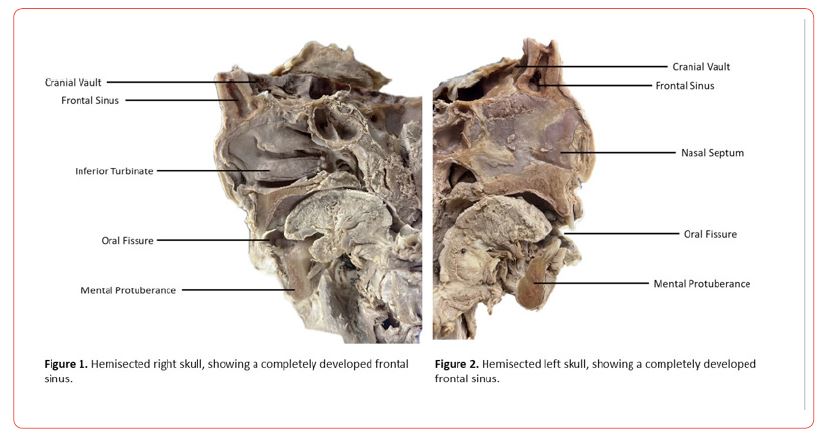
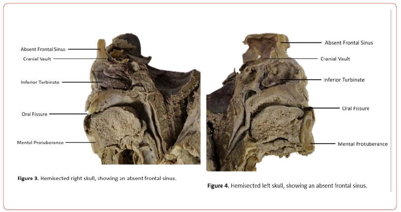
After data was collected from the cadavers, it was determined that three of the forty-eight cadaveric specimens exhibited bilateral agenesis of the frontal sinus leaving forty-five of the forty-eight cadavers to exhibit unilateral or bilateral present frontal sinuses. This correlates to a prevalence of bilateral frontal sinus absence in 6.25% of the specimen population.


Introduction
Embryology of Agenesis of Frontal Sinus Development
The frontal sinuses begin to develop around gestation week four and continue to develop into early Adulthood [2]; hypoplasia of these sinuses can occur at any point throughout that time frame. Agenesis would have to happen early on in the process and develop from a complete failure of pneumatization to occur. The frontal sinuses develop independently of the maxillary and ethmoid sinuses [11]. In a recent study, 100% of infantile frontal sinuses were absent while the anterior sphenoid, posterior sphenoid, and maxillary sinuses were already developed [11]. In a separate study conducted on children under the age of 15 months, MRI imaging revealed that the frontal sinuses were the last to develop [5]. 100% of the newborns imaged had present ethmoid sinuses, while only 45.7% of them had maxillary sinuses. By the end of the first year of life, 97.8% of the infants showed pneumatization of their maxillary sinuses. The sphenoid sinuses were present in 86% of the children aged 4 months to 24 months. The frontal sinuses were the last to develop with only 8% of the population showing pneumatization of the frontal sinuses by age 2 [5]. It wasn’t until age 15 that 97.8% of the children showed signs of present frontal sinuses.
It is theorized that the frontal sinuses begin to form around gestation week four through week sixteen as either the elongation of the nasal infundibulum into the frontal bones or due to the migration of the anterior ethmoidal cells into the frontal bones [2]. Within the frontal bones, primary pneumatization begins until around age two with secondary pneumatization occurring shortly thereafter [2]. The sinuses continue to grow via the resorption of the frontal bones until age 18 [2]. This growth occurs independently within each frontal bone, as the metopic suture joins the frontal bones together but does not inhibit the growth of the frontal sinuses [12]. Similarly, it has been theorized that persistence of the metopic suture (metopism) can affect the development of the frontal sinus. A 2017 study revealed that 17 out of 245 dry skulls presented metopism. Researchers then radiographically analyzed the selected 17 skulls for frontal sinus development [8]. Only one skull of the 17 showed frontal sinus agenesis, exhibiting a population prevalence of 6.25%. 12.5% of the skulls exhibited aplasia of the frontal sinus, while 25% exhibited hyperplasia8. The researchers concluded that metopism was found to not have a role in frontal sinus development based on the data collected.
Abnormalities in pneumatization can lead to variations such as single conjoined frontal sinuses, a unilateral frontal sinus, or bilateral agenesis of the frontal sinuses. Our study specifically focused on a bilateral agenesis of the frontal sinus.
Comparisons between variants
In a recent study performed on Oman patients, bilateral agenesis was also found in 2% of the Population [13]. This varies from our data where we found 6.25% of the population having agenesis of the frontal sinuses. However, in a separate study performed by Erdem et al. on Turkish individuals, the frontal sinus was bilaterally absent in 3.8% of the population. This prevalence is closer to that revealed in the Oman study. Both of these countries are located in a similar geographical area [7]. We hypothesize that our data differs from that of other various studies based on the population examined. Our sample population was from Texas in the United States where the predominant race is White, with the second most being Black. Genetic composition likely plays a role in the pneumatization of the frontal sinus.
Impacts of a lack of frontal sinus
While it is debated what exact functions the paranasal sinuses play in physiological processes andwhy they developed evolutionarily, the sinuses are believed to boast a variety of functions including decreasing the weight of the skull. They also are theorized to act as a buffer against facial trauma, providing an insulative feature to internal structures of the cranial cavity, and humidifying, heating, and filtering incoming air [14]. It should be noted that any of the paranasal sinuses, including the frontal sinus, can harbor infections or become inflamed, resulting in a condition known as sinusitis [15].
Bilateral agenesis of the frontal sinuses is thought to be benign in most cases. However, in a study investigating the sense of smell, Zinreich et al. concluded that complete opacification of the sinuses does diminish olfaction. They also concluded that the size of the sinus had no impact on olfaction, prompting the conclusion that hypoplasia would have no impact on smell but that complete agenesis of the frontal sinus would [9]. In a 2023 study conducted in Germany, patients with primary ciliary dyskinesia (PCD) were found to have much higher rates of bilateral frontal sinus agenesis which resulted in exacerbated respiratory symptoms [16]. It was concluded that individuals with agenesis of the frontal sinuses should be considered for diagnostic evaluation of PCD [16]. Due to the frontal sinuses being lined with mucus producing cells that help lubricate the nasal tissue and prevent it from drying out [8], it could be hypothesized that individuals without frontal sinuses may experience increased rates of infection, report complaints of dry nose, or other chronic respiratory symptoms. It has also been proposed by experts that agenesis of the frontal sinus can have surgical implications during a sinusotomy [10]. Hypothesized implications of frontal sinus agenesis could be explored by investigating the prevalence of expected symptoms in various populations with differing agenesis rates.
Conclusion
To conclude, this study contributes valuable insights into variations and developmental sequences of the frontal sinus. Literature is consistent regarding the prevalence of frontal sinus agenesis in various populations, ranging from 2% to 8% depending on the country of origin; the prevalence of bilateral frontal sinus absence in the sample population of this study was 6.25%, contributing corresponding evidence to current data established in the literature.
Highlighted in this study was the dynamic nature of sinus growth, with the frontal sinus exhibiting higher rates of variation in forms such as agenesis, hypoplasia, and aplasia due to the prolonged period required for complete pneumatization. Absence of the frontal sinuses typically presents in adolescence; although most patients remain asymptomatic, symptoms may begin to arise after the failure of pneumatization in early adulthood implicating the possible need for medical interventions and specialized patient management. Consideration of sinus anomalies such as bilateral frontal sinus absence in patient presentations will allow for a more comprehensive patient medical history which highlights the potential implications for patient care, such as the need for screening individuals with bilateral frontal sinus agenesis for conditions like primary ciliary dyskinesia (PCD).
Furthermore, our findings contribute to current data highlighting prevalence rates, which contribute to the importance of noting frontal sinus abnormalities in patient histories, ensuring appropriate care during surgeries, and future treatments of conditions such as chronic sinusitis. We believe that additional steps should be taken to further research regarding symptomology exhibited by patients with bilateral frontal sinus absence to better understand patient presentations.
Acknowledgement
The authors would like to thank the Willed Body Program at UNTHSC, the donors, and their families for their contribution to advancing medicine and anatomical knowledge.
Disclaimers
None.
Financial Support
None.
- Fahrioglu SL, VanKampen N, Andaloro C (2023) Anatomy, head and neck, sinus function and development. Stat Pearls - NCBI Bookshelf.
- Ma YX, Zhang WJ, Yan ZH, He JW, Cheng XJ, etal. (2013) Normal pneumatization time of paranasal sinuses in 799 children: evaluation with magnetic resonance imaging. CMAPH 93(11): 816-818.
- Cardesa A, Alos L, Nadal A, Franchi A (2017) Nasal Cavity and Paranasal Sinuses. (2nd Edition), Springer, Berlin, Pathology of the Head and Neck pp. 49-127.
- Štoković N, Trkulja V, Čuković-Bagić I, Lauc T, Grgurević L (2018) Anatomical variations of the frontal sinus and its relationship with the orbital cavity. Clin Anat 31(4): 576-582.
- Aydinlioğlu A, Kavakli A, Erdem S (2003) Absence of frontal sinus in Turkish individuals. Yonsei Med J 44(2): 215-218.
- Marino MJ, Riley CA, Wu EL, Weinstein JE, Emerson N, McCoul ED (2020) Variability of Paranasal Sinus Pneumatization in the Absence of Sinus Disease. Ochsner journal 20(2): 170-175.
- Çakur B, Sumbullu MA, Durna NB (2011) Aplasia and agenesis of the frontal sinus in Turkish individuals: a retrospective study using dental volumetric tomography. Int J Med Sci 8(3): 278-282.
- “Definition of Frontal Sinus - NCI Dictionary of Cancer Terms.” National Cancer Institute.
- Hong SC, Leopold DA, Oliverio PJ, Benson ML, Mellits D, etal. (1998) Relation between CT scan findings and human sense of smell. Otolaryngol Head Neck Surg 118(2): 183-186.
- Ozgursoy OB, Comert A, Yorulmaz I, Tekdemir I, Elhan A, etal. (2010) Hidden unilateral agenesis of the frontal sinus: human cadaver study of a potential surgical pitfall. Am J Otolaryngol 31(4): 231-234.
- Liao D, Duan C (2011) Computer-assisted anatomical evaluation of the nasal sinuses in infants. J Clin Otorhinolaryngol Head Neck Surg 25(23): 1057-1059.
- Batista SL, Viandelli Mundim-Picoli MB, Fortes Picoli F, Rodrigues LG, Bueno JM, etal. (2017) Prevalence of agenesis of frontal sinus in human skulls with metopism. J Forensic Odontostomatol 35(2): 20-27.
- Al Hatmi AS, Al Ajmi E, Albalushi H ,et al. (2023) Anatomical variations of the frontal sinus: A computed tomography-based study. F1000 Research 12:71.
- Cappello ZJ, Minutello K, Dublin AB (2023) Anatomy, Head and Neck, Nose Paranasal Sinuses. In Stat Pearls. Stat Pearls Publishing.
- Keir J (2009) Why do we have paranasal sinuses? J Laryngol Otol 123(1): 4-8.
- Schramm A, Raidt J, Gross A, Böhmer M, Beule AG (2023) Molecular Defects in Primary Ciliary Dyskinesia Are Associated with Agenesis of the Frontal and Sphenoid Paranasal Sinuses and Chronic Rhinosinusitis. Front Mol Biosci 10:1258374.
-
Sarah Brunson and Shaolin Liu*. Immunohistochemistry and its Applications in Neuroscience. Anat & Physiol Open Access J. 1(5): 2024. APOAJ.MS.ID.000522.
-
Frontal, Sinuses, Agenesis, Pneumatization, Prevalence, Cadavers, Embryology, Surgery, Symptoms, Sinusitis
-

This work is licensed under a Creative Commons Attribution-NonCommercial 4.0 International License.


