


 Research Article
Configuration of the Accessory Pancreatic Duct by the Injection-Radioscopy Method: Anatomical Study and Clinical Applications About 30 Cases
Received Date:February 17, 2026; Published Date:March 16, 2026
Summary
Objective: the purpose of our work was to study the morphology of the accessory pancreatic duct (APD) and to review the literature.
Material and Method: It was a fundamental observational study for descriptive purposes. It was about 30 pieces of fresh pancreas taken from
anatomical subjects during forensic autopsy. The average age was 39.43 years. The configuration of the APD was studied by the radioscopy-injection
method. The parameters studied were morphological and morphometric.
Results: the presence of a APD was noted in 11 anatomical rooms, i.e. in 36.7% of cases. They were classically born from the Main Pancreatic
Duct (MPD) at the junction of the isthm and the head of the pancreas. It all led to a permeable Minor Duodenal Papilla (mDP) after injection at the
CPP level upstream of the APD abuchement. Thus, the average distance between the Major Duodenal Papilla (MDP) and the PDm was 11.11 mm
[2.3mm-20mm]. The APD was born above the MPD in 6 cases (20%): it seemed to prolong the initial direction of MPD in 4 cases among which there
was 1 case where the APD was larger than the MPD which described loops around the latter; and in the 2 remaining cases it was generally oblique
at the top and towards the 2nd portion of the Duodenum (D2) while receiving canalicules of the upper and lower cephalic parts. The APD was born
below the MPD in 5 cases (16.7%); it described a lacross with a concavity superior to the lower cephalic part and then oblique at the top towards D2
at the upper cephalic part. The average diameter of the APD is 1.55 mm [0.9mm-2.3mm].
Conclusion: The APD’s anatomical study in the population is interesting in the sense that it makes it possible to identify or estimate groups at
risk regarding the occurrence of certain pathologies of the bilio-pancreatic pathways.
Keywords:Accessory pancreatic duct; Minor duodenal papilla; Injection-radioscopy
Introduction
The pancreas is a voluminous gland that is both exocrine and endocrine, odd, appendage of the digestive tract [1,2]. Traditionally, the exocrine pancreas is drained mainly by the main pancreatic duct (MPD) and inconsistently by the accessory pancreatic duct (APD). However, when the latter exists, it has, at the same time or in isolation, multiple anatomical variations of origin, path, termination and/or dimensions [3]. In addition, it can become the main or only drainage duct of the gland in certain particular anatomical circumstances. His study is interesting in that it plays an important role in obstructions of the main pancreatic duct regardless of the cause. Thus, its absence is a risk factor for the occurrence of certain more or less serious pancreatic pathologies [4-6]. The objective of this study was to describe the configuration of the accessory pancreatic duct by the radioscopy injection method and to review the literature..
Materials and Methods
This is a fundamental observational study for descriptive purposes. It concerned 30 pieces of fresh pancreas taken from anatomical subjects during forensic autopsy (numbered from P1 to P30). The average age was 39.43 years (Type deviation=13.9). The sex ratio was 3.3. The average size of the subjects was 175.10 cm [extreme 163cm-187cm]. The average length of the pancreas was 22.48 cm +/-2.4. In the laboratory, the transverse length of the pancreas was measured with a tape measure, from the right edge of the head from the middle of the descending portion of the duodenum to the end of the tail. Then the choledoc duct was spotted on the posterior face of the room and then catheterized with an intranulle of adapted caliber. Subsequently, the pancreas was sectioned sagittally over its entire height using a scalpel at the body-tail junction. The main pancreatic duct was spotted in the section and catheterized. Then the room was sent to the imaging department to take x-ray shots using a versatile digital radio-fluoroscopy device with a tilting table. First of all, a first shot without injection was taken.
Then, the contrast agent was injected at constant pressure, under radioscopic control, into the main pancreatic duct (1st step), then into the choledoch (2nd time) and finally both at the same time (3rd time). The intra-pancreatic ducts were spotted until they were dislodged at the duodenal mucosa. When the injection was deemed sufficient, front incidence shots are taken for the different times. For each preparation, photographs were taken. The assessment of the permeability of the duodenal papillae was judged on the injection clichés. The actual dimensions (length and diameter of the ducts) were taken at the PACS (Picture Archiving and Communication System). The parameters studied were: the size of the anatomical subject; the length of the pancreas; the existence and configuration of the accessory pancreatic duct; the permeability of the major and minor duodenal papillae; the overall path as well as the dimensions of the main and accessory pancreatic ducts. The data have been analyzed by SPSS software version 25.0.
Results
The presence of an accessory pancreatic duct was noted in 11 anatomical parts, in 36.7% of cases. They were born classically from the main pancreatic duct at the junction of the isthm and the head of the pancreas. It all led to a minor duodenal papilla (PDm) permeable after injection at the main pancreatic duct upstream of the opening of the accessory pancreatic duct. Thus, the average distance between the major duodenal papilla (PDM) and the PDm was 11.11 mm with extremes of 2.3 mm and 20 mm. The APD was born above the MPD in 6 cases (20%):
• It seemed to extend the initial direction of MPD in 4 cases
among which there was 1 case where the APD was larger than
the MPD which described loops around the latter (Figure 1).
• And in the 2 remaining cases it was globally oblique at the
top and towards the 2nd portion of the duodenum (D2) while
receiving canalicles from the upper and lower cephalic parts
(Figures 2&3).
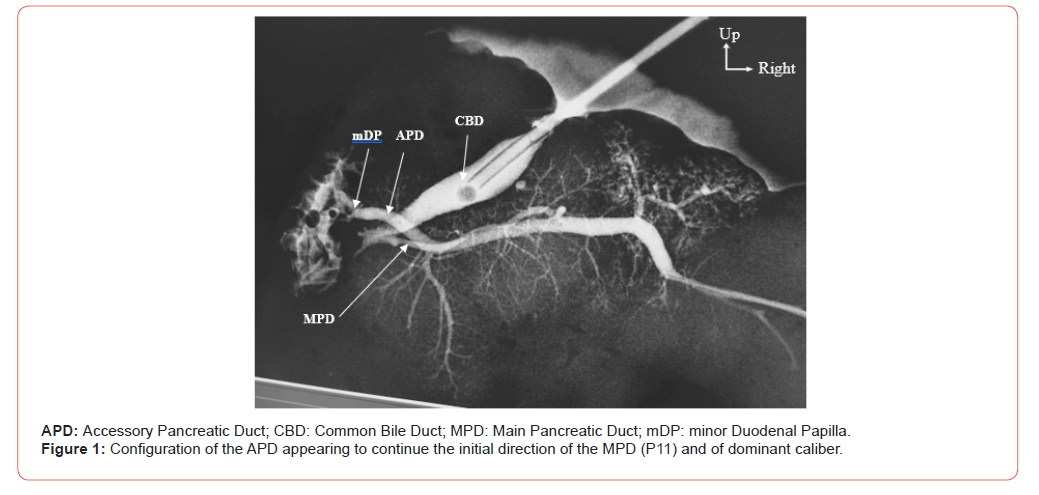
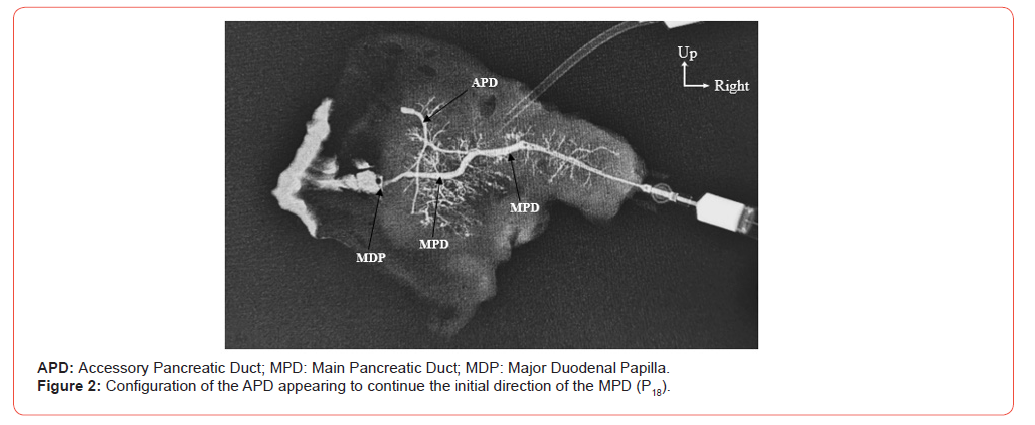
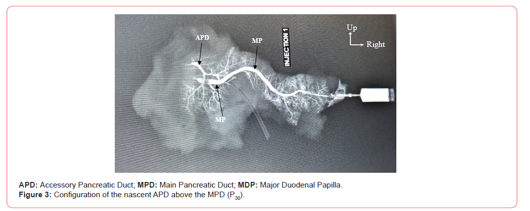
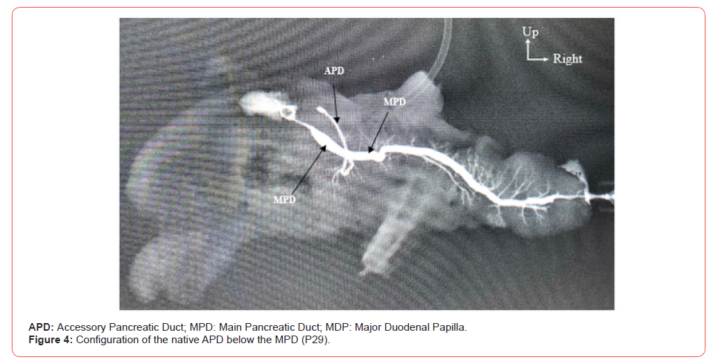
The APD was born below the MPD in 5 cases (16.7%); it described a cross with a concavity superior to the lower cephalic part and then oblique to the top towards D2 to the upper cephalic part (Figure 4). The average diameter of the APD is 1.55 mm with extremes of 0.9 mm and 2.3 mm.
Discussion
Methodology
The anterograde injection-radioscopy method is a means of seeing almost all the ramifications of the pancreatic ducts. This method allowed many authors to provide new data on pancreatic ducts [7-10]. It is an easily feasible exploration method, already proven for years. Indeed, dissection makes it easy to follow the duct that drains the tail and body of the pancreas during its relatively straight path to the head of the pancreas [11,13]. But it becomes much more difficult to follow the duct, to identify its tributaries and its communications once it has entered the head of the gland. Thus, the rise of endoscopic retrograde pancreatography, as a clinical procedure, has prompted some authors after a tunneling of the choledoc and main pancreatic ducts to obtain pancreatograms in post-mortem anatomical subjects [9,10,14]. In 1911, Baldwin was already providing convincing results by an injection-radioscopy method that the pancreatic duct dissection method [15]. In addition, the anterograde injection, giving similar results, is a much more physiological test by providing more or less details of the anatomy of the pancreatic ducts.
Morphology of the Accessory Pancreatic Duct
An accessory pancreatic duct that generally drains the anterior and upper parts of the head of the pancreas is present between 50% and more than 70% of the subjects [16,17]. This duct is located more ventrally than the main pancreatic duct. And in most cases, there is a communication between the two ducts in the head of the pancreas. The accessory pancreatic duct may have its own duodenal orifice on the minor duodenal papilla [15,16,18]. If an accessory pancreatic duct has both a permeable duodenal orifice and a communication with the main pancreatic duct, there is at least one anatomical arrangement for an alternative pathway by which pancreatic secretion could reach the duodenum if the orifice of the main pancreatic duct is blocked. According to Nowak’s work, the absence of an accessory pancreatic duct is a factor of pancreatitis [17]. He reported that among people with pancreatitis, only 17% of patients had an accessory pancreatic duct, compared to 69% in the control group.
Schmitt noted the existence of a much higher accessory pancreatic duct (94.1%) [19]. The advantage of the presence of the accessory pancreatic duct and its permeability is that it forms a safety valve by reducing the pressure in the main pancreatic duct [20]. In our study, the accessory pancreatic duct existed in 11 cases (36.7%) and it all led to a permeable minor duodenal papilla. These ratios are similar to those of the literature. Our rate of permeable minor duodenal papillae is close to that of Millbourn, namely 33% on a series of 182 cases [16]. Kamisawa finds 43% of minor duodenal papillae permeable by making a relevant observation [18]. He considers that a minor duodenal papilla impermeable during a retrograde endoscopic pancreatography using radiopaque products could be permeable with the retrograde injection of tinted products such as indigocarmine; in this case, it can be said that the levels of permeable minor duodenal papillaes found using radiopaque products are slightly below the levels found in vivo.
However, we can remember in the light of the various studies that the proportion of subjects within a population with a permeable minor duodenal papilla oscillates between a third and half of this population with one exception: Wilasrusmee finds 8.74% of minor duodenal papilla permeable to methylene blue injection on 103 pancreases taken from Thai subjects [21]. The ducts that reach the minor duodenal papilla, from the origin to the duodenal end, can be one-eyed. Thus, the accessory pancreatic duct then acts entirely as a major tributary of the main pancreatic duct [5,6,22,23]. In total, there seems to be at least one agreement that in about 90% of pancreas, the only or main excretory duct is the main pancreatic duct that opens to the major duodenal papilla. In about 50 to 70% of the pancreas, an accessory pancreatic duct is present, and can transmit various amounts of secretion (none, part or all) into the duodenum through the minor duodenal papilla [3-16]. In a small number of cases, the main pancreatic duct has undergone atrophy and the secretion of the entire gland reaches the duodenum through the minor duodenal papilla.
Thus the accessory pancreatic duct constitutes the main duct system [3]. When the accessory pancreatic duct is well developed, its path through the head of the gland is in a straight line without the angles. And the main pancreatic duct usually has, in the path, loops around the latter [3]. This configuration was noted in a case in our series (3.3%). Two accessory pancreatic ducts with two minor papillae and with a communication with the major duct system were found in one case by Berman et al. (1960) in their series of 130 specimens [11]. The presence of several small accessory pancreatic ducts has been described by Cross (1956) as being frequent. Indeed, after examining 400 pancreases, he concluded that in most cases, the choledoc duct in its intracranial and intraduodenal segments is connected to many small ducts of pancreatic origin. In addition, many small pancreatic ducts penetrate directly into the lumen of the intestine, especially in the hepatopancreatic ampoule and in the second portion of the duodenum.
It is true that when the heterotopic pancreatic tissue, disconnects with the main pancreas, is present in the wall of the duodenum, the small ducts of the heterotopic glandular tissue discharge directly into the duodenum [3]. But the literature review shows that the conduits of this type described by Cross have not been recognized by most authors [24-26]. In some very rare situations, the main pancreatic duct and the accessory pancreatic duct can coexist with separate duodenal orifices without communications in the gland. Thus, there are two parts within the pancreatic parenchyma, each with a distinct and autonomous drainage canal system. In such cases, the drainage system of the accessory pancreatic duct may be larger and more important. This anatomical arrangement was first described in 1849 by Rokitansky as a double pancreas. But recently, it is recognized as the pancreas divisum [3]. An absence of fusion of the main and accessory pancreatic ducts was found, also by magnetic resonance cholangiopancreatography, by Adibelli et al. in 90 cases, i.e. 5.5% of the 1628 pancreatograms performed.
And they concluded that the existence of a divisum pancreas associated with anatomical variations in the biliary tract would be a risk factor for the development of pancreatic biliary tumors and should be closely monitored by magnetic resonance cholangiopancreatography [27]. It has been reported by some authors that in the pancreas divisum, inadequate drainage via the accessory pancreatic duct can predispose to pancreatitis [28]. This pathology was also found frequently in all series where this anatomical variant was observed [28-30]. Thus, the authors concluded that the absence of fusion of the pancreatic ducts predisposes to pancreatitis [4,31,32]. The minor duodenal papilla is usually located about 2 cm above the major duodenal papilla on the duodenal mucosa and on a ventral plane in relation to the major papilla [15]. It is much easier to recognize the major duodenal papilla. But the minor duodenal papilla, although sometimes easy to find, can be very discreet. However, it could be identified in each of the 100 specimens studied by Baldwin [15]. It is not uncommon for there to be a certain variation in the frequency of the different types of canal morphology found by different authors.
Conclusion
The anatomical study of the accessory pancreatic duct within the population is interesting in the sense that it makes it possible to identify or estimate the groups at risk regarding the occurrence of certain pancreatic pathologies. These anatomical data have important clinical applications and open up new medical-surgical perspectives, including the prevention of the occurrence of pancreatitis. This prevention is based on the early detection of anatomical variants of the accessory pancreatic duct.
Conflits of Interests
None
Ethics Committee Approval
Approved
Authors’ Contribution
All authors conbributed to the writting and profreading of this manuscript.
- Kamina P (2004) Précis danatomie clinique. Maloine, Tome 3 Paris PP: 313-321.
- Netter FH (2017) Atlas danatomie humaine. Masson, 7ème éd, Paris PP: 340-353.
- Cruickshank A H, Alan H (1986) Pathology of the pancreas. Springer-Verag, New-York, 1e édition, PP: 288.
- Jagielski M, Smoczyński M, Adrych K (2018) The anatomical variations of pancreatic duct in the patients with pancreatic diseases. Eur J Transl Clin Med 1: 63-66.
- Prasanna LC, Rajagopal KV, Thomas HR, Bhat KM (2015) Accessory pancreatic duct patterns and their clinical implications. J Clin Diagn Res 9(3): AC05-AC07.
- Vinay Kumar N, Lokanadham S (2019) A Study on Incidence of Accessory Pancreatic Duct and its Clinical Importance. Maedica (Bucur) 14(1): 22-25.
- Darnis E, Cronier P, Mercier P, Moreau P, Pillet J, Boyer J (1984) A radio-anatomical study in vivo on pancreatic ducts. Anat Clin 6(2): 109-116.
- Kasugai T, Kuno N, Kobayashi S, Hattori K (1972) Endoscopic pancreatocholangiography. Gastro enterology 63(2): 217-226.
- MacCarty RL, Stephens DH, Brown AL Jr, Carlson HC (1975) Retrograde pancreatography in autopsy specimens. Am J Roentgenol Radium Ther Nucl Med 123(2): 359-366.
- Sigfusson BF, Wehlin L, Lundström CG (1983) Variant of pancreatic duct system of importance in endoscopic retrograde cholangiopancreatography observations on autopsy specimen. Acta Radiol Diagn (Stockholm) 24(2): 113-128.
- Berman LG, Prior JT, Abramow SM, Ziegler DD (1960) A study of the pancreatic duct system in man by the use of vinyl acetate casts of postmortem preparations. Surg Gynecol Obstet 110: 391-403.
- Govindraj N, Shabna C (2017) Variations in the duct system of pancreas: a cadaveric study. Int J Anat Res 5(3.1): 4136-4143.
- Shahriah S, Nurunnabi ASM, Johora F, Siddiqua D, Ara S (2014) Cadaveric study of the anatomical variations of the accessory pancreatic duct. J Bangladesh Soc Physiol 9(2): 83-88.
- Prasad M, Rout S, Putta T, Kurien RT, Chowdhury SD, et al (2019) Anatomical patterns of the pancreatic ductal system-a cadaveric and magnetic resonance cholangiopancreatography study. J Morphol Sci 36: 279-285.
- Baldwin WM (1911) The pancreatic ducts in man, together with a study of the microscopical structure of the minor duodenal papilla. Anat Rec 5(5): 197-228.
- Millbourn E (1950) On the excretory ducts of the pancreas in man, with special reference to their relations to each other, to the common bile duct and to the duodenum. Acta anat 9(1-2): 1-34.
- Nowak A, Nowakowska-Dutawa E, Rybicka J (1990) Patency of the Santorini duct and acute biliary pancreatitis. A prospective ERCP study. Endoscopy 22(3): 124-126.
- Kamisawa T (2004) Clinical significance of the minor duodenal papilla and accessory pancreatic duct. J Gastroenterol 39(7): 605-615.
- Schmitt F, Maignan A, Ploteau S, Hamel A, Lagier S, et al. (2010) New anatomical data on the drainage patterns of the uncinate process of the pancreas. Surg Radiol Anat 32(8): 777-781.
- Pina LN (2016) Does the accessory pancreatic duct have any protective role in acute biliary pancreatitis? Anatomical findings from cadaver dissections. MOJ Anat Physiol 2(7): 180.
- Wilasrusmee C, Pongchairerks P (1999) Panreaticobiliary ductal anatomy in thai people. J Hepatobiliary Pancreat Surg 6(1): 79-85.
- Arora AK, Piplani ML, Kapoor SS, Bhatia BS, Singh ARK, et al. (2011) A research study of santorini duct. J Clin Diagn Res 5(8): 1510-1513.
- Mchonde GJ, Gesase AP (2014) Termination pattern of the main and accessory pancreatic ducts among Tanzanians. Anat J Afr 3: 223-227.
- Ishaque I, Ilyas A, Khan O, Kazmi QH, Ladak AH, et al. (2020) Role of magnetic resonance cholangiopancreatography (MRCP) in learning anatomical variations in pancreatic duct. J Pak Med Assoc 70(3): 472-476.
- Ojo AS (2020) Pancreatic Duct Variations and the Risk of Post-Endoscopic Retrograde Cholangiopancreatography Pancreatitis. Cureus 12(9): e10445.
- Aube C, Hentati N, Tanguy JY, Fournier HD, Papon X, et al. (2003) Radio-anatomic study of the pancreatic duct by MR cholangiopancreatography. Surg Radiol Anat 25(1): 64-69.
- Adibelli ZH, Adatepe M, Isayeva L, Esen OS, Yildirim M (2017) Pancreas divisum: a risk factor for pancreaticobiliary tumors - an analysis of 1628 MR cholangiography examinations. Diagn Interv Imaging 98(2): 141-147.
- Covantsev S, Chicu C, Mazuruc N, Belic O (2022) Pancreatic ductal anatomy: more than meets the eye. Surg Radiol Anat 44(9): 1231-1238.
- Covantev S (2018) Pancreas divisum: a reemerging risk factor for pancreatic diseases. Rom J Intern Med 56(4): 233-242.
- Renzulli M, Pagano N, Golfieri R (2020) Pancreas divisum inversus. Clin Anat 33(5): 646-652.
- De Jong DM, Stassen PM, Poley JW, Fockens P, Timmer R, et al. (2021) Clinical outcome of endoscopic therapy in patients with symptomatic pancreas divisum: a Dutch cohort study. Endosc Int Open 9(7): E1164-E1170.
- Jirasiritham J, Wilasrusmee C, Poprom N, Larbcharoensub N (2016) Pancreaticobiliary Ductal Anatomy in the Normal Population. Asian Pac J Cancer Prev 17(9): 4363-4365.
-

This work is licensed under a Creative Commons Attribution-NonCommercial 4.0 International License.