

 Case Report
Cardiovascular Anatomy of the Huemul Deer (Hippocamelus bisulus)
Received Date:August 19, 2026; Published Date:September 08, 2026
Abstract
Hearts from a neonatal death case, from another young fawn and an adult male resulted in a first description of the cardiac anatomy in huemul deer (Hippocamelus bisulcus). Dextrocardia, a patent ductus arteriosus, and a congenital atrial septal defect were confirmed in the neonate. In this species, the anterior descending coronary artery (anterior interventricular artery) is entirely intramyocardial: defense or parietal stress under certain conditions can lead to severe ventricular arrhythmia and ‘sudden death’. The cardiac conduction system follows the general pattern found in mammals, particularly ungulates. The mitral valve presented striated cardiac muscle within its leaflets - a feature found in some species but never as prominently as in the huemul. The heart of the neonate had a structural congenital heart disease (non-lethal), but it died right after birth due to intestinal volvulus caused by congenital jejunoileal junction stenosis. Both issues add to the already known harm caused by inbreeding. These significant findings underscore the need to intensify efforts to prevent such occurrences likely stemming from inbreeding.
Keywords:Cervid; huemul; cardiac anatomy; pathology; congenital; genetics; inbreeding
Introduction
The huemul (Hippocamelus bisulcus) is one of the most threatened deer species on the planet [1]. Although it was persecuted rather than studied for much of the 20th century, the study and preservation of this species have gained significant prominence in the latter part of that century and in the present one. Accordingly, recent studies confirmed that the artificial all-year permanence in summer ranges explains the high prevalence of pathologies and concomitant very short live spans [2-7]. Initiated in 2012, the project “Huemul in danger: adaptive management proposal” was approved to Flueck and Smith-Flueck, and together with the Fundacion Shoonem resulted in the establishment of the Breeding Station Shoonem. By 2022, and in collaboration with the Fundacion Temaiken, the first huemul were captured and translocated to the Breeding Station, which has been operating since then.
Within this framework of individual protection and monitoring, a huemul fawn died shortly after birth; a necropsy was subsequently performed, along with a detailed analysis of the cause of death and tissue histology, in accordance with established protocols. In the present article, we describe the cardiac anatomy of this neonatal fawn and of additional huemul cases. One case stem from a specimen that was one or two weeks old, while the third case stems from an adult male. Based on our literature review, these are the first such descriptions made available. We present a study of the heart from a specimen that was a few days old and exhibited dextrocardia, a patent ductus arteriosus (also called the ductus Botalli), and an atrial septal defect. Additionally, the coronary anatomy of the huemul and the atrioventricular cardiac conduction system (bundle of His and its branches) are also described here for the first time.
Case Presentation
The heart of a huemul that died as a neonate due to intestinal volvulus caused by congenital jejunoileal junction stenosis was dissected. The necropsy was performed at the Pasteur Institute in Buenos Aires (Argentina), supervised by the head of Anatomic Pathology and the facility’s medical director, with the participation of members from the University of Buenos Aires (UBA), and the Temaikén Foundation. 29 Regarding the heart inspection: a supersaturated sodium solution and 1% formalin were used for preservation, after which the coronary tree was injected with prevulcanized latex following the laboratory’s standard technique.
The dissection was initially performed using the nakedeye technique, subsequently supplemented by magnification. A Newton® microscope with 6x to 10x magnification was used for the detailed dissection. Cardiovascular surgical forceps and scissors were used to dissect the delicate structures. It is important to specify the use of these particular instruments, as standard tools for general surgery, necropsy, or routine dissection are unsuitable. The method used to inject the coronary tree is also significant, given that the coronary vessels of the newborn huemul barely exceeded one millimeter in diameter.
Results
The results are rather surprising. Although inbreeding leads to
an increased incidence of congenital defects, we did not expect to
find three structural congenital heart anomalies, namely:
a) dextrocardia: isolated, with “situs solitus” and
atrioventricular concordance, concordant venous return, and
L-aortic arch.
b) patent ductus arteriosus: Ostium secundum-type
interatrial connection. It should be noted that this finding is
unequivocal; it is not a simple patent oval foramen, but a clear
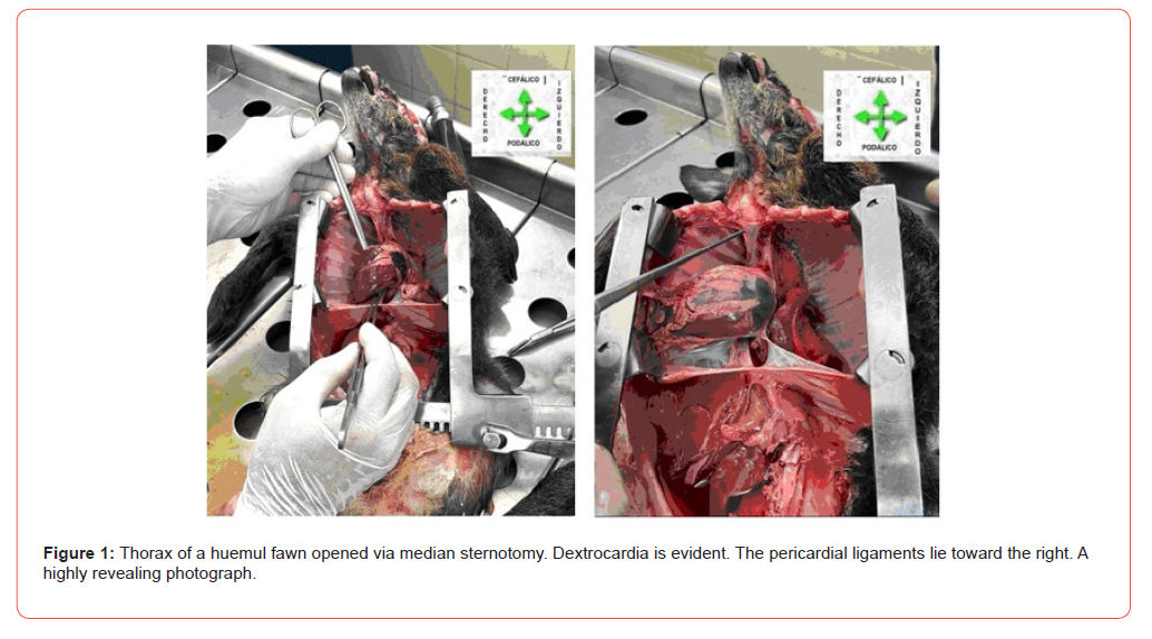
atrial septal defect, as highlighted in the photographs (Figure
1).
General External Configuration
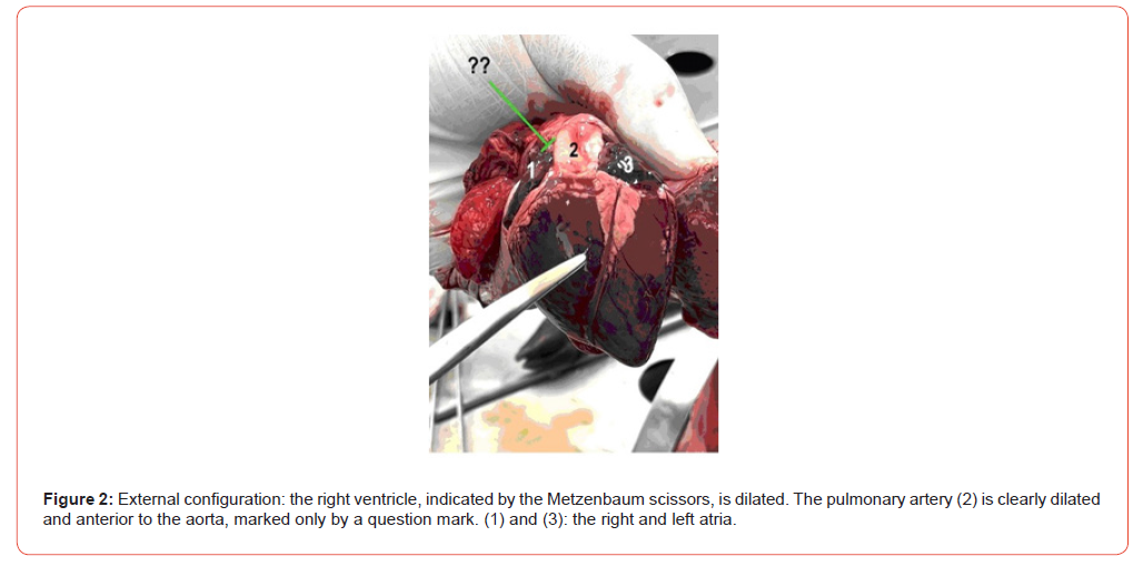
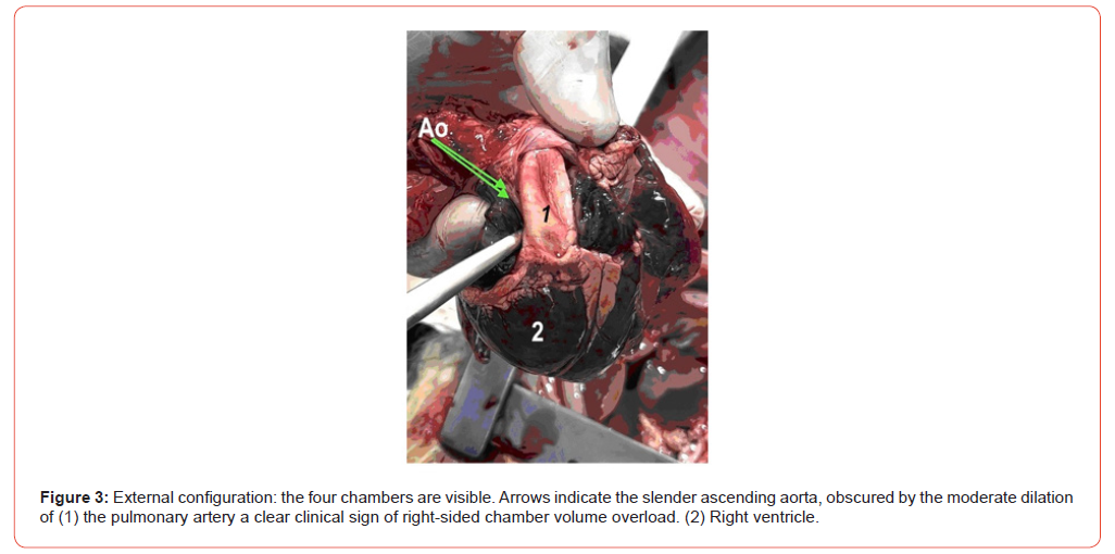
In this case, a dilated right heart chamber was remarkable. This prompted consideration of right-sided hyperflow conditions, leading to the discovery of an atrial septal defect and a patent ductus arteriosus (Figure 2,3).
Ductus Arteriosus
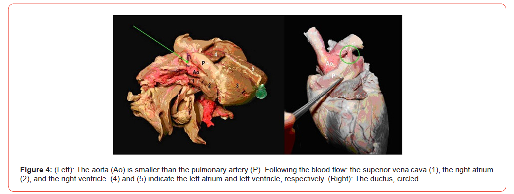
We have found a patent ductus in two huemul hearts: one from an adult and the fawn case detailed here. In the adult, the patent tract had a negligible lumen, though its diameter was not insignificant. In the fawn case at hand, however, the ductus was clearly patent and of considerable size. Figure 4 shows an adult heart (right) alongside the heart from the fawn. Ligatures were applied to the great vessels of the heart from the fawn in order to inject prevulcanized latex into the coronary arteries - and only the coronary arteries (Figure 4).
Coronary Arteries
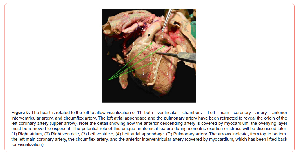
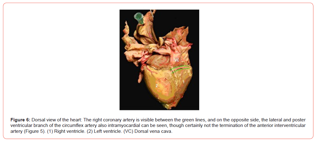
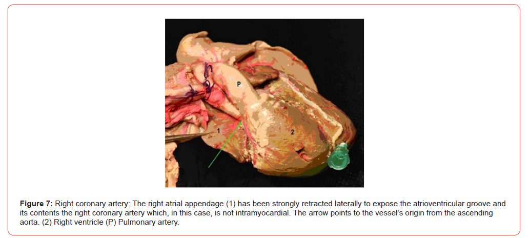
The coronary arteries in the huemul (based on two fully dissected specimens: an adult and the fawn of this report) follow the general mammalian pattern. There are two coronary arteries designated right and left along with their branches. It is worth noting that we observed a configuration in the huemul that is uncommon in mammals (though found in marsupials and some rodents). Specifically, the anterior descending artery (anterior interventricular artery) is entirely intramyocardial. The anterior descending artery is intramyocardial in both the adult and fawn specimens of this report. In the commentary, we will discuss the significance of this finding in relation to sudden death (Figure 5-7).
Cardiac Conduction System
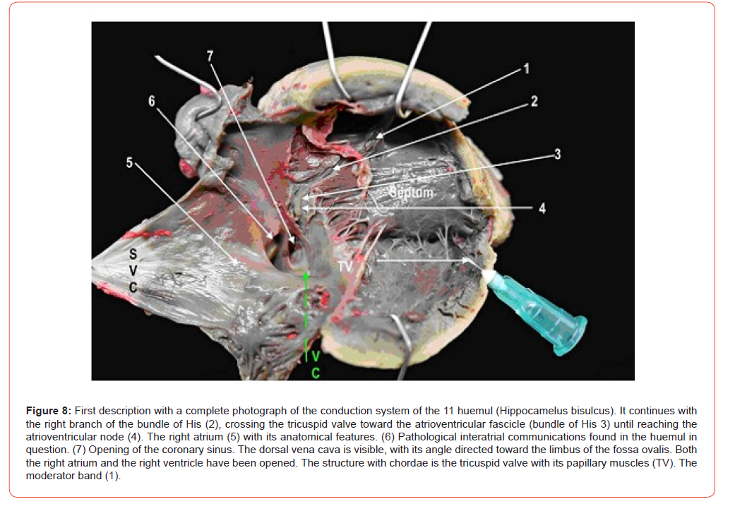
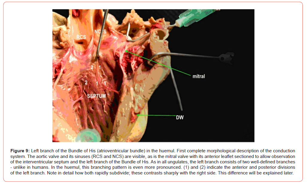
The cardiac excitation-conduction system-or the Tawara-His- Purkinje system, discovered over three decades of human effort to understand how the orderly cardiac impulse is transmitted–is composed of myocardial cells (not nerves) isolated within collagen. One of the authors has dissected specimens from more than 40 animal species (including whales), successfully demonstrating the atrioventricular node (Tawara), the atrioventricular bundle, and its branches in the huemul. This is the first description featuring photographs of this system, which follows the general structural plan found in mammals, particularly ungulates (Figure 8,9).
Associated Congenital Heart Disease
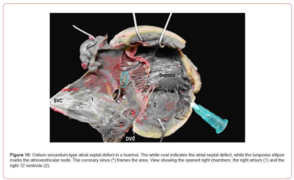
Ostium secundum atrial septal defect and patent ductus arteriosus were confirmed. As illustrated in Figures 4 and 8, this specimen presented a patent ductus arteriosus and an atrial septal defect. The atrial septal defect is described in further detail (Figure 10,11).
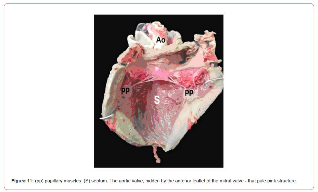
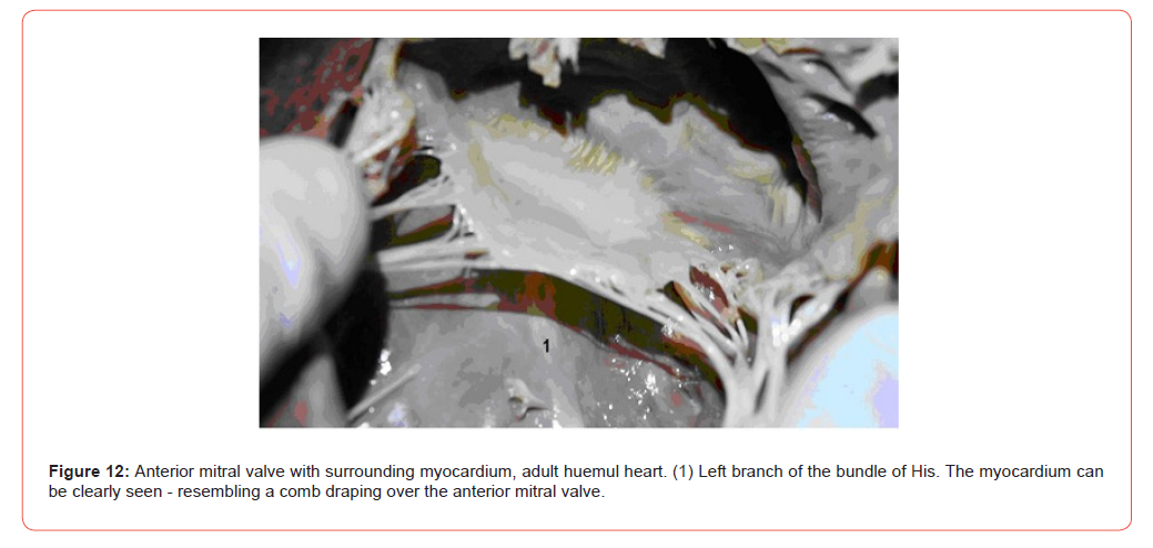
Figure 12 reveals the muscle within the veils an anatomical feature rarely visualized with such clarity. Although the photograph shows the adult specimen, the fawn mentioned in this publication also possesses this trait; the image is included solely to provide further visual evidence (Figure 12).
Discussion
Sudden Death
South American camelids - and the huemul in particular were studied only anecdotally until recent decades, when genuine efforts to protect the species began to be undertaken. This is one such effort: gaining an understanding of the huemul species. Herbivores in general, and ungulates in particular, tend to exhibit a poorly understood phenomenon when threatened: sudden deaths. Theories have been proposed that lack any physiological or morphological basis. It is reasonable to hypothesize a vagal reaction - or any other mechanism one might choose – such that the Divine Providence, in the face of imminent death, spares the animal by bringing about a painless, premature end. While we may poetically subscribe to that elegant intellectual explanation, the findings in two huemul hearts with identical structures draw our attention to a specific fact: the anterior interventricular artery is completely intramyocardial.
Although this finding is observable in rodents, humans, marsupials, and cetaceans, in this instance, the vessel in question – which supplies blood to 55% of the left ventricle and the septum - is entirely covered by a layer of myocardium. In this morphological study, we document this clearly, proposing that rather than a vagal reflex or some other event that is difficult to verify, it is an isometric effort (such as a struggle or parietal stress caused by arterial hypertension resulting from a catecholamine surge) which triggers a contraction across the ENTIRE left ventricular wall AND the myocardial bridge. Under certain conditions, this can lead to severe ventricular arrhythmia and SUDDEN DEATH. We would certainly not contradict Sir William Harvey, who as early as 1617 began to say that “until every aspect of morphology is clarified, no one should speak of physiology.” Indeed, more than 400 years later, we remain aligned with his views. It is also our duty to report this morphological finding as another step forward in our understanding, and to at least hypothesize about its function.
Structural Congenital Heart Disease and Digestive Tract Disease
The finding of a structural congenital heart disease (non-lethal), along with congenital jejunoileal junction stenosis in the huemul (lethal in this case), adds to the already known harm caused by inbreeding. These morphological findings reinforce the concept of crossbreeding huemul from different areas - with an appropriate program - to mitigate inbreeding.
Conduction System
This is the first detailed (and generic) description of the atrioventricular bundle in this cervid. It is worth noting its differences from that of humans and other species. The structure of the conduction system has already been dissected in another adult huemul with identical findings.
The Mitral Valve Is Supported by The Usual Collagen System and By Striated Muscle
This finding, which is not exclusive to the huemul deer as it is also found in humans and other mammals, is very prominent in the huemul. It is so prominent that it can be seen with the naked eye, something that is not so clearly visible in other mammals. The increase in intracavitary pressure during isometric exertion, as well as the unknown connections of these myocardial fibers, may be a source of arrhythmogenic activity.
Conclusions
The cardiac anatomy of the huemul has been delineated in great detail, covering its external and internal configuration, coronary vessels, and cardiac conduction system; the descriptions of the latter two represent first case descriptions in the international literature. A non-immediately lethal congenital structural heart defect was identified in this fawn, as well as an intestinal malformation (the cause of death), both possibly linked to the inbreeding affecting the species. This significant finding underscores the need to intensify efforts to prevent such occurrences. The presence of intramyocardial vessels suggests a potential cause of sudden death in these animals.
Declarations
Acknowledgments
The research was done as part of the project “Huemul in danger: adaptive management proposal” approved by the authorities of the Province of Chubut, and the corresponding transit permits.
Conflict of Interests
The authors declare no conflict of interest.
Ethics Approval
This does not apply in the current article.
- Jiménez J, Guineo G, Corti P, Smith JA, Flueck W, et al. (2008) Hippocamelus The IUCN Red List of Threatened Species. e.T10054A3157381.
- Flueck WT, Smith-Flueck JM (2008) Age-independent osteopathology in skeletons of a south American cervid, the Patagonian huemul (Hippocamelus bisulcus). J Wildl Dis 44(3): 636-648.
- Flueck WT, Smith-Flueck JM (2011) Recent advances in the nutritional ecology of the Patagonian huemul: implications for recovery. Animal Prod Sci 51(4): 311-326.
- Flueck WT, Smith-Flueck JM (2017) Troubling disease syndrome in endangered live Patagonian huemul deer (Hippocamelus bisulcus) from the Protected Park Shoonem: Unusually high prevalence of osteopathology. BMC Res Notes 10: 739.
- Flueck WT (2020) Nutrition as an etiological factor causing diseases in endangered huemul deer. BMC Res Notes 13(1): 276.
- Flueck WT, Smith-Flueck JM, Escobar ME, Zuliani M, Fuchs B, et al. (2022) Loss of migratory traditions makes the endangered Patagonian huemul deer a year-round refugee in its summer habitat. Conservation 2(2): 322-348.
- Flueck WT, Smith-Flueck JM, Escobar ME, Zuliani M, Fuchs B, et al. (2023) Review of Historical and Zooarchaeological Data to Trace Past Biogeographic Distribution of Endangered Huemul (Hippocamelus bisulcus) to Enhance Conservation Strategies. Conservation 3(4): 569-594.
-

This work is licensed under a Creative Commons Attribution-NonCommercial 4.0 International License.