


 Review Article
Biomechanical Changes in Cardiac Structure and Activation of Metabolic Pathways in Aortic Stenosis
Received Date:March 12, 2026; Published Date:April 09, 2026
Abstract
Degenerative Aortic Stenosis (AS) is the most prevalent valvular heart disease in the Western world and represents a paradigmatic model of chronic pressure overload. Sustained increases in left ventricular wall stress induce concentric hypertrophy, interstitial fibrosis, and alterations in the extracellular matrix. In parallel, metabolic reprogramming occurs, characterized by shifts in substrate oxidation, mitochondrial dysfunction, and activation of signaling pathways such as AMPK, mTOR, Notch1, transforming growth factor-β (TGF-β), and Sphingosine-1-Phosphate (S1P). In addition, the natriuretic peptide system modulates the initial adaptive response. This review integrates the biomechanical and metabolic mechanisms involved in the progression from compensated hypertrophy to heart failure in aortic stenosis.
Introduction
Calcified Aortic Stenosis (AS) is currently recognized as an active and complex disease characterized by chronic inflammatory processes, osteogenic differentiation of valvular interstitial cells, and progressive remodeling of the aortic valve [1,2]. This process leads to narrowing of the valvular orifice and, consequently, to progressive obstruction of the left ventricular outflow tract. From a hemodynamic perspective, this obstruction produces a chronic increase in left ventricular afterload, resulting in elevated myocardial wall stress. According to Laplace’s law, wall stress is directly proportional to intracavitary pressure and ventricular radius and inversely proportional to wall thickness [3]. As a compensatory mechanism to counteract this sustained increase in afterload, the myocardium develops concentric Left Ventricular Hypertrophy (LVH), characterized by increased wall thickness without significant dilation of the ventricular cavity [4]. Initially, this process is adaptive, as it reduces wall stress and helps maintain cardiac output. However, over time, sustained hypertrophy becomes associated with structural, molecular, and metabolic alterations that drive the transition from a compensated state to myocardial dysfunction and the development of heart failure [5].
Ventricular Biomechanics in Aortic Stenosis
Pressure Overload and Mechanotransduction
Chronic elevation of ventricular afterload induces a complex mechanotransduction response in car-diomyocytes. This process involves the conversion of mechanical stimuli, such as stretch or increased wall tension, into intracellular biochemical signals that regulate gene expression and cellular growth. In this context, several mechanoreceptors located in the sarcolemma and cytoskeleton, including integrins and focal adhesion complexes, play a key role in detecting changes in the mechanical environment of the myocardium [6,7]. Activation of these mechanical sensors triggers multiple intracellular signaling cascades. Among the most relevant are the MAPK/ERK and calcineurin-NFAT pathways, which promote cardiomyocyte hypertrophy through increased protein synthesis and regulation of genes associated with cellular growth [8,9]. In addition, mechanosensitive ion channels, particularly those belonging to the Transient Receptor Potential Canonical (TRPC) family, contribute to stretch-induced calcium influx, thereby facilitating activation of hypertrophic signaling programs and modulating excitation–contraction coupling [10].
Structural Remodeling and Fibrosis
As pressure overload persists, left ventricular hypertrophy is accompanied by deeper structural alterations in myocardial tissue. One key process during this stage is remodeling of the extracellular matrix, primarily mediated by the activation of cardiac fibroblasts and their differentiation into myofibroblasts. This process is largely regulated by profibrotic cytokines, particularly transforming growth factor-β (TGF-β) [11,12]. Activation of these pathways leads to increased synthesis and deposition of collagen, particularly type I collagen, within the myocardial interstitium. Progressive accumulation of extracellular matrix components increases myocardial stiffness and alters ventricular mechanical properties, promoting the development of diastolic dysfunction [13]. Histological studies in patients with severe AS have demonstrated diffuse interstitial fibrosis even at early stages of the disease, before a significant reduction in left ventricular ejection fraction becomes evident [14]. These findings suggest that structural myocardial alterations precede the most overt clinical manifestations of functional deterioration.
Metabolic Reprogramming in Pressure-Overload Hypertrophy
Changes in Energy Metabolism
The healthy adult myocardium primarily generates ATP through fatty acid β-oxidation, a highly efficient process under physiological conditions [15]. However, in the setting of pressure-overloadinduced hypertrophy, as occurs in aortic stenosis, metabolic reprogramming takes place, characterized by a shift toward increased glucose utilization as the primary energy substrate. This phenomenon resembles the metabolic profile of the fetal heart and is accompanied by reexpression of fetal genes involved in energy metabolism [16]. Among the molecular mechanisms contributing to this metabolic transition is reduced activity of peroxisome proliferator-activated receptor-α (PPAR-α), together with alterations in transcriptional coactivators such as PGC-1α, which play a central role in regulating mitochondrial biogenesis and fatty acid oxidation [17].
AMPK and Cellular Energy Sensors
AMP-Activated Protein Kinase (AMPK) functions as a central sensor of cellular energy status. When the ATP/AMP ratio decreases, AMPK becomes activated to restore energy balance by stimulating catabolic processes and inhibiting anabolic pathways [18]. In the context of pressure overload, early AMPK activation may represent an adaptive mechanism. It promotes glucose uptake, improves metabolic efficiency, and stimulates autophagic processes that facilitate removal of damaged organelles [19]. However, in more advanced stages of disease, AMPK regulation may become disrupted, leading to disorganization of cellular energy metabolism and reduced mitochondrial oxidative phosphorylation capacity [20].
Mitochondrial Dysfunction and Oxidative Stress
Chronic cardiac hypertrophy is also associated with alterations in mitochondrial structure and function. In particular, dysfunction of the mitochondrial electron transport chain has been described, reducing the efficiency of ATP production and promoting generation of Reactive Oxygen Species (ROS) [21]. Increased oxidative stress has multiple pathological consequences. Among these is activation of proinflammatory transcription factors such as NF-κB, which promote a state of chronic low-grade inflammation within myocardial tissue [22]. This inflammatory environment contributes to perpetuation of adverse cardiac remodeling, promoting both hypertrophy and interstitial fibrosis.
Specific Molecular Pathways in Aortic Stenosis
Sphingosine-1-Phosphate (S1P)
Sphingosine-1-Phosphate (S1P) is a bioactive sphingolipid involved in the regulation of cellular pro-liferation, survival, and fibrogenic processes [23]. In experimental models of pressure overload, activation of sphingosine kinase-1 increases S1P production, contributing to cardiac hypertrophy through activation of S1P receptors S1PR1-3 [24,25]. In addition to its direct effects on cardiomyocytes, S1P also enhances TGF-β-mediated signaling, promoting fibroblast activation and extracellular matrix deposition within the myocardium [26].
Atrial Natriuretic Peptide (ANP)
Increased intracardiac pressure and atrial stretch stimulate the release of Atrial Natriuretic Peptide (ANP) [27]. This peptide exerts multiple physiological effects, including diuretic, natriuretic, and vasodilatory actions. At the cellular level, ANP activates particulate guanylate cyclase, leading to increased intracellular Cyclic Guanosine Monophosphate (cGMP) levels [28]. cGMPmediated signaling exerts antihypertrophic and antifibrotic effects in the myocardium, suggesting a compensatory role in response to pressure-overload-induced remodeling. Clinically, circulating levels of ANP and B-type Natriuretic Peptide (BNP) correlate closely with the severity of aortic stenosis and patient prognosis [29].
Notch1 and Valvular Calcification
The Notch signaling pathway plays a crucial role in cardiovascular development and tissue homeostasis. Mutations in the NOTCH1 gene have been associated with earlyonset valvular calcification [30]. Loss or reduction of Notch signaling promotes osteoblastic differentiation of valvular interstitial cells through activation of osteogenic transcription factors such as Runx2 [31]. Beyond its role in the valve, Notch signaling also influences myocardial biology by modulating hypertrophic responses and promoting cellular survival mechanisms under hemodynamic stress [32].
PI3K-Akt-mTOR
The PI3K-Akt-mTOR pathway is one of the principal regulators of cellular growth, protein synthesis, and cardiac hypertrophy [33]. Under conditions of pressure overload, this signaling cascade becomes activated and contributes to cardiomyocyte growth and ventricular remodeling. Experimental models have demonstrated that pharmacological inhibition of mTOR can attenuate pressureoverload- induced cardiac remodeling, reducing both hypertrophy and myocardial fibrosis [34,35]. These findings suggest that this pathway may represent a potential therapeutic target in diseases characterized by pathological ventricular hypertrophy.
TGF-β
Transforming growth factor-β (TGF-β) is a central mediator of both valvular and myocardial fibrosis [36]. Its activation may occur in response to mechanical stimuli associated with pressure overload. Once activated, TGF-β promotes the transition of fibroblasts into myofibroblasts-cells highly specialized in extracellular matrix production-leading to progressive collagen deposition and increased tissue stiffness [37].
RISK and SAFE Pathways
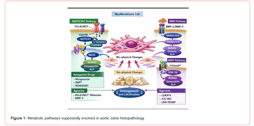
The signaling pathways known as RISK (Reperfusion Injury Salvage Kinase) and SAFE (Survivor Activating Factor Enhancement) play important roles in cellular protection against various stress conditions. The RISK pathway primarily involves PI3K-Akt activation, whereas the SAFE pathway is mediated through STAT3 signaling [38,39]. Under acute stress conditions, activation of these pathways may exert cardioprotective effects. However, in the context of chronic pressure overload, sustained activation may lose its protective efficacy and may even contribute to pathological hypertrophy and adverse myocardial remodeling [40-50]. We show a schematic drawing where the possible metabolic pathways involved in bone differentiation and calcification can be seen in (Figure 1).
Clinical Implications
The clinical course of aortic stenosis and the response to treatment depend largely on the degree of myocardial remodeling present at the time of intervention. In particular, the reversibility of ventricular remodeling after aortic valve replacement is strongly influenced by the presence and extent of preexisting myocardial fibrosis. In this context, the identification of biomarkers capable of reflecting the metabolic and molecular processes involved in cardiac remodeling represents an important area of investigation. Molecules such as sphingosine-1-phosphate and natriuretic peptides may provide relevant information regarding the activity of these pathological pathways. Furthermore, the study of modulators of signaling routes such as Notch or TGF-β opens new perspectives for the development of targeted therapies aimed at intervening earlier in the remodeling process associated with aortic stenosis.
- Otto CM, Kuusisto J, Reichenbach DD, Gown AM, O’Brien KD (1994) Characterization of the early lesion of degenerative valvular aortic stenosis. Histological and immunohistochemical studies. Circulation 90(2): 844-853.
- Lindman BR, Clavel MA, Mathieu P, Iung B, Lancellotti P, et al. (2016) Calcific aortic stenosis. Nat Rev Dis Primers. 2: 16006.
- Grossman W, Jones D, McLaurin LP (1975) Wall stress and patterns of hypertrophy in the human left ventricle. J Clin Invest 56(1): 56-64.
- Hill JA, Olson EN (2008) Cardiac plasticity. N Engl J Med 358(13): 1370-1380.
- Doenst T, Nguyen TD, Abel ED (2013) Cardiac metabolism in heart failure: implications beyond ATP production. Cir Res 113 (6): 709-724.
- Carabello BA (2002) Evaluation and management of patients with aortic stenosis. Circulation 105(15): 1746-1750.
- Lyon RC, Zanella F, Omens JH, Sheikh F (2015) Mechanotransduction in cardiac hypertrophy and failure. Circ Res 116(8): 1462-1476.
- Frey N, Olson EN (2003) Cardiac hypertrophy: the good, the bad, and the ugly. Annu Rev Physiol 65: 45-79.
- Molkentin JD (2004) Calcineurin-NFAT signaling regulates the cardiac hypertrophic response in coordination with the MAPKs. Cardiovasc Res 63(3): 467-475.
- Eder P, Molkentin JD (2011) TRPC channels as effectors of cardiac hypertrophy. Circ Res 108(2): 265-272.
- Bujak M, Frangogiannis NG (2007) The role of TGF-beta signaling in myocardial infarction and cardiac remodeling. Cardiovasc Res 74(2): 184-195.
- Weber KT (1989) Cardiac interstitium in health and disease: the fibrillar collagen network. J Am Coll Cardiol 13(7): 1637-1652.
- Hein S, Arnon E, Kostin S, Schonburg M, Elsasser A, et al. (2003) Progression from compensated hypertrophy to failure in the pressure-overloaded human heart: structural deterioration and compensatory mechanisms. Circulation 107(7): 984-991.
- González A, Ravassa S, Lopez B, Moreno MU, Beaumont J, et al. (2018) Myocardial remodeling in Hypertension. Hypertension 72(3): 549-558.
- Lopaschuk GD, Folmes CDL, Stanley WC (2007) Cardiac energy metabolism in obesity. Circ Res 101(4): 335-347.
- Peterzan MA, Lygate CA, Neubauer S, Rider OJ (2017) Metabolic remodeling in hypertrophied and failing myocardium: a review. Circulation 313(3): H597-H616.
- Lehman JJ, Kelly DP (2002) Transcriptional activation of energy metabolic switches in the developing and hypertrophied heart. Clin Exp Pharmacol Physiol 29(4): 339-345.
- Hardie DG et al. (2012) AMPK: a nutrient and energy sensor that maintains energy homeostasis. Nat Rev Mol Cell Biol 13(4): 251-262.
- Zaha VG, Young LH (2012) AMP-activated protein kinase regulation and biological actions in the heart. Circ Res 111(6): 800-814.
- Rosca MG, Hoppel CL (2010) Mitochondria in heart failure. Cardiovasc Res 88(1): 40-50.
- Tsutsui H, Kinugawa S, Matsushima S (2011) Oxidative stress and heart failure. Am J Physiol Heart Circ Physiol 301(6): H2181-H2190.
- Mann DL (2002) Inflammatory mediators and the failing heart: past, present, and the foreseeable future. Circ Res 91(11): 988-998.
- Spiegel S, Milstien S (2003) Sphingosine-1-phosphate: an enigmatic signaling lipid. Nat Rev Mol Cell Biol 4(5): 397-407.
- Takuwa Y, Okamoto Y, Yoshioka K, Takuwa N (2008) Sphingosine-1-phosphate signaling and biological activities in the cardiovascular system. Biochim Biophys Acta 1781(9): 483-488.
- Means CK, Brown JH (2009) Sphingosine-1-phosphate receptor signaling in the heart. Cardiovasc Res 82(2): 193-200.
- Gellings Lowe N, Swaney JS, Moreno KM, Sabbadini RA (2009) Sphingosine-1-phosphate and sphingosine kinase are critical for transforming growth factor-β-stimulated collagen production by cardiac fibroblasts. Cardiovasc Res 82(2): 303-312.
- Palluk R, Gaida W, Hoefke W (1985) Atrial natriuretic factor. Life Sci 36(15): 1415-1425.
- Potter LR, Abbey-Hosch S, Dickey DM (2006) Natriuretic peptides, their receptors, and cyclic guanosine monophosphate-dependent signaling functions. Endocr Rev 27(1): 47-72.
- Bergler-Klein J (2009) Natriuretic peptides in the management of aortic stenosis. Curr Cardiol Rep 11(2): 85-93.
- Garg V, Muth AN, Ransom JF, Schluterman MK, Barnes R, et al. (2005) Mutations in NOTCH1 cause aortic valve disease. Nature 437(7056): 270-274.
- Nigam V, Srivastava D (2009) Notch1 represses osteogenic pathways in aortic valve cells. J Mol Cell Cardiol 47(6): 828-834.
- Croquelois A, Domenighetti AA, Nemir M, Lepore M, Rosenblatt-Velin N, et al. (2008) Control of the adaptive response of the heart to stress via the Notch1 receptor pathway. J Exp Med 205(13): 3173-3185.
- Shioi T, McMullen JR, Tarnavski O, Converso K, Sherwood MC, et al. (2003) Rapamycin attenuates load-induced cardiac hypertrophy in mice. Circulation 107(12): 1664-1670.
- McMullen JR, Sherwood MC, Tarnavski O, Zhang L, Dorfman AL, et al. (2004) Inhibition of mTOR signaling with rapamycin regresses established cardiac hypertrophy induced by pressure overload. Circulation 109(24): 3050-3055.
- Sciarretta S, Forte M, Frati G, Sadoshima J (2018) New Insights into the Role of mTOR Signaling in the Cardiovascular System. Circ Res 122(3): 489-505.
- Border WA, Noble NA (1994) Transforming growth factor beta in tissue fibrosis. N Engl J Med 331(19): 1286-1292.
- Bujak M, Frangogiannis NG (2007) The role of TGF-β signaling in myocardial infarction and cardiac remodeling. Cardiovascular Research 74(2): 184-195.
- Hausenloy DJ, Yellon DM (2004) New directions for protecting the heart against ischaemia-reperfusion injury: targeting the Reperfusion Injury Salvage Kinase (RISK)-pathway. Cardiovasc Res 61(3): 448-460.
- Lecour S (2009) Multiple protective pathways against reperfusion injury: a SAFE path without Action? J Mol Cell Cardiol 46(5): 607-609.
- Nakamura M, Sadoshima J (2018) Mechanisms of physiological and pathological cardiac hypertrophy. Nat Rev Cardiol 15(7): 387-407.
- Ritterhoff J, Young S, Villet O, Shao D, Neto FC, et al. (2020) Metabolic Remodeling Promotes Cardiac Hypertrophy by Directing Glucose to Aspartate Biosynthesis. Circ Res 126(2): 182-196.
- Treibel TA, Lopez B, Gonzalez A, Menacho K, Schofield RS, et al. (2018) Reappraising myocardial fibrosis in severe aortic stenosis: an invasive and non-invasive study in 133 patients. J Am Coll Cardiol 39(8): 699-709.
- Neubauer S (2007) The failing heart--an engine out of fuel. N Engl J Med 356(11): 1140-1151.
- Liao Y, Liu C, Xiong T, Zhao M, Zheng W, et al. (2020) Metabolic Modulation and Potential Biomarkers of the Prognosis Identification for Severe Aortic Stenosis after TAVR by a Metabolomics Study. Cardio Res Pract PP: 3946913.
- Herum KM, Choppe J, Kumar A, Engler A, McCulloch AD, et al. (2017) Mechanical regulation of cardiac fibroblast profibrotic phenotypes. Mol Biol Cell 28(14): 1871-1882.
- Kaelin WG, McKnight SL (2013) Influence of metabolism on epigenetics and disease. Cell 153(1): 56-69.
- Sundaresan NR, Vasudevan P, Zhong L, Kim G, Samant S, et al. (2012) The sirtuin SIRT6 blocks IGF-Akt signaling and development of cardiac hypertrophy by targeting c-Jun. Nat Med 18(11): 1643-1650.
- Papait R, Greco C, Kunderfranco P, Latronico MVG, Condorelli G (2013) Epigenetics: a new mechanism of regulation of heart failure? Basic Res Cardiol 108(4): 361.
- Wehbe N, Nasser SA, Pintus G, Badran A, Eid AH, et al. (2019) MicroRNAs in cardiac hypertrophy. Int J Mol Sci 20(19): 4714.
- Villar AV, Garcia R, Merino D, Liano M, Cobo M, et al. (2013) Myocardial and circulating levels of microRNA-21 reflect left ventricular fibrosis in aortic stenosis patients. Int J Cardiol 167(6): 2875-2881.
-

This work is licensed under a Creative Commons Attribution-NonCommercial 4.0 International License.