




 Research Article
Phylogenetic Analysis of the Chandipura Virus Identifies Emerging Human Lineages, Vector Dominance, and Geographic Clustering
Amity Institute of Public Health & Hospital Administration, Amity University, Uttar Pradesh, India
Received Date: March 09, 2026; Published Date: March 23, 2026
Abstract
Background and objectives: Chandipura virus is an emerging arthropod-borne virus responsible for recurring outbreaks of acute encephalitis,
especially in children in India. The necessity to investigate its evolutionary dynamics and transmission ecology has been brought to light by recent
outbreaks. In order to record the establishment of novel lineages, host connections, and geographic clustering analysis, the current work examines
phylogenetic analysis among all accessible isolates of the Chandipura virus from humans and non-human animals.
Methods: The NCBI GenBank database yielded 29 whole and partial CHPV genome sequences from human, vector, and animal hosts. The
sequences are typical of Africa, India, and surrounding areas. MAFFT was used to align the sequences, and TrimAI was then used to trim them.
GTR model in FastTree was used to construct the maximum-likelihood phylogenetic trees and visualized in iTOL, comprising of metadata on host,
geography, and year of isolation.
Results: A clear separation of CHPV isolates by host and geography was shown by phylogenetic analysis. Phylogenetic study revealed a
distinct separation of CHPV isolates by host and region. Genomes originated from humans were only found in India. Rather than human-to-human
transmission, high genetic diversity was observed, indicating frequent spillover. However, isolates from Senegal and Kenya that were obtained from
sandflies indicate a robust vector-driven transmission cycle since they are grouped into closely related lineages. The establishment of divergent
lineages was shown in the different clustering of recent isolates from India in 2024 compared to the older pandemic strains. Significant gaps in
surveillance were reflected in the limited genetic continuity across time.
Interpretation and Conclusions: The results show that humans serve as incidental hosts and that vector ecology significantly influences the
evolution of CHPV. This emphasises the necessity of ongoing genomic surveillance. Early warning systems urgently require an integrated One Health
approach to monitor vector–human–environment interactions.
Keywords: One Health; Phylogeny; Chandipura Virus; Encephalitis; Human
Introduction
Chandipura virus (CHPV), an arthropod-borne neurotropic virus, belongs to the family Rhabdoviridae and the genus Vesiculovirus, which also includes Vesicular stomatitis Indiana and New Jersey viruses [1]. It displays the characteristic bulletshaped morphology of rhabdoviruses and carries an enveloped, helical nucleocapsid enclosing an ~11 kb negative-sense singlestranded RNA genome [2]. The genome encodes five structural proteins—N, P, M, G, and L—transcribed in a gradient from a single promoter at the 3′ end, mediating encapsidation, replication, transcription, assembly, and host-cell entry. Although CHPV shares many molecular and biological features with vesiculoviruses, its pronounced neurotropism and ability to cause acute, often fatal encephalitis in children distinguishes it as a pathogen of major public health relevance [3]. This molecular profile becomes more meaningful when examined in light of the virus’s emergence, reemergence, and spread across India and beyond.
Chandipura virus (CHPV) was first isolated in 1965 from a febrile case in Chandipura village, Maharashtra, following the isolation of another strain from a hedgehog in Nigeria [5]. Later isolations from human encephalitis cases in Madhya Pradesh in 1980 established its neurovirulent properties. Over the years, CHPV activity was reported from several Indian states, but its epidemic importance gained momentum after the devastating “Epidemic Brain Attack” in Andhra Pradesh in 2003 [6]. Severe outbreaks were recorded in Gujarat (2004, 2005) and Maharashtra (2005), bringing to the fore its killing potential, especially among children. Sporadic and outbreak-associated encephalitis continued into the next decade, with confirmed outbreaks in Odisha (2009), Vidarbha (2012), and clusters in Bihar (2018) cumulatively accounting for hundreds of deaths among children [7-9]. Very recently, CHPV has resurfaced as a major public health concern: in July 2024, Gujarat reported an unprecedented rise in encephalitis cases due to which CHPV accounted for ≈23.5% of the confirmed cases, while more cases were found in Rajasthan [10]. Serological evidences in the past from Gujarat (2011–2012) and repeated outbreaks in Andhra Pradesh, Gujarat, Maharashtra, and Odisha indicated continued viral circulation within endemic foci [11-13]. This seen epidemiology underlines the importance of understanding how the CHPV infection presents clinically and what drives its rapid progression.
It is primarily transmitted by the infected sandflies Phlebotomus papatasi, Phlebotomus argentipes, and Sergentomyia spp., although earlier evidence implicated Aedes aegypti mosquitoes. Phlebotomus argentipes, with its high susceptibility to CHPV under laboratory models and widespread distribution throughout endemic regions, is considered to be the primary vector [14]. Transmission is CHPV is due to housing and environmental factors that promote sandfly development, such as un-plastered mud or brick walls, moisture retention, low indoor lighting, and water stagnation. Climatedriven factors including extended monsoons, higher temperatures, and increased humidity boost vector density and viral circulation, and seasonal amplification of CHPV takes place during monsoon and post-monsoon periods. Although high-altitude events show increasing ecological appropriateness, outbreaks happen in lowaltitude and semi-arid areas. adjacent human-animal contact, raising livestock, animal shelters adjacent to residences, and children’s high biological vulnerability are additional risk factors. The transmission dynamics of CHPV are driven by a combination of vectorial, environmental, geographic, and host factors, which further solidify the virus’s status as a chronic danger in tropical and semi-arid regions [15,16].
The Chandipura virus replicates inside endothelial cells after entering a human host by endocytosis, causing viremia that promotes systemic spread and disruption of the blood–brain barrier, ultimately leading to the development of severe neurological diseases [17]. Early detection by pattern recognition receptors, which triggers initial release of cytokines, type I interferons, complement activation, and NK cell recruitment, which indicates aggressive pathogenesis. A cytokine storm results from ineffective early clearance, which raises T-cell and antigen-presenting cell activation. After a peripheral phase marked by monocyte infection, CHPV enters the central nervous system (CNS) through either a “Trojan horse” mechanism or direct infection of Brain Microvascular Endothelial Cells (BMECs). This invasion is likely caused by haematogenous dissemination and clathrin-mediated endocytosis. Infection of neurones, astrocytes, pericytes, and microglia inside the central nervous system results in production of NO, TNF-α, IFN-γ, IL-6, IL-10, MCP-1, MMPs, and virus-induced apoptosis leading to increased activity of the viral matrix protein [18].
Clinically, the CHPV infection manifests as an acute febrile illness that progresses quickly, with an incubation period of two to seven days. Within twenty-four to thirty hours after the onset of fever, neurological impairment occurs. high-grade fever, vomiting, tachycardia, and malaise leads to seizures, altered sensorium, photophobia, coma, hemiparesis, neck stiffness, and elevated intracranial pressure. Children under the age of fifteen are disproportionately affected by these symptoms, which are linked to a significant mortality [15].
The evolutionary dynamics of the Chandipura virus will be examined in this manuscript utilising thorough sequencing studies based on the present knowledge of its ecology, pathogenicity, and epidemiology. In order to characterise patterns of viral divergence, evaluate lineage-specific mutations, and find possible indicators of host adaptability or geographic clustering, this work combines all available genomic sequences from humans and animals. Such data is essential for enhancing monitoring, guiding future research on the mechanisms underlying the genesis and spread of CHPV, and informing the improvement of diagnostics.
Materials and Methods
Data Retrieval
From the NCBI GenBank database (https://www.ncbi.nlm.nih. gov/), we extracted 29 complete and partial genome sequences (1966-2024) of the Chandipura virus (CHPV) in November 2025. These sequences were exceeding than 11000 nucleotides and included metadata on its geography, data and purpose of sequencing. The selected genomes encompassed various regions, hosts, and temporal contexts to facilitate a comprehensive analysis of CHPV evolutionary dynamics. Table 1 shows the 29 genomes with their characteristic features. The retrieved nucleotide sequences were saved in FASTA format for subsequent multiple sequence alignment and phylogenetic analysis.
Multiple Sequence Alignment and Trimming
The downloaded nucleotide sequences were aligned using MAFFT algorithm (v7.520) [19]. We used the --auto option to automatically select the optimal alignment strategy that fits best to sequence length and similarity. Manual check was also performed on the aligned file to ensure effective alignment of the conserved regions. To retain the high-quality alignment of phylogenetically informative sites, the divergent and poorly aligned regions that could negatively impact the phylogenetic inference were eliminated using trimAI (v1.4) using automated1 mode to appropriately select the most suitable trim strategy based on the alignment characteristics [20].
Phylogenetic Tree Construction
Using the high-quality trimmed alignment file, we constructed the maximum-likelihood phylogenetic tree employing the Generalized Time-Reversible (GTR) model with discrete gamma correction for handling different mutation rates in the FastTree (v2.1.11) using the nucleotide (-nt) option which gives an effective tree structure for large genomic datasets efficiently [21]. The resulting tree was saved in Newick format. The resulting tree exhibited clear branching patterns, indicating genetic divergence among Chandipura virus isolates. Bootstrap support values generated by FastTree were generally high (>85%) for major internal nodes, confirming the reliability of the inferred topology
Tree Visualization and Annotation
The Newick-formatted tree file was uploaded to visualize and annotate the phylogenetic tree using the Interactive Tree of Life (iTOL) web server (https://itol.embl.de/) [22]. The associated metadata that includes accession numbers, geographic origin, and year of isolation were integrated to produce a color-coded and annotated phylogenetic representation that facilitates the interpretation of evolutionary relationships and clustering patterns among Chandipura virus isolates.
Results
Genome Sequence Retrieval and Alignment
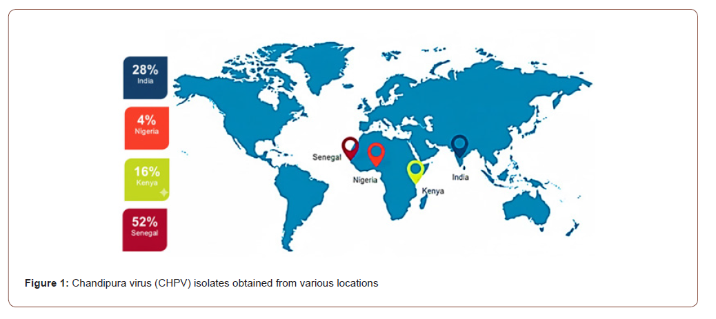
From the NCBI GenBank database, we obtained a set of 29 complete and partial genome sequences of the Chandipura virus (CHPV) that varied from approximately 6.7 kb to 11 kb and includes isolates obtained from various locations and decades across India as well as neighbouring countries like Senegal, Nigeria, and Kenya (Figure 1). The MAFFT algorithm for multiple sequence alignment produced a coherent alignment that showed both localized areas of variation and conserved genomic regions among isolates, especially in the glycoprotein (G) and polymerase (L) gene segments. These variable regions are known to contribute to host adaptation and viral evolution within vesiculoviruses.
Alignment Refinement
To refine the raw alignment file obtained and to remove poorly aligned and divergent sites from the trimAI, we used the automated1mode which resulted in high quality of ~10,400bp in length eliminating approximately 3-5% of the ambiguous positions. As a result, the noise from the non-informative regions was reduced and the refined dataset preserved the conserved motifs critical for Rhabdoviridae classification. This quality alignment served as the input for downstream phylogenetic inference.
Phylogenetic Analysis
The phylogenetic analysis of the Chandipura virus (CHPV) genome sequences was conducted using a Newick-formatted tree file (chpv_tree.nwk). The constructed phylogenetic tree revealed a distinct clustering pattern among the CHPV isolates, indicating genetic diversity within the population. The phylogenetic dataset comprised seven Chandipura virus (CHPV) genomes isolated from Homo sapiens, seventeen from phlebotomine sandflies (Phlebotomus spp.), and one from a hedgehog (Figure 2(a)).
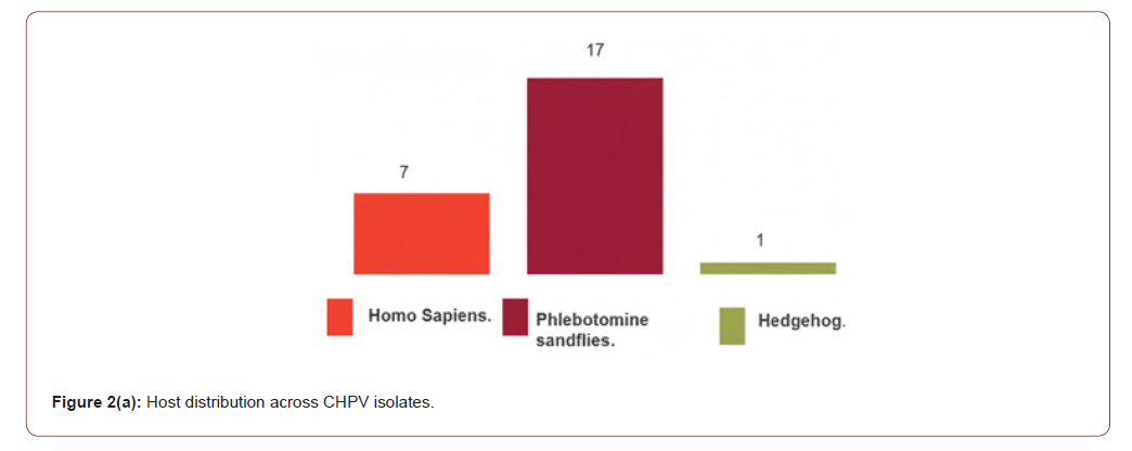
All human-derived CHPV genomes were exclusively obtained from India, spanning isolates collected between 2003 and 2024, including both complete and partial genomes. The genomes derived from non-human hosts, on the other hand, displayed a noticeably wider geographic distribution. Senegal accounted for 76% (13/17) of the isolates derived from sandflies, while Kenya accounted for 24% (4/17). In 1966, a single genome derived from hedgehogs was isolated in Nigeria. Within the available CHPV genomes, this hostlocation distribution highlights distinct geographic and ecological segregation. Different host- and region-specific clustering was found in preliminary phylogenetic reconstruction, indicating the circulation of several evolutionary lineages of CHPV influenced by both vector ecology and regional transmission dynamics (Figure 2(b)).
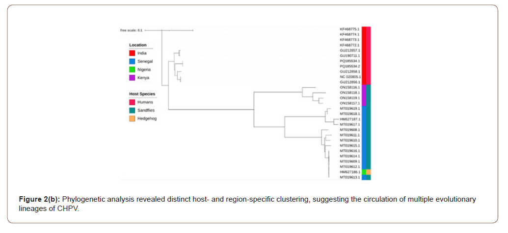
The Chandipura virus has undergone limited but noticeable genetic diversification over time, corresponding to the iTOL visualization, which clearly showed phylogenetic clustering consistent with known Rhabdoviridae taxonomy and geographic distribution.
Discussion
In this study, the genomic landscape, evolutionary process, and epidemiological path of Chandipura virus have been outlined using publicly available sequence information. The authors have used the phylogenetic approach along with host-specific distribution, temporal distribution, and country-wise distribution to shed new light on the ecology, transmission process through vectors, and the scope of cross-species transmission of CHPV. The findings highlight significant gaps in understanding the correct distribution and evolution of the newly reported vesiculovirus, along with the fact that the genomic surveillance process is uneven.
Senegal has a disproportionately high percentage of CHPV genomes (52% of the total sample), followed by India (28%), Kenya (16%), and Nigeria (4%), according to our country-level analysis. This trend is supported by our phylogenetic analysis, which revealed that Senegalese strains formed a highly supported, temporally clustered lineage corresponding to samples collected primarily between 1992 and 1997. Tight phylogenetic clustering and the frequent identification of genetically related strains over several years point to the possibility of exclusive endemic circulation within sandfly populations in Barkedji, Senegal. The limited viral introductions from external sources, ecological stability in vector populations, or possible overwintering mechanisms that sustain the virus within the sandfly population could all contribute to this lineage’s relative homogeneity.
Table 1: Selected 29 CHPV genomes with their characteristic features.

These observations are consistent with earlier studies demonstrating endemic circulation of Chandipura virus within sandfly populations in Barkedji, Senegal, characterized by repeated isolation of closely related strains over multiple years and limited evidence of external viral introductions [23,24]. Similar spatially restricted maintenance cycles governed primarily by vector–host ecology have been widely reported for vesiculoviruses and other rhabdoviruses [25].
Indian genomes, on the other hand, were phylogenetically more diverse and dispersed across several branches because they were solely obtained from human cases [26,27]. Despite the reduced sample size, this variability might point to independent, recurring spillover occurrences from vector reservoirs rather than ongoing human-to-human transmission [28,29]. The recent development of 2024 isolates in Gujarat contrasts sharply with the epidemics that occurred between 2003 and 2007. Rather than a genuine lack of CHPV activity, the lack of genomic data for almost 20 years is probably due to surveillance gaps [30,31,27]. The concept of either cryptic transmission within vector populations or reintroduction of divergent viral lineages is supported by the phylogenetic diversity of the 2024 isolates, which seem different from previous Indian strains. These results emphasize the critical necessity for ongoing genetic surveillance in areas where pediatric encephalitis has been linked to CHPV.
Phlebotomine sandfly-derived genomes make up 68% of the dataset, which emphasizes the vector’s crucial role in shaping CHPV evolution [24]. Our phylogenetic tree 25 shows that sandfly sequences span multiple clades, including the oldest lineages from Senegal and a variety of contemporary lineages from Kenya [25]. Kenya’s genomes from 2016 to 2017 stand out as a separate clade, indicating evolutionary paths unique to the region. The ecological variations in sandfly species composition, host feeding habits, and environmental factors affecting vector abundance are probably the causes of this heterogeneity [24,25].
The genomes from 2016 to 2017 from Kenya stand out as a distinct clade, suggesting regional evolutionary paths. This heterogeneity is likely caused by ecological differences in the species composition of sandflies, host feeding patterns, and environmental factors influencing vector abundance [24,25]. These results demonstrate that CHPV is essentially a vector-maintained infection, with humans serving only as incidental hosts.
The discovery of a historical hedgehog-derived CHPV isolate from Nigeria in 1966 broadens the host range and calls into doubt the multi-host ecology of the virus. Although hedgehogs are not thought to be key reservoirs, their infection suggests that vertebrate hosts other than humans might be involved in the cycle of transmission. Vesiculoviruses have a well-established cross species spillover, and our results suggest that vertebrates may be more widely involved in the ecology of CHPV [25].
Even overall genome lengths were mostly conserved (≈11,061– 11,120 bp), diversity in completeness presents considerable difficulties for fine-scale molecular studies, especially among genomes produced from sandflies. However, the phylogenetic structure was strong enough to identify country-specific clusters and deduce probable routes of transmission. Field based vector sampling and metagenomic recovery techniques, which frequently produce incomplete sequences, may be the cause of the prevalence of partial genomes from Senegal and Kenya. The true genetic diversity within endemic areas may be understated by this incomplete depiction [25].
Numerous lines of evidence point to a combination of sampling bias and biological realism in the observed patterns. The larger representation of Senegal and Kenya is probably due to their intensive entomological monitoring programs, which systematically trap sandflies. In contrast, there is very little vector monitoring data in public archives, and India’s genetic landscape is strongly skewed toward human sequences linked to outbreaks [25]. Reconstructing transmission channels and comprehending inter-epidemic viral maintenance in the Indian subcontinent are hampered by this imbalance.
There are several limitations to this study. The lack of genomic sequences from other CHPV endemic South Asian regions, like Nepal, makes the dataset geographically biased [32]. Deeper analyses, such as estimating mutation rates and identifying changes that define a lineage, are limited by the incomplete nature of many genomes. Accurate reconstruction of transmission pathways is hampered by metadata gaps, such as missing vector species, clinical information, and ecological context. Long temporal gaps also make it difficult to track viral evolution and dispersal reliably, especially the 17-year absence of Indian sequences [33].
These results clearly have public health implications, despite these limitations. Clustering of the Senegalese and Kenyan strains emphasizes the need for increased vector surveillance and regular whole-genome sequencing throughout Africa. The detection of divergent 2024 isolates in India heralds the possibility of a reemergence and so calls for systematic genomic monitoring in sandfly populations and outbreak-prone areas. This will facilitate early detection and response through a coordinated One Health framework that incorporates vector, human, and possible reservoir surveillance. This work provides an immediate but extensive global genomic and evolutionary perspective on CHPV. Our study reinforces the urgent need for continuous genomic surveillance and forms the foundation for future molecular epidemiology and early warning systems for CHPV and related vesiculoviruses by exposing its vector-driven ecology, the unevenness in geographic sampling, and episodic spillover into humans [25,26,30].
Acknowledgment
Authors acknowledge Amity University.
Financial Support & Sponsorship
None.
Conflicts of Interest
None.
Use of Artificial Intelligence (AI)-Assisted Technology for Manuscript Preparation
The authors confirm that they used ChatGPT (https://chat. openai.com/chat) for improving language and readability at the initial draft sections and no images were manipulated using AI.
References
- Ghosh S, Dutta K, Basu A (2013) Chandipura virus induces neuronal death through Fas-mediated extrinsic apoptotic pathway. J Virol 87(22): 12398-12406.
- Pradhan P, Mukherjee T, Mohanty S (2025) Chandipura Virus: A Growing Public Health Threat in India and Beyond. Infect Disord Drug Targets 25(6): e18715265351833.
- Cherian SS, Gunjikar RS, Banerjee A, Kumar S, Arankalle VA (2012) Whole genomes of Chandipura virus isolates and comparative analysis with other rhabdoviruses. PLoS One 7(1): e30315.
- Bhatt PN, Rodrigues FM (1967) Chandipura: A new arbovirus isolated in India from patients with febrile illness. Indian J Med Res 55: 1295–1305.
- Giovanetti M, Micheli V, Mancon A, Mileto D, Rizzo A (2025) Phylogenetic Analysis of Chandipura virus: Insights from a Preliminary Genomic Study. Int J Mol Sci 26(3): 1021.
- Sudeep AB, Gurav YK, Bondre VP (2016) Changing clinical scenario in Chandipura virus infection. Indian J Med Res 143(6): 712-721.
- Brisse ME, Ly H (2024) Chandipura Virus Causing Large Viral Encephalitis Outbreaks in India. Pathogens 13(12): 1110.
- Dwibedi B, Sabat J, Hazra RK, Kumar A, Dinesh DS, et al. (2015) Chandipura virus infection causing encephalitis in a tribal population of Odisha in eastern India. Natl Med J India 28(4): 185-187.
- Kanabar B, Malek S, Piparva K (2024) Temporal Trends in Outbreaks of Chandipura Viral Infection in India: A Systematic Review. Cureus 16(8): e68097.
- Kumar N, Bondre VP (2024) Re-emergence of Chandipura virus infection in India. Virulence 15(1): 2421218.
- Tandale BV, Tikute SS, Arankalle VA, Sathe PS, Joshi MV, et al. (2008) Chandipura virus: a major cause of acute encephalitis in children in North Telangana, Andhra Pradesh, India. J Med Virol. 80(1): 118-124.
- Alhajj M, Zubair M, Farhana A (2023) Enzyme-linked immunosorbent assay (ELISA). British Society for Immunology. 80: 118–124.
- Damle RG, Sankararaman V, Bhide VS, Jadhav VK, Walimbe AM, et al. (2018) Molecular Evidence of Chandipura Virus from Sergentomyia species of Sandflies in Gujarat, India. Jpn J Infect Dis 71(3): 247-249.
- Akingbola A, Adegbesan A, Adegoke K, Chuku J, Ojo O, et al. (2025) Chandipura Virus Resurgence in India: Insights into Diagnostic Tools, Antiviral Development, and Public Health Implications. Glob Health Epidemiol Genom 2025: 1015031.
- Lodha L, Swaminathan A, John A, Kunhikannan S, Sundar B, et al. (2025) Chandipura virus: A comprehensive review. Infect Dis Now 55(8): 105179.
- Adnan H, Salomon I, Ahmed M (2025) Chandipura virus outbreak: a growing public health crisis that demands immediate action. Ann Med Surg (Lond) 87(1): 24-26.
- Debnath R, Chawla PA (2024) Chandipura virus: The 2024 resurgence and Gujarat outbreak. Asian Pac J Trop Biomed 14(12): 507–513.
- Ghosh S, Basu A (2017) Neuropathogenesis by Chandipura virus: An acute encephalitis syndrome in India. Natl Med J India 30(1): 21-25.
- Rozewicki J, Li S, Amada KM, Standley DM, Katoh K (2019) MAFFT-DASH: integrated protein sequence and structural alignment. Nucleic Acids Res 47(W1): W5-W10.
- Capella-Gutiérrez S, Silla-Martínez JM, Gabaldón (2009) trimAl: a tool for automated alignment trimming in large-scale phylogenetic analyses. Bioinformatics 25(15): 1972-1973.
- Price MN, Dehal PS, Arkin AP (2010) Fast Tree 2--approximately maximum-likelihood trees for large alignments. PLoS One 5(3): e9490.
- Letunic I, Bork P (2021) Interactive Tree of Life (iTOL) v5: an online tool for phylogenetic tree display and annotation. Nucleic Acids Res 49(W1): W293-W296.
- Mallick D, Yadav U, Gupta M, Kumar D, Kumar R (2025) The evolving landscape of Chandipura virus: A comprehensive account of outbreaks to recent advances. Virology 608: 110541.
- Pawar N, Seth AK (2025) Chandipura virus in India: A comprehensive epidemiological review. J Vector Borne Dis 62(4): 391-397.
- Dietzgen RG, Kondo H, Goodin MM, Kurath G, Vasilakis N (2017) The family Rhabdoviridae: mono- and bipartite negative-sense RNA viruses with diverse genome organization and common evolutionary origins. Virus Res 227: 158-170.
- Damle RG, Sankararaman V, Bhide VS, Mahamuni SA, Walimbe AM, et al. (2017) Genetic characterization of the glycoprotein G of Chandipura viruses in India with emphasis on an outbreak of 2015. Infect Genet Evol 55: 112-116.
- Basak S, Mondal A, Polley S, Mukhopadhyay S, Chattopadhyay D (2007) Reviewing Chandipura: a vesiculovirus in human epidemics. Biosci Rep 27(4-5): 275-298.
- Van Ranst M (2004) Chandipura virus: an emerging human pathogen? Lancet 364(9437): 821-822.
- Chadha MS, Arankalle VA, Jadi RS, Joshi MV, Thakare JP, et al. (2005) An outbreak of Chandipura virus encephalitis in the eastern districts of Gujarat state, India. Am J Trop Med Hyg 73(3): 566-570.
- Mourya DT, Yadav PD, Ullas PT, Bhardwaj SD, Sahay RR, et al. (2019) Emerging/re-emerging viral diseases & new viruses on the Indian horizon. Indian J Med Res 149(4): 447-467.
- Mehand MS, Al-Shorbaji F, Millett P, Murgue B (2018) The WHO R&D Blueprint: 2018 review of emerging infectious diseases requiring urgent research and development efforts. Antiviral Res 159: 63-67.
- Grubaugh ND, Ladner JT, Lemey P, Pybus OG, Rambaut A, et al. (2019) Tracking virus outbreaks in the twenty-first century. Nat Microbiol 4(1): 10-19.
- World Health Organization (2023) Global genomic surveillance strategy for pathogens with pandemic and epidemic potential 2022–2032: progress report on the first year of implementation [Internet]. Geneva: World Health Organization.
-

This work is licensed under a Creative Commons Attribution-NonCommercial 4.0 International License.


 a Creative Commons Attribution 4.0 International License. Based on a work at www.irispublishers.com.
Best viewed in

 | Above IE 9.0 version | Privacy Policy