




 Case Report
Urgent Open Cholecystectomy in a 7-Year-Old Male with Idiopathic Acute Calculous Cholecystitis: A Case Report
Received Date: January 07, 2026; Published Date: January 20, 2026
Abstract
Introduction: Gallstone disease is rare in children, but its incidence is increasing due to widespread use of ultrasonography.
Symptoms and Clinical Findings: A 7-year-old male presented with postprandial colicky abdominal pain and vomiting. Physical examination showed deep tenderness in the right hypochondrium without guarding or Murphy’s sign.
Management: Abdominal ultrasonography revealed a distended gallbladder with multiple small stones and a thickened wall, leading to a diagnosis of acute calculous cholecystitis. The patient underwent open cholecystectomy without complications and was discharged on postoperative day three.
Conclusion: Although rare in children, idiopathic acute calculous cholecystitis should be considered in pediatric patients with unexplained abdominal pain and vomiting.
Keywords: Pediatric gallstones; Acute calculous cholecystitis; Idiopathic cholelithiasis; Open cholecystectomy; Case report
Abbreviations: CBD: Common Bile Duct; IV: Intravenous; BMI: Body Mass Index; RBC: Red Blood Cells; WBC: White Blood Cells
Introduction
Gallstone disease in children is uncommon, comprising only 0.1–0.2% of cases, but its prevalence is increasing due to improved diagnostic imaging such as ultrasonography [1]. Pediatric patients often present with atypical symptoms, and in many cases, the etiology remains unclear [2]. This case report describes a 7-year-old male with idiopathic acute calculous cholecystitis who required urgent open cholecystectomy, highlighting the importance of considering this diagnosis in children with unexplained abdominal pain.
Case Presentation
A 7-year-old male (20 kg, normal BMI) presented with postprandial colicky umbilical abdominal pain, acute in onset, intermittent, and associated with vomiting and anorexia. There was no constipation, abdominal distention, jaundice, altered stool or urine color, pruritus, bleeding tendencies, or fever. His medical history was unremarkable, with no chronic illnesses, family history of gallstones, diabetes, or hemolytic disorders.
On examination, he was stable, with deep tenderness in the right hypochondrium but no guarding or Murphy’s sign.
Routine laboratory tests, including a complete blood count, were within normal limits, showing normal red blood cell morphology, white blood cell count, platelets, and reticulocytes. Abdominal ultrasonography revealed a distended gallbladder containing three small stones (~5 mm) with a mildly thickened, edematous wall (5 mm), while the common bile duct was normal in caliber. No additional abnormalities were detected, and the findings confirmed acute calculous cholecystitis. No diagnostic challenges were encountered.
Following appropriate preoperative investigations, the patient underwent open cholecystectomy under general anesthesia. Intraoperatively, the gallbladder was partially distended with changes consistent with acute cholecystitis. The anatomy of Calot’s triangle was normal, with no lymphadenopathy. The cystic duct and artery were identified, ligated, in and divided, and the gallbladder was successfully dissected from the liver and removed (figure 1). Postoperatively, the patient was managed with IV fluids, antibiotics, and analgesia. Bowel sounds returned within 12 hours, allowing diet initiation without nausea or vomiting (Figure 1).
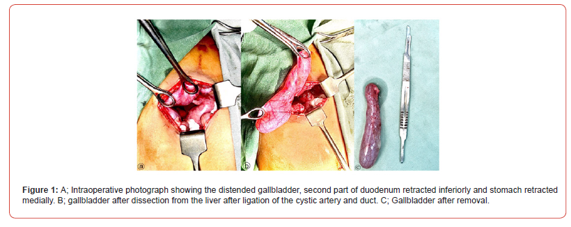
The postoperative course was uneventful, and the patient was discharged on postoperative day three in stable condition. He was referred to the pediatric department for further hematologic evaluation to rule out underlying etiologies. No complications or recurrence of symptoms were reported at follow-up (Table 1).
Table 1:Timeline of Events.

Discussion
Gallstones in pediatric patients are rare, with the incidence in Sudan remaining largely unstudied [3]. Unlike adults, children often present with atypical or nonspecific symptoms, and Murphy’s sign is not always positive [4]. Ultrasonography remains the gold standard for diagnosis [5].
Risk factors vary by age; neonates often develop gallstones due to prematurity or parenteral nutrition, whereas adolescents are more likely to have hemolytic disorders or obesity-related cholelithiasis [6]. In our patient, no identifiable risk factors were found, suggesting an idiopathic etiology, which accounts for 20– 65% of pediatric cases [7].
Although laparoscopic cholecystectomy is the standard of care [8], open cholecystectomy remains a viable option, particularly in urgent settings. Conservative management may be attempted in infants, but children aged 2–12 years are less likely to respond [9].
This case underscores the importance of considering cholelithiasis in pediatric patients presenting with unexplained abdominal pain.
Conclusion
Idiopathic acute calculous cholecystitis is rare in children but should be considered in pediatric patients with unexplained abdominal pain and vomiting. Early diagnosis and surgical intervention can lead to excellent outcomes.
Patient Perspective
The patient’s family expressed relief after the procedure, noting a complete resolution of symptoms.
Ethical Considerations
Written informed consent was obtained from the patient’s guardian for participation and publication of this case report, including the use of relevant clinical details and images. Ethical approval for this study was obtained from the Omdurman Islamic University, under approval number .
Competing Interests
The authors declare that they have no competing interests, whether financial, personal, academic, or ideological, that could influence the content of this case report.
Availability of Data and Material
All data supporting the findings of this case report are included in the manuscript. Additional data or materials can be provided upon reasonable request to the corresponding author.
Funding
No external funding was received for the preparation of this case report.
References
- Abascal JV, Alvarez PB, Rosales FM, Ramirez AF, Khalil MIA, et al. (2014) First case with Huntington's disease in The Gambia. Journal of the Neurological Sciences 338: 238-240.
- Aiyesimoju AB, Osuntokun BO, Bademosi O, Adeuja AO (1984) Hereditary neurodegenerative disorders in Nigerian Africans. Neurology 34: 361-362.
- Andrew SE, Goldberg YP, Kremer B, et al. (1994) Huntington disease without CAG expansion: phenocopies or errors in assignment? Am J Hum Genet 54: 852-863.
- Assami S, Azzedine H, Nouioua S, Mundwiller E, Mahoui S, et al. (2011) Pantothenate Kinase–Associated Neurodegeneration: Clinical Description of 10 Patients and Identification of New Mutations. Movement Disorders 26 (9): 1777-1779.
- Baine FK, Kay C, Ketelaar ME, et al. (2013) Huntington disease in the South African population occurs on diverse and ethnically distinct genetic haplotypes. Eur J Hum Genet 21(10): 1120-1127.
- Baine F, Krause A, Greenberg L (2016) The frequency of Huntington disease and Huntington disease-like 2 in the South African population. Neuroepidemiology 46(3): 198-202.
- Baine FK, Peerbhai N, Krause A (2018) A study of Huntington disease-like syndromes in black South African patients reveals a single SCA2 mutation and a unique distribution of normal alleles across five repeat loci. Journal of the Neurological Sciences 390: 200-204.
- Bardien S, Abrahams F, Soodyall H, Merwe LVD, Greenberg J, et al. (2007) A South African Mixed Ancestry Family with Huntington Disease-Like 2: Clinical and Genetic Features. Movement Disorders 22(14): 2083-2089.
- BATES G, HARPER PS, JONES L (editors) (2002) Huntington’s disease, 3rd ed. Oxford: Oxford University Press.
- Bouhouche A, Regragui W, Lamghari H, Khaldi K, Birouk N, et al. (2015) Clinical and genetic data of Huntington disease in Moroccan patients. Afri Health Sci 15(4): 1232-1238.
- Coulibaly T, Karambe M, Guinto CO, Traore M (2012) Huntington's disease confirmed by genetic testing in three Malian families. Mov Disord; 27(Suppl.1): 171.
- Donaldson I, Marsden CD, Schneider SA, Bhatia KB (2012) Clinical approach to movement disorders. In Marsden's Book of Movement Disorders. Donaldson I, Marsden CD, Schneider SA, Bhatia KB. Editors. Oxford, United Kingdom: Oxford University Press 140-141.
- Gregory A, Polster BJ, Hayflick SJ (2008) Clinical and genetic delineation of neurodegeneration with brain iron accumulation. J Med Genet 46(2): 73-80.
- Kaboré J, Ouédraogo A (2000) Huntington disease in Burkina Faso. Rev Neurol (Paris) 156(12): 1157-1158.
- Krause A, Mitchell C, Essop F, Tager S, Temlett J, et al. (2015) Junctophilin 3 (JPH3) expansion mutations caus- ing Huntington disease like 2 (HDL2) are common in South African patients with African ancestry and a Huntington disease phenotype. Am J Med Genet B Neuropsychiatr Genet 168(7): 573-585.
- Krause A, Temlett J, Van Der Meyden K, Ross CA, Callahan C, Margolis RL (2002) CAG/CTG repeat expansions at the HDL2 locus are a common cause of Huntington disease in black South Africans. Am J Hum Genet 71(4): 528-528.
- Kremer B, Goldberg P, Andrew SE, et al. (1994) A Worldwide Study of the Huntington’s Disease Mutation: the sensitivity and specificity of measuring CAG repeats. N Engl J Med 330: 1401-1406.
- Kumar N, Leonzino M, Hancock-Cerutti W, Horenkamp FA, LI P, et al. (2018) VPS13A and VPS13C are lipid transport proteins differentially localized at ER contact sites. J Cell Biol 217(10): 3625-3639.
- Landoure G, Mochel F, Meilleur K, Ly M, Sangare M, et al. (2013) Novel mutation in the ATM gene in a Malian family with ataxia telangiectasia. J Neurol 260: 324-326.
- Magazi DS, Krause A, Bonev V, Moagi M, Iqbal Z, et al. (2008) Huntington's disease: genetic heterogeneity in black African patients. S Afr Med J 98: 200-203.
- Park JS, Thorsness MK, Policastro R, et al. (2016) Yeast Vps13 promotes mitochondrial function and is localized at membrane contact sites. Mol Biol Cell 27: 2435-2449.
- Ross CA, Tabrizi SJ (2011) Huntington's disease: from molecular pathogenesis to clinical treatment. Lancet Neurol 10: 83-98.
- Stevanin G, Fujigasaki H, Lebre A-S, et al. (2003) Huntington’s disease-like phenotype due to trinucleotide repeat expansions in the TBP and JPH3 genes. Brain 126:1599-1603.
- Triki C, Feki I, Meziou M, Turki H, Zahaf A, et al. (2000) Clinical, biological and genetic study of 24 patients with ataxia-telangiectasia from southern Tunisia. Rev Neurol 156: 634-637.
- Velayos Baeza A, Dobson-Stone C, Rampoldi L, Bader B, Walker RH, et al. (2002) Chorea-Acanthocytosis. In: Adam MP, Ardinger HH, Pagon RA, Wallace SE, LJH B, Stephens K, Amemiya A, editors. Gene Reviews. Seattle: Seattle (WA): University of Washington: 1993-2019.
- Wagstaff LA, Joffe R (1969) Ataxia-telangiectasia in a South African Bantu child. S Afr Med J 43: 662-664.
-

This work is licensed under a Creative Commons Attribution-NonCommercial 4.0 International License.




