




 Review Article
Subcortical Vascular Dementia due to Binswanger Disease: A Diagnostic Challenge
Received Date: May 25, 2026; Published Date: June 16, 2026
Abstract
Binswanger disease (BD) is a progressive neurological disease caused by atherosclerosis and thrombo-embolism affecting the white matter and blood vessels affecting the deep brain structures (basal ganglia and thalamus). Depending on the vascular etiology, the symptoms and physical effects associated with Binswanger stroke may suddenly worsen, stabilize, and then improve briefly, but the patient’s general condition continues to improve as they continue to enter contact with the occluded vessel. High blood pressure, smoking, high cholesterol, heart disease, and diabetes are risk factors for Binswanger’s disease. Rare genetic disorders such as autosomal dominant cerebral artery disease (CADASIL) and subcortical infarct and leukoencephalopathy also cause Binswanger disease. Diagnosing Binswanger disease necessitates using a multimodal approach. More specific tests will become accessible in the coming years when novel pathophysiological mechanisms are revealed. It is a complex syndrome caused by variety of factors rather than a single disease.
Keywords:Binswanger disease, white matter, basal ganglia, thalamus
Introduction
Carotid endarterectomy (CEA) remains a cornerstone for preventing recurrent ischemic stroke in symptomatic carotid artery stenosis (CAS) of 50-99%. Meta-analyses of pivotal trials (NASCET, ECST, VA) demonstrate that CEA combined with best medical therapy (BMT) reduces 3-year stroke risk from approximately 20% to 3%, with maximal benefit in:The term “leukoencephalopathy” refers to a heterogeneous group of disorders characterized by the degeneration of the white matter of several etiologies: vascular, toxic, infectious, and genetic. The last group includes the so-called leukodystrophies [1]. The term Binswanger’s disease was given by Alois Alzheimer in 1902 in honor of his professor, Otto Binswanger, who first described the clinical and pathological aspects of the disease in 1884 [2]. Binswanger’s disease, or “subcortical arteriosclerotic encephalopathy,” as Olszewski called it 60 years after its first discovery, [3] refers to a type of leukoencephalopathy linked to circulatory and vascular factors with significant clinical consequences frequently associated with arterial hypertension, arteriosclerosis, and strokes [4]. Binswanger’s disease represents one of the causes which lead to vascular cognitive impairment alongside cerebral lacunes, amyloid angiopathy, and some forms of Alzheimer diseases, and it may coexist with any of these disorders [5].
High-grade stenosis (70-99%), particularly when performed ≤2 weeks post-symptoms [1,2]While vascular dementia is generally considered the second most common subtype of dementia, after Alzheimer disease, accounting for roughly 15%-20% of dementia cases in North America and Europe [6], the epidemiology of Binswanger’s disease is still not well studied [5]. Louis Caplan established in 1995 the first criteria for that are required for diagnosis, and they are subdivided into three categories which we will enunciate briefly in the following paragraphs [2]. These criteria still hold to the present day and have been adapted through the course of time along with a better understanding of the physiopathological and morphopathological characteristics.
Case report
A 70 years old retired non-diabetic, hypertensive gentleman with the past history of ischemic stroke (two years back) with residual right sided hemiparesis presented to our medicine department with the complaints of inability to walk for 1 year, but able to feel sensation associated with no numbness, dribbling of urine, urgency, frequency for 6 months. Patient also presented with dysphagia, dysarthria, progressive memory and cognitive decline, apathy and abulia for 6 months. Patient was found to have no headache, loss of consciousness, seizures, trauma, ENT bleed. There was no significant family history. Known case of CAD for eight years on antiplatelets, known case of dyslipidemia for the same duration on statins, known case of systemic hypertension for two years on antihypertensives, known case of Parkinsonism for 2 years on combination levodopa-carbidopa. The patient was government service holder and admitted to being a cigarette smoker (1 pack of cigarettes per day for over 40 years) and occasional alcohol consumers.
The general examination revealed that the patient was conscious and aware and had normal body temperature, no signs of recent trauma, and a BMI (Body Mass Index) of 22.7 kg/m2. The blood pressure was 150/85mm Hg, and the heart rate 94 beats per minute. Neurological examination revealed pyramidal tract signs characterized by hemiparesis regarding the right limbs with a score of 4/5 (on the MRC—Modified Research Council scale). Extensor plantar reflex was objectified in the right leg. The patient also had central face palsy on the same side. Extrapyramidal signs were present characterized by slowness, right upper limb rigidity, hypomimia and a low-volume, monotonous speech. Mild cognitive impairment on MMSE testing (a score of 26/30). The abilities affected in our patient were executive functions, short-term memory, and mathematical functions.
Complete bloodwork was done. No notable laboratory changes regarding the hematological profile, liver and kidney functions, and coagulation parameters were observed. No biological inflammatory syndrome was present on admission and during hospitalization. ECG was within normal range. HIV and syphilis serology was negative. A carotid Doppler ultrasonography was performed which showed bilateral non-stenotic atheromatous plaques, with heterogenous echogenicity, irregular surface, and a thickness ranging from 3.7 mm in the left common carotid artery to 4.1 mm in the right common carotid artery were observed.
MRI of brain was done which showed old lacunar infarct in left internal capsule, hyperintensities in the deep white matter, particularly in the periventricular regions, bilaterally and symmetrically and enlargement of the lateral ventricles which is consistent with subcortical atrophy (Figure 1).
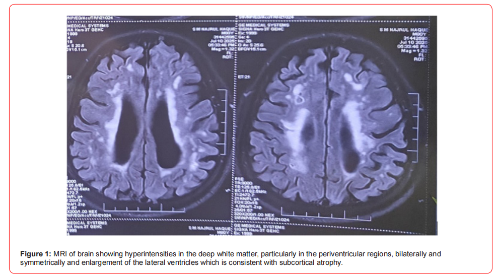
A positive diagnosis of Binswanger’s disease, lacunar stroke in posterior limb of the left inner capsule associated with pure motor hemiparesis regarding the right side of the body, and stage II arterial hypertension at presentation was established.
The therapeutic management targeted lowering of blood pressure in accordance with the actual guidelines and secondary stroke prevention using antiplatelet monotherapy and statins. Rosuvastatin 20 mg per day, levodopa with carbidopa and clopidogrel 75mg per day were continued. Perindopril and cilnidipine was started as antihypertensive medications. The patient was also evaluated in the first 48 hours after admission by a physiotherapist, and daily sessions of active and passive mobilization were performed. Regarding the clinical and neurological evolution, the motor deficit, fine motor control, and prehension improved during admission and during the first month after discharge from hospital.
Through the progressive nature of BD, the prognosis is generally poor. However, controlling vascular risk factors may slow disease progression. The patient was regularly followed up closely to monitor for further cognitive and functional decline.
Discussion
A diagnosis of Binswanger’s disease was proposed for our patient based on the clinical features regarding the risk factors, general and neurological examination, the white matter changes observed on MRI. More than 20 years have passed since Benett and Caplan proposed a diagnostic criterion for Binswanger’s disease.2 Since then, the pathophysiology of the disease has improved, and in complex instances, auxiliary tests can be employed in addition to the normal clinical features and imaging [7]. Changes in CSF biochemistry may indicate the neuroinflammation that occurs in small vessel disease [8]. Neuroinflammation induces a breakdown in the blood-brain barrier, resulting in increased permeability on the one hand and dramatic alterations in glial cell protein and cytokine expression patterns on the other. Increased albumin levels in the blood may reflect these vascular dementias having higher levels than those with huntingtons disease or other types of dementia. The clinician can use MRI diffusion tensor imaging (to assess white matter tract integrity) or dynamic contrast enhancement MRI (to detect blood-brain barrier disruption) to make a diagnosis. These imaging techniques are unproven and untested in the real world. It’s vital to remember that a diagnosis can’t be made merely on the basis of CT or MRI imaging; it needs to be backed up by a comprehensive clinical evaluation. Binswanger disease requires a multi modal approach to diagnosis. None of the biomarkers are sufficient to diagnose the condition on their own, but in patients with [9] cognitive impairment and neurological indications of questionable or unknown aetiology, using clinical data in conjunction with imaging and auxiliary testing can be helpful. There is a scarcity of information about the disease’s prognosis. Binswanger’s disease is currently incurable, and there is no cure. There are no particular clinical research addressing therapeutics for Binswanger disease, despite the American Heart Association (AHA) publishing therapy guidelines for people with vascular cognitive impairment. Blood pressure control, antiplatelet medication, and statins all aid in the prevention of secondary strokes and the slowing of white matter lesions. Dietary adjustments (salt reduction), physical exercise, physical therapy, and rehabilitations all play a key role in the treatment of these people, resulting in improved quality of life, as well as health advantages. It is often difficult to differentiate between Binswanger disease and Alzheimer’s disease clinically. Memory impairment and dysfunction in cognition takes place in both, but predominantly in the initial phase of the disease strokes, hypertension, and asymmetric motor and sensory deficits, points to BD. Predominant impairment in memory without associated apathy, confusion, or character change was uncommon in analysis of our case report. It is helpful in differentiating Alzheimer’s disease and BD by looking for the presence of hypodensity in white matter with infarcts in cornices and lacunar region [10]. Normal pressure hydrocephalus [NPH] presenting along with mental instability, when not associated with incontinence and gait disorder, simulates BD. Mild white matter low density, sometimes seen surrounding the enlarged frontal horns in NPH,46 simulates the white matter hypodensity of BD. Nevertheless, enlargement of ventricles is generally less pronounced and hypodensity in white matter more extensive in BD. A more detailed [11] clinicopathologic correlation is required to find out the exact cause of the characteristic white matter lesions, with serial sectioning of small vessels supplying the gliotic regions, experimental investigation of chronically hypertensive animals, and perhaps PET scan of patients with postulated BD.
A recent biomarker identified as having larger levels is patients with Binswanger’s disease is lipocalin 2 (LCN2). LCN2, also known as oncogene 24p3, a glycoprotein involved in NVU damage in patients with vascular disease. It had promising results and was found having larger levels in patients with vascular dementia as opposed to Alzheimer’s disease or other types of dementia [12].
Various imaging studies such as MRI diffusion tensor imaging (to evaluate white matter tracts integrity) or dynamic contrast enhancement MRI (to reveal disruption of the bloodbrain barrier) can aid the clinician in establishing the diagnosis. These imaging techniques and many others have unknown reproducibility and lack validation for Binswanger disease in larger populations. It is important to mention that the diagnosis cannot be given solely on CT or MRI imaging and requires a careful clinical examination [5,13,14].
Establishing the diagnosis for Binswanger’s disease requires a multimodal approach. None of the biomarkers alone are adequate to diagnose the disease, but using clinical data alongside imaging and ancillary tests can prove helpful in patients with cognitive impairment and neurological signs with uncertain or unknown etiology [5,13,14,15].
The literature is scarce regarding the prognosis of the disease. Binswanger disease is progressive, and there is currently no cure available. There are no specific clinical studies targeting therapies for Binswanger’s disease, although the American heart association (AHA) published treatment guidelines for patients with vascular cognitive impairment [25]. blood pressure control, antiplatelet therapy, and statins play a central role in secondary stroke prevention and lead to slowing the progression of white matter lesions. Dietary changes (especially reducing the intake of salt), physical activity, physical therapy, and rehabilitations are an important part in the management of these individuals, leading to improvement of the functional status and the quality of life, besides other wellknown health benefits [5,16,17].
Conclusion
Binswanger’s disease is a complex neuropsychiatric disease,and its pathophysiology is only partially understood. Treatment options available are only symptomatic which includes antidepressant to treat depression, anti-platelet to eradicate thromboembolism, statins to reduce atherosclerosis, anti-hypertensives to treat hypertension. As new pathophysiological mechanisms are revealed, other tests will become available in the years to come and also novel therapies will specifically target these mechanisms (inflammation, arterial stiffness, and clearance of cerebral waste) in order to better treat these patients.
Acknowledgement
None.
Conflict of Interest
None declared.
References
- Vanderver A, Prust M, Tonduti D, et al. (2015) Case definition and classification of leukodystrophies and leukoencephalopathies. Mol Genet Metab 114(4): 494-500.
- Caplan LR (1995) Binswanger's disease–revisited. Neurology 45(4): 626-633.
- Loeb C (2000) Binswanger's disease is not a single entity. Neurol Sci 21: 343-348.
- Ropper AH, Adams RD, Victor M, Brown RH, Victor M (2005) Adams and Victor’s principles of neurology, 8th edn. New York, NY: McGraw-Hill Medical Pub. Division: 831-832.
- Huisa BN, Rosenberg GA (2014) Binswanger's disease: toward a diagnosis agreement and therapeutic approach. Expert Rev Neurother 14(10): 1203-1213.
- Wolters FJ, Ikram MA (2019) Epidemiology of vascular dementia. Arterioscler Thromb Vasc Biol 39(8): 1542-1549.
- Branko N Huisa, Gary A Rosenberg (2025) Binswanger's disease: toward a diagnosis agreement and therapeutic approach.
- Branko N Huisa, Gary A Rosenberg (2025) Binswanger's disease: toward a diagnosis agreement and therapeutic approach.
- Philip B Gorelick, Scott E Counts, David Nyenhuis (2015) Vascular cognitive impairment and dementia.
- Jacobs L, Kinkel W (1976) Computarized axial transverse tomography in normal pressure hydrocephalus. Neurology 26: 501-507
- Tibor Kovács, Imre Szirmai, Mátyás Papp (2025) Clinico-pathology and differential diagnosis of Binswanger's disease.
- Llorens F, Hermann P, Villar-Piqué A, et al. (2020) Cerebrospinal fluid lipocalin 2 as a novel biomarker for the differential diagnosis of vascular dementia. Nat Commun 11: 619.
- Rosenberg GA (2016) Matrix metalloproteinase-mediated neuroinflammation in vascular cognitive impairment of the binswanger type. Cell Mol Neurobiol 36(2): 195-202.
- Huisa BN, Caprihan A, Thompson J, Prestopnik J, Qualls CR, Rosenberg GA (2015) Long-term blood-brain barrier permeability changes in binswanger disease. Stroke 46(9):2413-2418.
- Wolters FJ, Ikram MA (2019) Epidemiology of vascular dementia. Arterioscler Thromb Vasc Biol 39(8): 1542-1549.
- Dufouil C, Chalmers J, Coskun O, et al. (2005) Effects of blood pressure lowering on cerebral white matter hyperintensities in patients with stroke: the PROGRESS (perindopril protection against recurrent stroke study) magnetic resonance imaging substudy. Circulation 112(11): 1644-1650.
- Weber R, Weimar C, Blatchford J, et al. (2012) Telmisartan on top of antihypertensive treatment does not prevent progression of cerebral white matter lesions in the prevention regimen for effectively avoiding second strokes (PRoFESS) MRI substudy. Stroke 43(9): 2336-2342.
-

This work is licensed under a Creative Commons Attribution-NonCommercial 4.0 International License.




