


 Review Article
Regulation On Neuronal Excitability and Epileptogenesis. Part II
Received Date: October 13, 2026; Published Date: October 26, 2026
Abstract
Introduction: The “state of the art” regarding intracellular signaling pathways linked to mGluRs. Methods: This is a narrative review paper based
on the fact that G-protein coupled receptors play critical roles in the modulation of neuronal excitability. Meanwhile scaffold proteins are abundant
and essential components of the postsynaptic density (PSD). They play a major role in many synaptic functions including the trafficking, anchoring,
and clustering of glutamate receptors and adhesion molecules. Moreover, they link postsynaptic receptors with their downstream signaling proteins.
mGlu5 binds to NMDARs via scaffolding proteins, both postsynaptic density protein 95 (PSD-95) and Homer protein, leading to phosphorylation of
NMDARs. The net result is potentiation of NMDAR currents. Results: As relevant facts we pointed out three challenging data:
• Reduction in GAIP interacting protein C-terminus 1 (GIPC1) expression in both TLE patients and in mice with KA-induced epilepsy. Moreover,
GIPC1 colocalized with mGluR7 and its overexpression counteracted epileptogenesis through mGluR7 upregulation.
• Up- regulation of postsynaptic Homer 1a could protect against neuronal injury by reducing the level of phosphorylated extracellular signal
regulated kinase ERK and then disrupting drug target in order to control seizures and prevent epileptogenesis.
• In kindled rats the overexpression of neuropeptide Y (NPY) acts as a protective factor. Neuropeptide Y has been identified as being synthesized
in GABAergic neurons and to act as a neurotransmitter during cellular communication and is expressed in interneurons. Conclusion: We found
out three interesting points in order to design new antiepileptic drug-developments, each alone or in potencial sinergistic association. The
down-stream proteins of mGluRs such as those in ERK/MAPK, mTOR, PLC, PKC and CaM pathways may be also promising therapeutic targets.
Keywords: epileptogénesis, glutamate receptors, intracellular signaling pathways.
Introduction
This section, part II, is a brief review on glutamate metabotropic
receptors studies
• The implications of G proteins
• RGS proteins
• And scaffold proteins
• In the excitatory impulses within the neurons through
intracelular signaling pathways
• Challenging future directions in order to counteract these
impulses preventing epileptic attacks.
Glutamate Metabotropic Receptors
Group I mGluR subtypes (mGluR1 and mGluR5) are primarily expressed postsynaptically but exist also in presynaptic terminals of GABA and glutamatergic neurons [1,3]
Group I GluRs regulate neuronal excitability through modulation of ionotropic glutamate receptors activity at postsynaptic density (PSD). Activation of mGluR1 by a specific agonist can maintain and prolong interictal discharge. [4] Overactivation can lead to epileptogenesis. Group I promote the activity of PLC (phospholipase C) increasing the intracellular concentrations of diacylglicerol and inositoltriphosphate [5], leading to intracellular Ca2+accumulation. mGluR1 can also be activated by increases in intracellular calcium and depolarization of hippocampal CA1 pyramidal cells, which are involved in inducing long-term potentiation (LTP) and long-term depression (LTD), a long-lasting synaptic plasticity, at multiple glutamatergic synapses [6].
It also amplifies ionotropic signals by inhibition of TASK and TREK K+ channels. [7] Additionally, protein–protein interaction is an important coupling mechanism for the action and regulation of metabotropic receptors. mGlu5 binds to NMDARs via scaffolding proteins, both postsynaptic density protein 95 (PSD-95) and Homer protein, leading to phosphorylation of NMDARs. The net result is potentiation of NMDAR currents. [8] Group I mGluRs, both of the mGluR1 and mGluR5 subtypes, co-localize with NMDA receptors on noradrenergic terminals of rat hippocampus and human neocortex. [9] By binding to their protein-protein interaction, mGluRs interact with enzymes, ion channels and other proteins to assemble in diverse signal complexes. Signal complexes include kinases, phosphatases, calmodulin, calcineurin inhibitor and norbin [10].
Metabotropic glutamate receptors (mGluRs) can dimerize and bind to a variety of proteins in addition to trimeric G-proteins. These newly identified protein interactions lead to the discovery of new regulatory mechanisms that are independent of, and sometimes synergistic with, the classical G-protein-coupled second messenger pathways. Notably, several of these mechanisms connect mGluR-mediated signaling to other receptor classes, thereby creating a network of different receptor types and associated signal cascades. The intracellular C-termini of mGluRs play a key role in the regulation of these networks. [11] The authors suppose that suppression of mGluR1 may be a novel way to counteract epileptogenesis.
Several studies corroborated that Group II and Group III mGluRs are located at glutamatergic neurons in presynaptic ends and reduce glutamate release. 2 which makes them potential targets for antiseizure drug development. Activation of group II / III receptors reduces the concentration of cAMP by inhibiting AC (adenyl ciclase). Theses receptors are linked to a G-protein (Gi) which in turn decrease presynaptic Ca2+ concentration therefore limiting the release of glutamate. [12, 13] Chinese authors detected a reduction in GAIP interacting protein C-terminus 1 (GIPC1) expression in both TLE patients and in mice with KA-induced epilepsy. They further showed that GIPC1 co-localized with mGluR7 and its overexpression counteracted epileptogenesis through mGluR7 upregulation [14].
G Proteins
G proteins, also known as guanine nucleotide-binding proteins, are a family of proteins that act as molecular switches inside cells, and are involved in transmitting signals from a variety of stimuli from outside into the cell. Their activity is regulated by factors that control their ability to bind and hydrolyze guanosine triphosphate (GTP) to guanosine diphosphate (GDP). When they are bound to GTP, they are activated, and when they are bound to GDP, they are turned off. G proteins are in fact GTPases.
There are two classes of G proteins. The first function as monomeric small GTPases (RAS superfamily) freely distributed in cytoplasm or nucleoplasm, while the second (large G proteins) function as heterotrimeric G protein complexes anchored to the cell membrane. The latter class of complexes are made up of alpha (α), beta (β), and gamma (γ) subunits. [15] The beta and gamma subunits can form a stable dimeric complex referred to as the betagamma complex. [16] Heterotrimeric G proteins located within the cell are activated by G protein-coupled receptors (GPCRs) spanning the cell membrane. Signaling molecules bind to a domain of the GPCR located outside the cell, and an intracellular GPCR domain then in turn activates a particular G protein.
GPCRs and G proteins transmit signals from hormones, neurotransmitters, and other factors. G proteins regulate metabolic enzymes, ion channels, transporter proteins, and other parts of the cell machinery, controlling transcription, motility, contractility, and secretion, which in turn regulate diverse systemic functions such as embryonic development, learning and memory, and homeostasis. [17] When a ligand activates the GPCR, it induces a conformational change in the receptor that allows it to function as a guanine nucleotide exchange factor (GEF), exchanging GDP for GTP. The GTP is bound to the Gα subunit. This exchange triggers the dissociation of the Gα subunit bound to GTP from the Gβγ dimer. Once this occurs, Gα-GTP and Gβγ can activate different second messenger pathways and effector proteins, allowing the receptor to activate the next G protein [18].
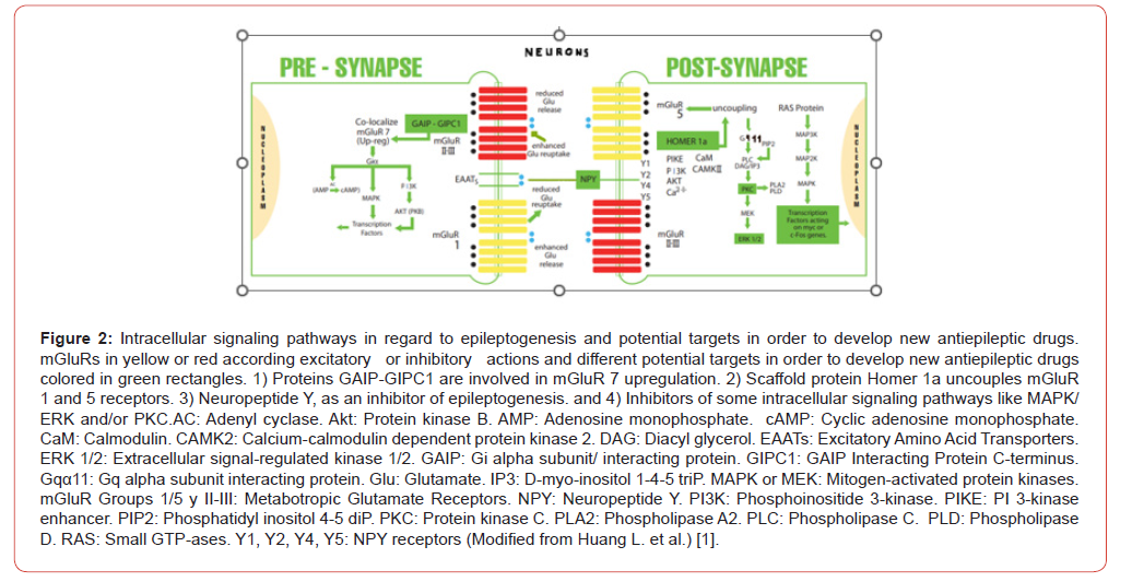
Gαq/11 stimulates the membrane-bound phospholipase C (PLC), which then cleaves into two second messengers: IP3 and diacylglycerol (DAG). The Inositol Phospholipid Dependent Pathway (PKC) is a signal transduction pathway for many downstream targets (Figure 1 and 2).
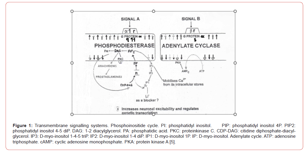
Gαs activates the cAMP-dependent pathway by stimulating the production of cyclic AMP (cAMP) from ATP. This is accomplished by direct stimulation of the membrane-associated enzyme adenylate cyclase. cAMP can then act as a second messenger that then interacts with, and activates protein kinase A (PKA). PKA can phosphorylate a myriad downstream targets (Figure 1). Gq, Go and Gs are stimulatory receptors whereas Gi is inhibitory. Gq and go activate the phospholipase C (PLC) and, subsequently, the protein kinase C (PKC) pathways. Gs activates the cAMP. Gi, on the other hand, inhibits several cellular signaling cascades.
GPCRs is the largest family of integral membrane proteins first identified as receptor proteins that couple via heterotrimeric G-proteins to regulate a vast variety of effector proteins modulating cellular functions. They interact with a myriad of proteins that not only function to improve or attenuate their signalling but also to couple these receptors to heterotrimeric G-protein-independent signalling pathways. In addition, intracellular and transmembrane proteins associated with GPCRs regulate their processing in the endoplasmic reticulum, trafficking to the cell surface, compartmentalization to plasma membrane microdomains, endocytosis and trafficking between intracellular membrane compartments.
G protein-coupled receptors (GPCRs) play key roles in intercellular signaling in the brain. Their effects on cellular function have been largely studied in neurons, but their functional consequences on astrocytes are less known. Durkee et al. found that while GqGPCR activation led to cellular activation in both neurons and astrocytes, Gi/oGPCR activation led to cellular inhibition in neurons and cellular activation in astrocytes. Astroglial activation by either Gq or Gi/o protein-mediated signaling stimulated gliotransmitter release, which increased neuronal excitability [19].
In order to evaluate the involvement of the stimulatory G-protein (Gs)-related transduction system in the basic mechanisms of epilepsy, Iwasa et al. [20, 21] examined the expression levels of Gsα mRNA and specific GTP-binding ability in the hippocampus of amygdaloid-kindled rats at various seizure stages. Northern blot analysis showed a significant increase in the Gsα mRNA expression level in the bilateral hippocampus at 24 h after the last generalized seizure. The [3H]-GTP-binding assay with isoproterenol (IPN), a β-receptor agonist, revealed a remarkable increase of Bmax values in the sham-operated control and partially kindled groups. However, the IPN-induced increase of Bmax values was abolished on both sides of the hippocampus at 24 h after and at 4 weeks after the last generalized seizure in fully kindled rats. This data suggests that alteration in the Gs function and β-adrenergic receptor-Gs coupling might be implicated in the neurobiological basis of the induction mechanisms of the generalization of seizures and the mechanisms of the maintenance of enduring epileptogenesis. Conversely, the Gs-related transduction system might have a lesser impact on the acquisition process of epileptogenesis. Authors suggest that a functional imbalance among various types of GTP binding proteins (Gs, Gi or Go) might be related to the acquisition of epileptogenesis in the kindling model of epilepsy [20].
In PTZ rats, both brain regions, hippocampus and neocortex displayed a marked increase in [35S] GTPγS incorporation stimulated by ACh, bethanechol and particularly oxotremorine. A generalized seizure in immature rats can entail a long-term and presumably permanent increase in the efficacy of G-protein coupling to muscarinic receptors in the hippocampus and neocortex when adult. By analogy, such a mechanism could account for the susceptibility to epilepsy of human adults having suffered from prolonged seizures in early life [22].
Although scaffold proteins are not strictly defined in their function, they are known to interact and/or bind with multiple members of a signalling pathway, linking them into complexes. In such pathways, they regulate signal transduction and help localize pathway components to specific areas of the cell such as the plasma membrane, cytoplasm, nucleus, Golgi apparatus, endosomes, and mitochondria. Scaffold proteins regulate signal trasduction cascades, by changing their molecular composition in the complexes.
Scaffold proteins act in at least four ways:
• Binding signaling components,
• Localizing these components to specific areas of the cell,
• Regulating signal transduction by coordinating positive
and negative feedback signals, and
• Insulating correct signaling proteins from competing
proteins. (34) In more detail:
Scaffold proteins cluster signaling components of a cascade into complexes. This assembly may be able to enhance signaling specificity by preventing unnecessary interactions between signaling proteins, and enhance signaling efficiency by increasing the proximity and effective concentration of components in the scaffold complex. A common example of how scaffold proteins enhance specificity is a scaffold that binds a protein kinase and its substrate, thereby ensuring specific kinase phosphorylation.
Scaffold proteins localize the signaling reaction to a specific area in the cell, a process that could be important for the local production of signaling intermediates. A particular example of this process is the scaffold A-kinase anchor proteins (AKAPs), which guide cyclic AMP-dependent protein kinase (PKA) to various sites in the cell. [35] This localization is able to locally regulate PKA and results in the local phosphorylation by PKA of its substrates.
Many hypotheses about how scaffold proteins coordinate positive and negative feedback come from engineered scaffolds into mathematical modeling. In three-kinase signaling cascades, scaffolds bind all three kinases, enhancing kinase specificity and restricting signal amplification by limiting kinase phosphorylation to only one downstream target. These abilities may be related to the stability of the interaction between the scaffold and the kinases, the basal phosphatase activity in the cell, scaffold location, and expression levels of the signaling components [36,37].
Signaling pathways are often inactivated by enzymes that reverse the activation state and/or induce the degradation of signaling components. Scaffolds have been proposed to protect activated signaling molecules from inactivation and/ or degradation. Mathematical modeling has shown that kinases in a cascade without scaffolds have a higher probability of being dephosphorylated by phosphatases before they are even able to phosphorylate downstream target. [37] Furthermore, scaffolds have been shown to insulate kinases from substrate- and ATPcompetitive inhibitors [38].
One example of a scaffold protein that modulates calcium signaling are the proteins of the Homer family. Homer proteins has been shown to positively regulate calcium signaling in neurons by linking the glutamate receptor with triphosphate receptors in the endoplasmic reticulum. [39] Downregulation of Homer1b/c could attenuate group I mGluR dependent Ca2+ signaling through the regulation of Ca2+ release in the endoplasmic reticulum thus protecting neurons from glutamate excitotoxicity after injury. [40] Upregulation of postsynaptic Homer 1a could protect against neuronal injury by reducing the level of phosphorylated extracellular signal regulated kinase ERK and then disrupting mGluR-ERK signaling. Homer 1a may be a promising therapeutic drug target in order to control seizures and prevent epileptogenesis (Figure 2) [10].
Homer 1 protein has an N-terminal EVH1 domain involved in protein interaction, and a C-terminal winded-coil domain involved in self-association. It consists of two major splice variants, short (1a) and long (1b, 1c) form. Homer 1a has only EVH1 monomeric domain while Homer 1b and c have both EVH1 and windedcoil domains and are tetrameric. The winded-coil can be further separated into N-terminal half and C-terminal half [36]. The N-terminal half of the winded-coil domain is predicted to be a parallel dimer while the C-terminal half is a hybrid of dimeric and antiparallel tetrameric winded-coil. As a whole, long Homer is predicted to have a dumbbell-like structure where two pairs of EVH1 domains are located on two sides of long (50nm) winded-coil domain.
Homer-1a/Ves11S is expressed during seizures and reduce seizure susceptibility. Cellular mechanisms for this feedback regulation were studied in neocortical pyramidal cells injecting Homer 1a protein intracellularly. The injection reduced membrane excitability. Resting potential was hyperpolarized by 5-10mV and mean frequency of spikes evoked by depolarizing currents injection was decreased [41].
Kindling by repeated electrical stimulation of the amygdala resulted in the differential expression of 264 genes in the hippocampus compared to controls. The most strongly induced gene was Homer 1A, an immediate early gene involved in the modulation of glutamate receptor function. The overexpression of Homer 1A in the hippocampus of kindled rats was confirmed by RT-PCR. Immunohistochemical characterization of mice showed a marked Homer 1A overexpression in glutamatergic neurons of the hippocampus. Kindling of Homer 1A overexpressing mice resulted in a retardation of seizure generalization compared to wild-type controls. These data demonstrate that kindlinginduced epileptogenesis leads to a striking overexpression of Homer 1A in the hippocampus, which may represent an intrinsic antiepileptogenic and anticonvulsant mechanism, that counteracts progression of the disease [42].
Avedissian et al. investigated differentially expressed genes by comparison of mRNAs from normal and epileptic rat hippocampus in the pilocarpine model of epilepsy. Using an Expressed Sequence Tags (EST) methodology, ORESTES epilepsy database revealed diverse protein family domains, such as cytoskeletal, cell signaling and PK kinase, which could be involved in processes underlying epileptogenesis. They showed that the expression of Homer 1a, known to be coupled to mGluR and NMDA synaptic transmission, is associated with pilocarpine induced status epilepticus [43].
Inversely, overexpression of Homer1b/c induced the overexpression of mGluR1 and mGluR5, which showed an increase in neuronal excitability [44].
Scaffold proteins are abundant and essential components of the postsynaptic density (PSD). They play a major role in many synaptic functions including the trafficking, anchoring, and clustering of glutamate receptors and adhesion molecules. Moreover, they link postsynaptic receptors with their downstream signaling proteins and regulate the dynamics of cytoskeletal structures. By definition, PSD scaffold proteins do not have intrinsic enzymatic activities but are formed by modular and specific domains deputed to form large protein networks [45].
The postsynaptic density (PSD) protein complex has long been a major target of proteomics in neuroscience. As the number of glutamate receptors on a synapse is one of the main determinants of synaptic efficacy, determining the absolute numbers of receptors in the PSD is necessary for estimating the amplitude of the excitatory postsynaptic current (EPSC) in individual synapses. Moreover, as the receptor molecules are embedded in a macromolecular complex within the PSD, stoichiometry between the receptors and other PSD proteins could help explain the functional and regional specialization of the synapses and their possible roles in synaptic functions [46].
Future Directions
Signal transduction is the starting point of the effector component of the signaling pathway. In this process, the signal, by interacting with the receptor, starts a series of molecular events within the cell leading to the final effect of the signaling process. Second messenger systems can amplify or modulate a signal, in which activation of a few receptors results in multiple secondary messengers being activated, thereby amplifying the initial signal (the first messenger). The downstream effects of these signaling pathways may include additional enzymatic activities such as proteolytic cleavage, phosphorylation, methylation, and/ or ubiquitination. Signaling molecules can be synthesized from various biosynthetic pathways and released through passive or active transports. Different receptor systems may share common signal transduction pathways with interacting proteins.
The multistep process of the transduction stage is often composed of the activation of proteins by addition or removal of phosphate groups or even the release of other small molecules or ions that can act as messengers.
Cystic fibrosis transmembrane conductance regulatorassociated ligand (CAL) through binding to adaptors proteins profoundly inhibits the ubiquitination of mGluR5a and enhances receptor expression at the protein level but not at the mRNA level. These findings reveal that mGluR5a protein expression is physiologically regulated via its interaction with CAL. These results also suggest a molecular mechanism by which mGluR5a protein expression may be regulated at the post-translational level by the CAL protein, possibly by blocking ubiquitination-dependent receptor degradation. [47] The key difference between adaptor and scaffold protein is that the adaptor proteins usually small proteins that binds only to two proteins involved in the signalling pathway, while scaffold proteins are large proteins that binds to a number of different proteins involved in the signalling pathway.
For example, the ligand epidermal growth factor (EGF) binds to the receptor (called EGFR). This activates the receptors phosphorylation. [48] The phosphorylated receptor binds to an adaptor protein (GRB2), which couples the signal to further downstream signaling processes. Many cellular proteins are activated downstream of the growth factor receptors (such as EGFR) that initiate this signal transduction pathway.
A more complex signal transduction pathway is the MAPK/ERK pathway, which involves changes of protein–protein interactions inside the cell, induced by an external signal. Several of these receptors are kinases that start their phosphorylation and of other proteins when binding to a ligand. This phosphorylation can generate a binding site for a different protein and thus induce protein–protein interaction. One of the signal transduction pathways that is activated is called the mitogen-activated protein kinase (MAPK) pathway. The signal transduction component labeled as “MAPK” in the pathway was originally called “ERK,” so the pathway is called the MAPK/ERK pathway. The MAPK protein is an enzyme, a protein kinase that can attach phosphate to target proteins such as the transcription factor MYC and thus, alter gene transcription (Figure 2), and ultimately, cell cycle progression. Finally, the end of a signal pathway leads to the regulation of cellular activity. This response can take place in the nucleus or in the cytoplasm of the cell. A majority of signaling pathways control protein synthesis by turning certain genes on and off in the nucleus. The signal that starts the MAPK/ERK pathway is the binding of extracellular mitogen to a cell surface receptor. This allows a Ras protein (a Small GTPase) to swap a GDP molecule for a GTP molecule, flipping the “on/off switch” of the pathway. The Ras protein can then activate MAP3K (e.g., Raf), which activates MAP2K, which activates MAPK. Finally, MAPK can activate a transcription factor, such as Myc. MAPK regulates the activities of several transcription factors. MAPK can phosphorylate C-myc (Figure 2). MAPK phosphorylates and activates MNK, which, in turn, phosphorylates CREB. MAPK also regulates the transcription of the C-Fos gene. By altering the level and activity of transcription factors, MAPK leads to altered transcription of genes that are important for the cell cycle. Merlo et al. studied the activation state of ERK1/2 in rat hippocampal slices during application of the K+ channel blocker 4-aminopyridine (4AP, 50 μm), a procedure that enhances synaptic transmission and leads to the appearance of epileptiform activity. Hippocampal slices superfused with 4AP-containing medium exhibited a marked activation of ERK1/2 phosphorylation that peaked within about 20 min. Application of the ERK pathway inhibitors U0126 (50 μm) or PD98059 (100 μm) markedly reduced 4AP-induced epileptiform synchronization, abolishing ictal discharges in the CA3 area. The data demonstrate that activation of ERK1/2 accompanies the appearance of epileptiform activity induced by 4AP and suggest a cause-effect relationship between the ERK pathway and epileptiform synchronization [49].
A rational strategy for preventing epilepsy is to target primary signaling pathways that initially trigger the numerous downstream mechanisms mediating epileptogenesis. The mammalian target of rapamycin (mTOR) pathway is a candidate, because mTOR regulates multiple cellular functions that may contribute to epileptogenesis, including protein synthesis, cell growth and proliferation, and synaptic plasticity. The importance of the mTOR pathway in epileptogenesis is best illustrated by tuberous sclerosis complex (TSC), one of the most common genetic causes of epilepsy. In mouse models of TSC, mTOR inhibitors prevent the development of epilepsy and underlying brain abnormalities associated with epileptogenesis. Accumulating evidence suggests that mTOR also participates in epileptogenesis due to a variety of other causes, including focal cortical dysplasia and acquired brain injuries, such as in animal models following status epilepticus or traumatic brain injury. mTOR inhibition may represent a potential antiepileptogenic therapy for diverse types of epilepsy, including both genetic and acquired epilepsies. [50] Recent studies in animal models have suggested that the mammalian target of rapamycin (mTOR) signaling pathway is involved in several features of mesio-temporal lobe epilepsy (MTLE), and that its inhibition could have therapeutic interests. However, controversies remain whether mTOR activation is cause or consequence of MTLE. Shima et al by intrahippocampally injections with kainate (1 nmol), found a biphasic increase of phospho-S6 (p-S6) ribosomal protein expression, the downstream product of the mTOR signaling pathway, in the dispersed granule cell layer (GCL) of the dentate gyrus with a second phase lasting up to 6 months. Chronic treatment with rapamycin suppressed p-S6 expression, granule cell dispersion and mossy fiber sprouting, but did not reduce cell loss, BDNF overexpression, glutamic acid decarboxylase (GAD)67 expression or the development of hippocampal paroxysmal discharges. [51] This data suggests that activation of the mTOR signaling pathway results from the increased neuronal excitation that develops in the GCL and may contribute to MTLE morphological changes. However, the data does not support the role of this pathway in the development of MTLE or its inhibition as a therapy for this form of epilepsy.
Cultured astrocytes exposed to oxygen and glucose deprivation (OGD) for 6 h presented significant reductions in the Glutamate- Aspartate Transporter (GLAST) expression and extracellular glutamate clearance. These reductions were effectively restored by mGluR2/3 activation with mGluR2/3 agonists, LY379268 or DCGIV, after the 6 h OGD insult. These mGluR2/3-mediated restorative effects were inhibited by selective mGluR2/3 antagonists LY341459 or EGLU. The mGluR2/3 activation also induced activations of signaling pathways including extracellular signal-regulated kinase (ERK), phosphatidylinositol 3-kinase (PI3K) and nuclear transcription factor-κB (NFκB). These activations were prevented by blocking mGluR2/3 with LY341459, an mGluR2/3 antagonist. Furthermore, blocking ERK, PI3K and NFκB signaling pathways with U0126, LY294002 and pyrrolidine dithiocarbamate, respectively, significantly inhibited the mGluR2/3-mediated restorative effects. These results suggest that application of mGluR2/3 agonists after OGD insults can effectively reverse the OGD-reduced expression of GLAST proteins and restore clearance of extracellular glutamate by serially activating ERK/PI3K/NFκB signaling pathways in cultured astrocytes [52].
Transient inhibition of TrkB-PLCγ1 signaling initiated after an isolated seizure limited progression of epileptogenesis, evidenced by the reduced severity and duration of subsequent seizures and reverted a subset of animals to an earlier state of epileptogenesis. Remarkably, inhibition of TrkB-PLCγ1 signaling in the absence of a recent seizure did not reduce severity of subsequent seizures. These results suggest a novel strategy for limiting progression or potentially ameliorating severity of TLE whereby transient inhibition of TrkB-PLCγ1 signaling is initiated following a seizure [53].
In the hippocampus, group I mGluR elicited epileptiform discharges through PLCβ1 signaling. [54] Cuellar et al. [55] indicated that “l-cysteine sulfinic acid (CSA), an agonist of phospholipase D (PLD)-coupled mGluRs, mediated its effect by PLD-driven activation of protein kinase C (PKC), which may desensitize PLClinked group I mGluRs and thereby prevent group I mGluR-induced epileptogenesis. Previous studies indicated that PKCβ1, PKCβ2, and PKCγ might be involved in mGluR1α-related excitoneurotoxicity and epileptogenesis, (56) and mGluR5-PLCβ4-PKCβ2/PKCγ pathway were involved in excitotoxicity against hippocampal CA1 pyramidal neurons leading to the loss of these neurons. [57] In vitro studtes also indicated that PKC induced prolongation of epileptiform bursts was dependent on changes specific to mGluR5 and not mediated simply by a generalized increase in transmitter release. [58] In pilocarpine model, mGluR5 may regulates the PKCζ activation in the hippocampal interneurons in epilepsy [59].
Increased PKCε may support the “epsilon theory” of epileptogenesis. [60] Alteration of the expression of cyclicAMP dependent protein kinase (cPKA) subtypes cPKAβ and cPKAγ in mouse hippocampus [60] suggests that hippocampal PKC and cPKA isoforms play different roles in neuronal hyperexcitability and epileptogenesis, and may be targets for development of anticonvulsive and antiepileptogenic drugs [61].
Brain-derived neurotrophic factor BDNF applied during the first week of hippocampal stimulation significantly delayed the progression of kindling, an effect that outlasted the end of the infusion by at least 7 days. Conversely, infusion of BDNF antisense oligodeoxynucleotides to reduce the expression of endogenous BDNF in the hippocampus aggravated the electroencephalographic expression of seizures. Chronic infusion of BDNF increased the expression of NPY in the hippocampus, with a time course similar to that of the protective effect of the neurotrophin on kindling. Finally, chronic infusion of NPY in the hippocampus delayed the progression of hippocampal kindling, whereas anti-NPY antibodies had an aggravating effect. These results suggest that the seizureinduced increase in BDNF expression in the hippocampus may constitute an endogenous protective mechanism able to counteract hippocampal epileptogenesis. This protective effect appears to be mediated at least in part through the regulation of NPY expression [62].
Accumulated data suggest that agonists or antagonists of mGluRs, especially group I mGluR antagonists are promising candidate drugs to control seizures and subsequent neurodegeneration, i.e., to prevent epileptogenesis. The interacting or down-stream proteins of mGluRs such as Homers, PKCs and CaM may be more promising therapeutic targets due to the fact that these proteins are shared by different neurotransmitter receptor signal transduction pathways, i.e, serve as common signal transduction molecules. For instance, inhibition of PKC may not only reduce mGluR5- related neuronal hyperactivity, but also induce GABAA receptor hypersensitivity. Therefore, PKC inhibitor may be more effective in controlling epileptogenesis than single mGluR5 antagonist or GABAA receptor agonist. However, because of there being so many PKC isoforms, activation of each one of them may produce different effects (10).
Conclusion
This review allows us to signal various possible targets in order to block epileptogenesis: 1) agonists of Group II-III mGluRs, for example GAIP interacting protein C-terminus 1 (GIPC1) colocalized with mGluR7 and its overexpression counteract epileptogenesis through mGluR7 upregulation. GIPC was originally identified binding specifically to the C terminus of RGS-GAIP, a protein involved in the regulation of G protein signaling. 2) Homer 1A in hippocampus seems to be an intrinsic antiepileptogenic and anticonvulsant mechanism in the course of epileptogenesis in kindled rats which counteracts the progression of the disease. The short form of Homer 1 uncouples mGluR 1 and 5 signaling as a part of a mechanism of homeostatic plasticity that dampens the neuronal responsiveness when input activity is too high. 3) also, in kindled rats the overexpression of neuropeptide Y (NPY) acts as a protective factor. Neuropeptide Y has been identified as being synthesized in GABAergic neurons and to act as a neurotransmitter during cellular communication and is expressed in interneurons. The receptor protein that NPY operates on is a G protein-coupled receptor and can act pre- or post-synaptically. 4) by inhibiting intracellular transduction signaling pathways like ERK, mTOR, PLC, PKC and or CaM, in order to prevent the epileptogenic effect of Group I mGluRs. It will probably be necessary to test a novel combination of drugs to add different strategies.
Acknowledgment
Morena Bargone, for the Art work in Figure 2.
Dr. Ure, Dr. Perassolo, Dr. Wainwright
University of Buenos Aires, Argentina
Funding
No funding source.
Conflict of Interest
No conflicts of interest
References
- Huang L, Xiao W, Wang Y, Li J, Gong J, et al. (2024) Metabotropic glutamate receptors (mGluRs) in epileptogenesis: an update on abnormal mGluRs signaling and its therapeutic implications. Neural Regen Res 19(2): 360-368.
- Luessen DJ, Conn PJ (2022) Allosteric modulators of metabotropic glutamate receptors as novel therapeutics for neuropsychiatric disease. Pharmacol Rev 74(3): 630-661.
- Ure J, Baudry M, Perassolo M (2006) Metabotropic glutamate receptors and epilepsy. J Neurol Sci 247: 1-9.
- Merlin LR, Wong RS (1997) Role of Group I metabotropic glutamate receptors in the patterning of epileptiform activities in vitro. J. Neurophysiol 78(1): 539–544.
- Ure JA, Perassolo M (2000) Update on the pathophysiology of the epilepsies. J Neurol Sci 177(1): 1-17.
- Mannaioni G, Marino MJ, Valenti O, Traynelis SF, P Jeffrey Conn (2001) Metabotropic glutamate receptors 1 and 5 differentially regulate CA1 pyramidal cell function. J Neurosci 21(16): 5925–5934.
- Chemin J, Girard C, Duprat F, Lesage F, Romey G et al. (2003) Mechanisms underlying excitatory effects of group I metabotropic glutamate receptors via inhibition of 2P domain K+ channels. EMBO J 22(20): 5403-5411.
- Hermans E, Challiss R A (2001) Structural, signalling and regulatory properties of the group I metabotropic glutamate receptors: Prototypic family C G-protein-coupled receptors. Biochem J 359(3): 465-484.
- Luccini E, Musante V, Neri E, Brambilla Bas M, Severi P et al. (2007) Functional interactions between presynaptic NMDA receptors and metabotropic glutamate receptors co-expressed on rat and human noradrenergic terminals. Br J Pharmacol 151(7): 1087-1094.
- Qian F, Tang FR (2016) Metabotropic Glutamate Receptors and Interacting Proteins in Epileptogenesis. Curr Neuropharmacol 14(5): 551-562.
- Enz R (2007) The trick of the tail: protein-protein interactions of metabotropic glutamate receptors. Bioessays 29(1): 60-73.
- Abdul-Ghani AS, Attwell PJ, Singh Kent N, Bradford HF, Croucher MJ et al. (1997) Anti-epileptogenic and anticonvulsant activity of L-2-amino-4-phosphonobutyrate, a presynaptic glutamate receptor agonist. Brain Res 755(2): 202-212.
- Pacheco Otalora LF, Skinner F, Oliveira MS, Farrell B, Arshadmansab MF et al. (2011) Chronic deficit in the expression of voltage-gated potassium channel Kv3.4 subunit in the hippocampus of pilocarpine-treated epileptic rats. Brain Res 1368: 308-316.
- Liu Y, Wang Y, Yang J, Xu T, Tan C et al. (2022) G-alpha interacting protein interacting et al., C terminus 1 regulates epilepto genesis by increasing the expression of metabotropic glutamate receptor 7. CNS Neurosci Ther 28(1): 126-138.
- Hurowitz EH, Melnyk JM, Chen YJ, Kouros-Mehr H, Simon MI et al. (2000) Genomic characterization of the human heterotrimeric G protein alpha, beta, and, gamma subunit genes. DNA Res 7(2): 111–120.
- Clapham DE, Neer EJ (1997) G protein beta gamma subunits. Ann Rev Pharmacol Toxicol 37: 167–203.
- Neves SR, Ram PT, Iyengar R (2002) G protein pathways. Science 296 (5573): 1636-1639.
- Yuen JW, Poon LS, Chan AS, Yu FW, Lo KH et al. (2010) Activation of STAT3 by specific Galpha subunits and multiple Gbetagamma dimers. Int J Biochem Cell Biol 42 (6): 1052–1059.
- Durkee CA, Covelo A, Lines J, Kofuji P, Aguilar J et al. (2019) Gi/o protein-coupled receptors inhibit neurons but activate astrocytes and stimulate gliotransmission. Glia 67(6): 1076-1093.
- Iwasa H, Hasegawa S, Kikuchi S, Watanabe K, Koseki K, et al. (991) Kindling-Induced Kindling-induced changes of [3H] GTP binding in the cerebral cortical membrane. Psychiatry Clin Neurosci 45(2): 303-307.
- Iwasa H, Kikuchi S, Mine S, Sugita K, Miyagishima H et al. (2000) Functional significance of stimulatory GTP-binding protein in hippocampus is associated with kindling-elicited epileptogenesis. Psychiatry Clin Neurosci 54(2): 191-197.
- Potier S, Sénécal J, Chabot JG, Psarropoulou C, Descarries L (2005) A pentylenetetrazole-induced generalized seizure in early life enhances the efficacy of muscarinic receptor coupling to G-protein in hippocampus and neocortex of adult rat. Eur J Neurosci 21(7): 1828-1836.
- Liu Y, Wang Y, Yang J, Xu T, Tan C et al. (2022) G-alpha interacting protein interacting et al., C terminus 1 regulates epilepto genesis by increasing the expression of metabotropic glutamate receptor 7. CNS Neurosci Ther 28(1): 126-138.
- Alberts B, Johnson A, Lewis J, Raff M, Roberts K et al. (2002) Molecular Biology of the Cell. Garland Science 4th edition: 877-ISBN 0815332181.
- Bourne HR, Sanders DA, McCormick F (1990) The GTPase superfamily: a conserved switch for diverse cell functions. Nature 348 (6297): 125-132.
- Cherfils J, Zeghouf M (2013) Regulation of small GTPases by GEFs, GAPs, and GDIs. Physiol Rev 93(1): 269-309.
- Bos JL, Rehmann H, Wittinghofer A (2007) GEFs and GAPs: critical elements in the control of small G proteins. Cell 129 (5): 865-877.
- Vetter IR, Wittinghofer A (2001) The guanine nucleotide-binding switch in three Science 294 (5545): 1299-1304.
- Schmidt A, Hall A (2002) Guanine nucleotide exchange factors for Rho GTPases: turning on the switch. Gen Dev 16 (13): 1587-609.
- Chardin P, Camonis JH, Gale NW, van Aelst L, Schlessinger J et al. (1993) Human Sos1: a guanine nucleotide exchange factor for Ras that binds to GRB2. Science 260 (5112): 1338-1343.
- Fernandez-Zapico ME, Gonzalez-Paz NC, Weiss E, Savoy DN, Molina JR et al. (2005) Ectopic expression of VAV1 reveals un unexpected role in pancreatic cancer tumorigenesis. Cancer Cell 7 (1): 39-49.
- Ross EM, Wilkie TM (2000) GTPase-activating proteins for heterotrimeric G proteins: regulators of G protein signaling (RGS) and RGS-like proteins. Annu Rev Biochem 69: 795-827.
- Seki T, Hayashi N, Nishimoto T (1996) RCC1 in the Ran pathway. J Biochem 120 (2): 207-214.
- Shaw AS, Filbert EL (2009) Scaffold proteins and immune-cell signalling. Nat Rev Immunol 9 (1): 47-56.
- Wong W, Scott JD (2004) AKAP signalling complexes: focal points in space and time. Nat Rev Mol Cell Biol 5 (12): 959-970.
- Levchenko A, Bruck J, Sternberg PW (2000) Scaffold proteins maybiphasically affect the levels of mitogen-activated protein kinase signaling and reduce its threshold properties. Proc Nat Acad Sci 97 (11): 5818-5823.
- Locasale JW, Shaw AS, Chakraborty AK (2007) Scaffold proteins confer diverse regulatory properties to protein kinase cascades. Proc Nat Acad Sci 104 (33): 13307-13312.
- Greenwald EC, Redden JM, Dodge-Kafka KL, Saucerman JJ (2014) Scaffold state switching amplifies, accelerates, and insulates protein kinase C signaling. J Biol Chem 289 (4): 2353-2360.
- Xiao B, Tu JC, Worley PF (2000) Homer: a link between neural activity and glutamate receptor Curr Opin Neurobiol 10 (3): 370-374.
- Lv MM, Cheng YC, Xiao ZB, Sun MY, Ren PC et al. (2014) Down-regulation of Homer1b/c attenuates group I metabotropic glutamate receptors dependent Ca²⁺ signaling through regulating endoplasmic reticulum Ca²⁺ release in PC12 cells. Biochem Biophys Res Commun 450(4): 1568-1574.
- Sakagami Y, Yamamoto K, Sugiura S, Inokh K, Hayashi T (2005) Essential roles of Homer-1a in homeostatic regulation of pyramidal cell excitability: a possible link to clinical benefits of electroconvulsive shock. Eur J Neurosci 21(12): 3229-3239.
- Potschka H, Krupp E, Ebert U, Gümbel C, Leichtlein C et al. (2002) Kindling-induced overexpression of Homer 1A and its functional implications for epileptogenesis. Eur J Neurosci 16(11): 2157-2165.
- Avedissian M, Longo BM, Jaqueta CB, Schnabel B, Paiva PB et al. (2006) Hippocampal gene expression analysis using the ORESTES methodology shows that homer 1a mRNA is upregulated in the acute period of the pilocarpineepilepsy model. Hippocampus 17(2): 130-136.
- Wang Y, Li Y, Wang G, Lu J, Li Z (2022) Overexpression of Homer1b/c induces valproic acid resistance in epilepsy. CNS Neurosci Ther 29(1): 331-343.
- Verpelli C, Schmeisser MJ, Sala C, Boeckers TM (2012) Scaffold proteins at the postsynaptic density. Adv Exp Med Biol 970: 29-61.
- Shinohara Y (2012) Quantification of postsynaptic density proteins: Glutamate receptor subunits and scaffoldingproteins. Hippocampus 22(5): 942-953.
- Cheng S, Zhang J, Zhu P, Ma Y, Xiong Y et al. (2010) The PDZ domain protein CAL interacts with mGluR5a and modulates receptor expression. J Neurochem112(3): 588-98.
- Tito C, Masciarelli S, Coletti G, Tali F (2025) EGF receptor in organ development tissue homeostasis and regeneration. J Biochem Sci 32(1): 24.
- Merlo D, Cifelli P, Cicconi S, Tancredi V, Avoli M (2004) 4-Aminopyridine-induced epileptogenesis depends on activation of mitogen-activated protein kinase ERK. J Neurochem 89(3): 654-659.
- Wong M (2010) Mammalian target of rapamycin (mTOR) inhibition as a potential antiepileptogenic therapy: From tuberous sclerosis to common acquired epilepsies. Epilepsia 51(1): 27-36.
- Shima A, Nitta N, Suzuki F, Laharie AM, Nozaki K et al. (2015) Activation of mTOR signaling pathway is secondary to neuronal excitability in a mouse model of mesio-temporal lobe epilepsy. Eur J Neurosci 41(7): 976-988.
- Lin CH, You JR, Wei KC, Gean PW (2014) Stimulating ERK/PI3K/NFκB signaling pathways upon activation of mGluR2/3 restores OGD-induced impairment in glutamate clearance in astrocytes. Eur J Neurosci 39(1): 83-96.
- Krishnamurthy K, Huang YZ, Harward SC, Sharma KK, Tamayo DL et al. (2019) Regression of Epileptogenesis by Inhibiting Tropomyosin Kinase B Signaling following a Seizure. Ann Neurol 86(6): 939-950.
- Chuang SC, Bianchi R, Kim D, Shin HS, Wong RK (2001) Group I metabotropic glutamate receptors elicit epileptiform discharges in the hippocampus through PLCbeta1 signaling. J Neurosci 21(16): 6387-6394.
- Cuellar JC, Griffith EL, Merlin LR (2005) Contrasting roles of protein kinase C in induction versus suppression of group I mGluRmediatedepileptogenesis in vitro. J Neurophysiol 94(5): 3643-3647.
- Tang FR, Lee WL, Gao H, Chen Y, Loh YT et al. (2004) Expression of different isoforms of protein kinase C in the rat hippocampus after pilocarpine-induced status epilepticus with special reference to CA1 area and the dentate gyrus. Hippocampus 14(1): 87-98.
- Liu JX, Tang YC, Liu Y, Tang FR (2008) mGluR5-PLCbeta4- PKCbeta2/PKCgamma pathways in hippocampal CA1 pyramidal neurons in pilocarpine model of status epilepticus in mGluR5+/+ mice. Epilepsy Res 82(2-3): 111-123.
- Fuortes MG, Faria LC, Merlin LR (2008) Impact of protein kinase C activation on epileptiform activity in the hippocampal slice. Epilepsy Res 82(1): 38-45.
- Toth M (2005) The epsilon theory: a novel synthesis of the underlying molecular and electrophysiological mechanisms of primary generalized epilepsy and the possible mechanism of action of valproate. Med Hypotheses 64(2): 267-272.
- Liu JX, Tang YC, Liu Y, Tang FR (2010) Status epilepticus alters hippocampal PKAbeta and PKAgamma expression in mice. 19(7): 414-420.
- Liu JX, Liu Y, Tang FR (2011) Pilocarpine-induced status epilepticus alters hippocampal PKC expression in mice. Acta Neurobiol Exp (Warsz.) 71(2): 220-232.
- Reibel S, Larmet Y, Carnahan J, Marescaux C, Depaulis A (2000) Endogenous Control of Hippocampal Epileptogenesis: A Molecular Cascade Involving Brain-Derived Neurotrophic Factor and Neuropeptide Y. Epilepsia 41(S6): S127-S133.
-

This work is licensed under a Creative Commons Attribution-NonCommercial 4.0 International License.