 Review Article
Is The Cerebellum Involved in Color Vision Pathway for Essential Tremor?
Anna Piro* and Daniel La Rosa
1National Research Council, Institute of Molecular Bioimaging and Physiology, Catanzaro, Italy
Anna Piro, National Research Council, Institute of Molecular Bioimaging and Physiology, Catanzaro, Italy
Received Date: October 04, 2023; Published Date: November 07, 2023
Abstract
Basing on some previous our results showing no involvement by cerebellum action on color vision pathway in Essential Tremor, in the present work we show as the granular cerebellum excitatory cells can, in some cases, modify the normal color vision pathway in Essential tremor, causing an abnormal color vision. Cerebellum is not only involved in the movement regulation, but its proprioceptive characteristics regulated by both not only inhibitory but also excitatory cells in its cortex, show particular consequences in Essential Tremor.
Keywords:Essential Tremor; Cerebellum; Glutamate; Color vision; Granular cerebellum cells
Visual Areas in Humans: Implications in Color Vision, and its Physiology and Pathophysiology
The primary visual cortex area V1 sends visual information to the extra-striate cortical areas for higher order visual processing. These extra-striate cortical areas are located anterior to the occipital lobe. The main ones are designated as visual areas V2, V3, V4, and V5. There are multiple areas that possibly have different roles in the ability to process color stimulus.
Anatomical and physiological studies have established that the color center begins in V1 and sends signals to extra-striate areas V2 and V4 for further processing, in particular because V4 has the strength of the color receptive fields in its neurons [1]. By MRI brain Imaging, three main areas stimulated by color have been found: V1, in the ventral occipital lobe, specifically the lingual gyrus, hV4, and αV4, anteriorly in the fusiform gyrus [2]. Two models were tested: one model had hV4 completely in the ventral side, and the second model had hV4 split into dorsal, and ventral sections. Other evidence, such as lesions in the ventral occipital lobe causing achromatopsia, suggested that ventral occipital area plays an important role in color vision [3].
The fusiform gyrus called above, also known as the lateral occipitaltemporal gyrus is part of the temporal lobe, and occipital lobe in Brodman area 37 [4], and it has been linked to synesthesia, dyslexia, prosopagnosia. In 2003, Ramachandran studied in order to identify the role of the fusiform gyrus within the color processing pathway in the brain. Examining the relationship within the pathway specifically in cases of synesthesia, Ramachandran found that synesthetes on average have a higher density of fibers surrounding the angular gyrus. The angular gyrus is involved in higher processing of colors [5]. The fibers relay shape information from the fusiform gyrus to the angular gyrus in order to produce the association of colors, and shapes in grapheme-color synesthesia [5]. Cross activation between the angular and fusiform gyrus has been observed in the average brain, implying that the fusiform gyrus regularly communicates with the visual pathway [6].
The study of color vision is an interdisciplinary subject which embraces aspects of physics, biochemistry, neurophysiology, and psychology. Light is absorbed by pigments in the photoreceptor layer of the eye’s retina initiating a photochemical reaction. By a transducer process, which is still largely a mystery, the various attributes of light energy are coded for transmission to the brain by neural signals; here the signals are later interpreted. The perception of color is a psychophysical experience which is dependent on the physiological coding, and processing that takes place in the eye and brain. The fault which gives rise to defective color vision, lies in the retina and/or visual pathway [7].
Cerebellum Structure: Excitatory Structure, Too
The stunning, intricate interaction between the visual, vestibular, and optical-motor systems ensures maintenance of orientation in space as well as visual recognition and target selection despite a host of sensory conflicts and adversary disturbances. Their main goals are to keep a target of interest on the fovea by either maintaining or shifting the direction of gaze in order to produce an accurate internal representation of the visual surrounding, in particular the selected target, and to continuously mirror the spatial relationship between these various visual elements and the self. Not surprising, the implementation of this host of elaborate neural networks encompasses almost every part of the brain, including the brainstem, cerebellum, extrapyramidal system and many areas of the cerebral cortex [8].
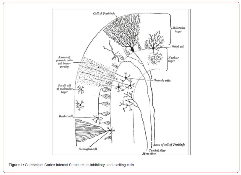
The excitatory amino acids, glutamate and aspartate, have been proposed as putative neurotransmitters in the mammalian central nervous system [9,10]. Initially they were suspected of being transmitters of primary afferent neurons and spinal cord interneurons, respectively, [9,11] and subsequently glutamate has seemed a strong candidate as the transmitter released by pyramidal tract neurons [12,13]. More recent work has supported the concept that glutamate may be released as the transmitter by many cerebral cortical-fugal neurons [14]. In all these cases a key factor in the assessment of the transmitter role of dicarboxylic amino acids has been the use of antagonist compounds. However, there not exists a good deal of indirect and circumstantial evidence favoring a transmitter role for glutamate at the synapses from granule cells via the parallel fibers onto Purkinje and other neurons in the cerebellar cortex but few direct studies on the effects of antagonists on this pathway have been made [15].
The existing evidence for a transmitter role of an amino acid from granule cells in cerebellar cortex is primarily neurochemical. In particular, it has been demonstrated that the conditions in which there is a substantially reduced number of granule cells in the cerebellum, as following certain viral infections, X-irradiation or in some mutant strains of nice, there is a pronounced reduction in the levels of glutamate. Probably glutamate could be the neurotransmitter released by granule cell parallel fibers in the cerebellar cortex [15] (Figure 1).

Essential Tremor: Really Color Vision is Always Normal?
Essential Tremor is a syndrome defined as a “bilateral upper extremity action tremor” and is among the most common movement disorder in adult [16], as are updated consensus statement from the task force on tremor of the International Parkinson and Movement Disorder Society defined for less than three years, worldwide, the crude prevalence rates of Essential Tremor in adults range from 0.4% to 6%, with a significant male predominance [17,18].
The historical practice of grouping all action tremors together may partially explain both the difficulties in identifying genetic causes and patients’ variable responses to treatment. But, although the pathophysiologic basis of this disorder is uncertain, it often has a familial basis with an autosomal dominant mode of inheritance. At least four gene loci have been implicated: three using linkage studies-hereditary essential tremor 1 (ETM1), ETM2, ETM3- and one using exome sequencing, ETM4; in some cases (ETM1), the disorder is related to a polymorphism in the D3 dopamine receptor gene (DRD3).
There is evidence of involvement of olival cerebellar, and cerebellum-thalamus-cortical pathways. Decreased levels of gamma-amino-butyric acid (GABAa) and beta-amino-butyric acid (GABAb) receptors have been found in the dentate nucleus [19]. The onset and progression of Essential Tremor sees the arm involvement by the kinetic tremor with or without postural tremor affecting both arms, and many patients develop head tremor, too. Head tremor will most commonly dissipate when the patient is supine, which can help in distinguishing Essential Tremor from other disease entities in which a resting head tremor is seen [20]. Some patients report balance difficulties, and tremors can involve muscles of the palate, pharynx, tongue, in addition to the larynx. Impairments of attention and working memory were commonly seen. The cognitive impairments are typically mild [21,22]. Patients with Essential tremor have a higher risk of developing Parkinson disease than the general population [19].
Present Study, and Discussion
Based on the previous manuscript by Anna Piro et al. [23], on the use of color vision as a biological marker to differentiate Parkinson’s Disease than Essential Tremor, the Author show, in the present manuscript, as the cerebellum structure influences color vision in Essential Tremor in some cases patients, negatively. Previously, we found that according with Hang [24], the influence of Parkinson’s disease is most noticeable in the pathway of a shortwave cones because the short-wave cones are widely separated. In the retina, the small bistratified ganglion cells [25] have much larger receptive fields than the midget ganglion cells and may be more dependent on dopamine. The abnormality can originate anywhere in the visual pathway from the retinal receptors to the visual cortex. The affection of the substantia nigra or the brain cortex in Parkinson’s Disease is the major cause for red/green, and/ or blue/yellow impair color vision in it.
But the involvement of an olive-cerebellar-rubral-thalamic loop did not seem to influence color vision in Essential Tremor, in that work. Instead, in the present manuscript, the presence of the patient with Essential Tremor showing an impair color vision both on red/ green, and blue/yellow axes, allowed us to assess the involvement of the cerebellum’s structure in the color vision pathway, too. Obviously, the impaired color vision diagnosis was made by the use of Ishihara test [26], Farnsworth D-15 test [27], City University test [28]. This result can be evidence of the favoring transmitter role for glutamate at the synapses from cerebellum granule cells via the parallel fibers onto Purkinje and other neurons in the cerebellar cortex but few direct studies on the effects of antagonists on this pathway have been made [29]. There is evidence that glutamate could be the neurotransmitter released by granule cell parallel fibers in the cerebellar cortex [29].
Based on the above statements, the cerebellum localization is surely very next to the visual area’s localization, at level of the occipital lobe. The excessive presence of glutamate as the most important neurotransmitter as in Essential Tremor, with a decreased GABA receptors in the cerebellum structure, can influence negatively color vision in the patient with it. In this way, we can confirm the spectacular, and first of all the optical motor system which can solve many amazing tasks. Other Authors confirm it [30]. All human elaborate neural networks encompass almost every part of the brain, including the brainstem, cerebellum, extrapyramidal system and many areas of the cerebral cortex.
Acknowledgment
The Authors thank Fondazione Cassa di Rosario di Calabria e Lucania for their contribution.

Conflict of Interest
No Conflict of Interest.
References
- Bartels A, Zeki S (1999) The clinical and functional measurement of cortical activity in the brain, with special reference to the two subdivisions (V4 and V4_) of the human colour centre. Royal Society: Phil Trans Royal Soc of London B series Biological Sciences 354(1387): 1371-1382.
- Murphey D, Yoshor D, Beauchamp M (2008) Perception matches selectivity in the human anterior color center. Curr Biol 18(3): 216-220.
- Winawer J, Horiguchi H, Sayres RA, Amano K, Wandell BA (2010) Mapping hV4 and ventral occipital cortex: the venous eclipse. J Vis 10(5): 1.
- Spector JM (2014) Conceptualizing the emerging field of smart learning environments. Sm Learn Envir 1: 2.
- Ramachandran V, Hubbart EM (2011) Synesthesia: A window into perception, thought and language. J Cons Stu 8(12): 3-34.
- Hubbard EM, Ramachandran VS (2005) Neurocognitive mechanisms of synesthesia. Neuron 48(3): 509-520.
- Fletcher R, Voke J (1985) Defective Color Vision, Fundamentals, Diagnosis and Management. Bristol: Adam Hilger.
- Schwarz N (2004) Metacognitive experiences in consumer judgment and decision making. J Cons Psychol 14(4): 332-348.
- Curtis DR (1974) Amino acid transmitters in the mammalian central nervous system. Ergeb Physiol 69(0): 97-188.
- Krujevic K (1970) Glutamate and gamma aminobutyric acid in brain. Nature 228: 119-124.
- Johnson HA (1972) Neuron survival in the aging mouse. Exp Gerontol, 2: 111-117.
- Stone TW (1973) Cortical pyramidal tract interneurons and their sensitivity to L glutamic acid. J Physiol 233(1): 211-225.
- Stone TW (1976) Blockade by amino acid antagonists of neuronal excitation mediated by the pyramidal tract. J Physiol, 257(1): 187-198.
- Spencer HJ (1976) Antagonism of cortical excitation of striatal neurons by glutamic acid diethylester: evidence for glutamic acid as an excitatory transmitter in the rat striatum. Brain Res 102(1): 91-101.
- Robbins TW (1984) Cortical, noradrenaline, attention and arousal. Psychol Med 14(1): 13-21.
- Bhatia KP, Peter Bain, Nin Bajaj, Rodger J Elble, Mark Hallett, et al. (2018) Consensus statement on the classification of tremors from the task force on tremor of the International Parkinson and Movement Disorder Society. Mov Dis 33(1): 75-87.
- Louis ED, K Marder, L Cote, S Pullman, B Ford, et al. (1995) Differences in the prevalence of essential tremor among elderly African Americans, Whites, and Hispanics in Northern Manhattan, NY. Arch Neurol 52(12): 1201-1205.
- Louis ED (2019) Essential tremor: a nuanced approach to the clinical features. Pract Neurol 19(5): 389-398.
- Aminoff MJ, Greenberg DA, Simon RP (2015) Clinical Neurology. New York: McGraw-Hill Edition.
- Agnew A (2012) Supine head tremor: a clinical comparison of essential tremor and spasmodic torticollis patients. J Neurol Neurosur Psych 83(2): 179-181.
- Azar M, Elodie Bertrand, Elan D Louis, Edward Huey, Kathleen Collins, et al. (2017) Awareness of cognitive impairment in individuals with essential tremor. J Neurol Sci 377: 155-160.
- Bermejo Pareja F, Contador, Rocío Trincado, David Lora, Álvaro Sánchez Ferro, et al. (2015) Prognostic significance of mild cognitive impairment subtypes for dementia and mortality: data from the NEDICES cohort. J Alzh Dis 50(3): 719-731.
- Piro A, Antonio Tagarelli, Giuseppe Nicoletti, Carmelina Chiriaco, Fabiana Novellino, et al. (2018) Color vision as a biological marker able to differentiate two phenotypically similar neurological diseases. Neurol Sci 39(5): 951-952.
- Haug BA, RU Kolle, C Trenkwalder, WH Oertel, W Paulus, et al. (1995) Predominant affection of the blue cone pathway in Parkinson’s disease. Brain 118(3): 771-778.
- Dacey DM, Lee BB (1994) The “blue on” opponent pathway in primate retina originates from a distinct bistratified ganglion cell type. Nature 367(6465): 731-735.
- Ishihara S (1990) Ishihara’s test for colorblindness. 38° Plates edition. Japan: Kanehara and Co.
- Farnsworth S (1943) The Farnsworth-Munsell 100 hue and dichotomous test for color vision. J Opt Soc Am 33(10): 568-578.
- Fletcher RJ (1975) The City University Color Vision test. London: Keeler Instruments.
- Stone TW (1979) Glutamate as the neurotransmitter of cerebellar granule cells in the rat: electrophysiological evidence. Br J Pharmacol 66(2): 291-296.
- Schwarz U (2004) Neuro-ophthalmology. A brief vademecum. Eur J Radiol 49(1): 31-63.
-
Anna Piro* and Daniel La Rosa. Is The Cerebellum Involved in Color Vision Pathway for Essential Tremor?. Arch Neurol & Neurosci. 16(1): 2023. ANN.MS.ID.000878.
-
Cerebellum, Color Vision, Essential Tremor, Molecular Bioimaging and Physiology, Cerebellum; Glutamate; Color vision; Granular cerebellum cells.
-

This work is licensed under a Creative Commons Attribution-NonCommercial 4.0 International License.


