 Research Article
Intravenous Laser Therapy in the Complex Treatment of Nervous System and Brain Diseases and the New Development Mechanism of the Diseases
Vladimir A Mikhaylov*
1Eternity Medicine Institute, KG Tower, PO Box 120618, Dubai Marina, UAE
Vladimir A Mikhaylov, Eternity Medicine Institute, KG Tower, PO Box 120618, Dubai Marina, UAE.
Received Date: October 07, 2021; Published Date: October 28, 2021
Abstract
Introduction: The first results of red laser influence on the nervous system were received in 1981. Extensive research had shown that laser irradiation increased the functional activity of the nervous system. We started using IFLT for treating nervous diseases in 2005. The accumulated experience has forced us to take a new look at the work of the vascular system. It became clear that disorders of blood supply in the nervous tissue lead to the development of many diseases of the nervous system and brain. We conducted mathematical calculations: the main role in the transportation of blood to the tissue is played by the artery, the power of the heart is only 0,49 - 0,027 % of the power needed to transport blood to the tissue. We have experience of using IFLT in complex treatment of patients with multiple sclerosis (21 patients), parkinsonism (17 patients), Alzheimer’s disease (4 patients), inflammatory and degenerative diseases of the peripheral nervous system (63 patients), different form of Encephalopathy (27 patients), and acute disorders of cerebral circulation (34 patients). The results of using IFLT on patients with Alzheimer’s disease were unsatisfactory. With all other diseases, a positive result was obtained. The period of observing some patients with multiple sclerosis (4 patients) and Parkinson’s disease (3 patients) was 15 years.
Background and Aims: The use of Intravenous Laser Blood Irradiation (ILBI) within the last 30 years has demonstrated high efficacy in the treatment of both vascular and nervous diseases.
Rationale: Laser energy at 630-640 nanometers is arguably most effective for irradiation of blood and the vascular wall. Photons at this wavelength are absorbed by oxygen, improve micro- circulation, can change the viscosity of the blood and affect the nerve and muscle elements of the vascular wall.
Conclusions: To sum up, more than 30 years of experience of using laser energy at 630-640 nm have shown that this waveband directly influences the parameters of all cells in the blood, blood plasma, and the coagulation process in the nerve and muscle elements of the vascular wall. Additionally, ILBI directly or indirectly affects the cells of the immune system, hormones, and exchange processes in an organism, thereby improving not only the function of the vascular system, but of the nervous system as well. Finally, it can lead to increased effectiveness in treatment of diseases of the nervous system and brain.
Keywords: Intravenous Frequency laser Therapy; Diseases of the nervous system and brain
Introduction
The Global Burden of Disease researchers now have published (Lancet Neurology, Vol. 16, November 2017) a more detailed study with estimates of the global, regional, and country-specific burden of neurological diseases - measured by prevalence, mortality, disability adjusted life-years, years of life lost, and years lived with disability during 1990–2015, and have explored variation in the burden by neurological disorder, age, sex, and overall country development. Since 1990, the number of deaths from neurological disorders increased by about 37% (from 6,87 million to 9,40 million), and the number of disability-adjusted life-years by about 7% (from 233,4 million to 250,7 million). These increases mean that in 2015 neurological disorders represented the largest cause of disability-adjusted life-years and the second-largest cause of global deaths from cardiovascular diseases [1]. The first results of red laser impact on the nervous system were collected in 1981. Extensive research had shown that laser irradiation increased the functional activity of the nervous system, as a result of the acceleration of metabolic processes [2]. According to our research the main role is played by improved microcirculation in damaged nerve tissue; it was especially important in brain damages [3,4]. The discovered new mechanism of vascular diseases development allowed us to take a fresh look at the development of various diseases of the nervous system and brain. The main role in their development is played by ischemic and dystrophic changes in the nervous tissue. These diseases occur both due to microcirculation disorders and damage to the arteries that provide normal functioning of the nervous tissue [4,5,6]. This point of view was confirmed by many fundamental studies of the physiology and histological structure of different types of arteries, and our mathematical calculations have confirmed all this too [7,8].
Materials And Methods
Equipment for ILBI:
The main laser therapy systems are used for ILBI:
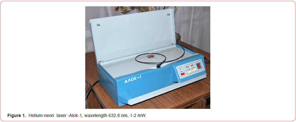
1. Helium-neon laser -Alok-1, wavelength 632.8 nm, 1-2 mW. (Figure 1).
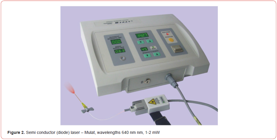
2. Semi conductor (diode) laser – Mulat, wavelengths 640 nm nm, 1-2 mW. (Figure 2)
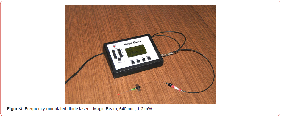
3. Frequency-modulated diode laser – Magic Beam, 640 nm, 1-2 mW. (Figure 3)
Technique of ILBI realization:
The puncture of an ulnar vein with one-time sterile catheter is performed. The catheter consist of a thin needle with a monofilament through which intravascular irradiation comes out (producer- Polironic). After each session of irradiation, the catheter is rejected.
Experimental Studies of the Red Waveband Efficacy
Influence on the Vascular Wall and Microcirculation
The majority of studies have related to the analgesic and antiischemic effect of LLLT and the positive reaction of the blood rheological properties and microcirculation to irradiation with 632.8 nm [9,10,11,12]. The influence of LLLT has been demonstrated with enhancement of the permeability of the vascular wall [13]. Musienko SM [14] showed that the He-Ne laser was used to treat skin injuries in patients with chronic venous insufficiency of the lower extremities: the pathological permeability of the vascular wall sufficiently decreased, simultaneously with the normalization of the transcapillary exchange. However, information regarding the influence of LLLT on microhemodynamics is contradictory [15,16,17]. Musienko SM [14] asserted that a microcirculatory link was a point of application in the mechanism of the therapeutic efficiency of laser radiation. The point has been made by other authors, confirmed by polarographic studies, that the activation of energetic and biosynthetic processes occurred following red light LLLT [18]. Furthermore, the major importance of microcirculation in the implementation of the biological effect of the red waveband, particularly of that around the He-Ne laser, has been confirmed by other studies [19]. A number of studies have confirmed the fact that ILBI stimulates the development of capillary tubes, eliminates vasospasm stasis and reverses the blood flow. Moreover, ILBI has been shown to decrease capillary edema and to increase the number of functioning capillary tubes [20].
Formation of New Capillary Tubes
It has been shown that exposure to low levels of laser radiation actively formed new capillary tubes, thereby augmenting oxygen delivery to tissues and optimizing tissue metabolism [19,21,22].
Influence on the Nervous System
Konovalov E.P. et al. [23] studied the influence of ILBI on the functional activity of a number of physiological systems. The analysis of the measurement of cardiac intervals showed that during the first two sessions of laser radiation increases were noticed in the variational scope (Ax), mode (Mo) and reduction mode (AMo) amplitude. Increase in pressure index (PI) was seen in 71.5 % of patients. These data pointed to an enhancement of parasympathetic nervous system activity and the development of auto-regulation of the cardiac rhythm. The studies of Rakhishev A.P. et al [24] showed ILBI-mediated changes in autonomic regulation towards parasympathetic tonus, an improvement in blood filling, decreased tonus, and decreased resistance in peripheral vessels. They showed that the intravascular laser therapy resulted in an increase of the parasympathetic nervous system tonus in 34% of patients, and in the appearance of normal tonus in the autonomic nervous system in 60% of cases.
In 1988 Serov V.N. et al. [25] revealed the ability of He-Ne LLLT to restore and then to stimulate the impaired function of external sexual organ’s receptors at kraurosis and vulvar leukoplakia. Other researchers noticed the activation of the myelination processes in neurons, thickening of axons and amplification of their regeneration. Increased amplitudes of action potentials in human forearm nerves following LLLT irradiation of the skin was observed [24,26]. The increased synthesis of particular proteins and an elevated rate of axonal motion in neurons concomitant with the enhanced plasma circulation in innervated organs was reported following ILBI with the He-Ne laser. Following laser irradiation, induction of the functional activity of the nervous system was observed, accompanied by detection of growth microtubules in nerve fibers as the result of acceleration of the metabolic processes.
Rochkind S. [27]described the LLLT-mediated preservation of the functional activity of severely crush-injured neurons in experimental rats and, further¬more, demonstrated a decrease of degenerative changes in motor neurons in the transection injury spinal cord model. LLLT irradiation of the spinal cord after severe injury and implantation of neural cells promoted fissile germination of axons in the injured area, resulting in partial recovery of locomotors function in injury-induced paraplegia.
Rochkind S, Nissan M., Alon M. et al. [28] applied transcutaneous LLLT to segments of the spinal cord after the crush injury of sciatic nerves in the rat model, immediately after the closure of the wound, with a 16 mW, 632.8 nm, He-Ne laser. The laser treatment was repeated for 30 minutes daily during 21 consecutive days. This study suggested that LLLT applied directly to the spinal cord at the appropriate dorsal root could improve recovery of injured peripheral nerves in a dermatomal-based approach. The results of the experiment strongly suggested that the process of neurons retrograde degeneration became stable in the central part of the crosscut nerve much closer to the injured area (1.2 - 1.5 cm) after local exposure to LLLT; whereas in control animals retrograde degeneration was tracked to within 2.5 cm from the level of the transected nerves.
Kositsyn R.S. et al. [29] considered that an increased disintegration rate of nervous elements and resorption enhancement of nerve fragments formed new condi¬tions for the acceleration of nerve regeneration. Thus, for example, the excretion of urine 5-hydro-indol- acetic acid has been studied as the metabolism of serotonin [30]. It was mentioned that increased excretion of this metabolite occurred under the influence of LLLT that proved the breakup of serotonin. The data indicated a significant laser effect, which was probably related to the laser effect on the humoral and inflammatory mechanisms of pain.
Another study evaluated the effect and mechanism of ILBI on brain injury. In this study, thirty-eight anesthetized Sprague Dawley rats underwent Feeney’s model of traumatic brain injury through a left lateral cranioectomy. It was found that ILBI-LLLT could improve posttraumatic memory deficits. Superoxide dismutase (SOD) activity, both as an antispasmodic and as an antioxidant, was higher in the treatment groups than in the control group, whereas the level of production of free radical-mediated malondialdehyde (MDA) was lower. These findings suggest that ILBI-LLLT produced a significant reduction in the damage to the brain caused by free radicals post-injury [31].
Influence on the Neuroendocrine System
Numerous studies on the effect of He-Ne LLLT on organisms have shown some functional and morphological changes in the hypothalamic-hypophysis adrenal system [32]. According to Serov V.N. et al. [33] laser irradiation at certain parameters is capable of achieving hormonal modifications of pathological homeostasis by means of increasing an afferent stimulation of the diencephalic patterns of the brain. These authors then studied the functional status of the central elements of the neuroendocrine system with experimental animal models and using histochemical and cytophotometric analysis. The helium-neon laser (LG-38) with an output power of 50 mW was used. The duration of the exposure was 3 min. daily for 15 days. The results showed an accumulation of secretory substances in the hypothalamic-epiphysis system and adenohypophysis. With the increase of laser irradiation up to 10 days the stimulation phenomena also increased, but on extending LLLT up to 15 days, overstimulation occurred, causing functional-morphological modifications, and demonstrating the development of exhaustion processes in neurosecretory elements and degeneration of neurosecretory cells. Exposure of a reflexogenic zone of that group of animals to LLLT for 5 days caused a considerable migration of neurohumoral products into the circulation system. The most significant reaction occurred in the supraorbital nucleus. An increase in both the cytoplasmic volume and the nucleus sizes in neurosecretory cells was registered. There was some decrease of delta basophils and the glycoprotein content in the adenohypophysis.
The epiphysis is recognized as the coordinating gonadotrophic activity organ of the hypothalamus, therefore the elucidation of the effect of LLLT on this organ is important. No changes were noted in the epiphysis structure after a single laser radiation. In the group that underwent 5 sessions of LLLT some substantial modifications in the organ structure were found; those modifications were considered to be an accumulation of hormone-positive substance (HPS) and nucleic acids. After 10 sessions of laser radiation some laxity of the HPS and nucleic color was observed; it was estimated as the beginning of the secretory activity of the gland. After 15 sessions the volume of the epiphysis, cytoplasm and nuclei of pinealocytes increased, and the color of the HPS had significantly decreased; considerable activation of the secretory function of the gland was noted. The authors came to the conclusion that the epiphysis reacted by the phase changes of its pattern with regard to the duration of exposure; on the fifth day, inhibition was seen, and on the tenth to fifteenth days, some activation of the organ function was noticed. The most notable reaction was seen in the trophictransport component of the gland.
The data obtained from the aforementioned experiment correlated with those obtained by Grishchenko L.V., [34] whose results showed that under clinical conditions laser radiation with exposure of the external orifice of the uterus for 1 min during 6-8 days caused a decrease in the production of melatonin, and that this promoted the restoration of the hypothalamic neurohormones production, as a result of which the recovery of menstrual function was in turn promoted. Peshev D.P. et al. [35] studied the pituitary body and ovaries of female rabbits, some of which were pregnant and others not, after exposure of a cervical reflexogenic zone to He- Ne LLLT at 20mW for 10 min during the period of pregnancy. After the LLLT session the analysis of the rabbits’ pituitary bodies showed high DNA and RNA contents in all zones of the pituitary bodies. Analysis of the pregnant rabbits failed to show any endocrine bodies-related pathology of the investigated functional organs.
Koshelev V.N. et al. [36] indicated an increase in catecholamine, serotonin, histamine concentration and activation of the hypothalamus-adrenal system following red light LLLT. These data have been confirmed by the works of other authors [37]. Laser therapy with red light normalizes hormonal pathological irregularities in the menstrual-ovarian cycle in women [38].
The Anatomical-Physiological and Mathematical Justification of the Development of Cardiovascular and Nervous Diseases
Our clinical experience has shown that IFLT affects not only the blood elements, but also the nerve elements of the vascular wall [2]. It became clear to us that the main role in the cardiovascular system is played by arteries. They play a major role in the delivery of blood to various tissues. Disturbance of their work is the starting point for the development of vascular and neurological diseases. Our mathematical studies have fully confirmed this assumption [6,7,8].
The mathematical model to determine the capacity of the heart to deliver blood to the capillaries
First, we determined the approximate length of the blood channel from the heart to the capillary bed according to Parashin V.B. and Itkin G.P [39]. The measurement was taken only of the artery that was present in the muscle fibers.
The results on total length of the arteries are presented in Table 1.
We calculated the power that is needed to deliver blood from the heart to the tissues of the body: [6,7,8]
L -The total length of all arteries vessels (cm):
Lmin- 2791791,3 (27917.91 m)
Lmax- 50459339,5 (504593,39 m)
The next step was to determine the capacity of the heart which is necessary for delivering blood to a capillary bed [39]:
Table 1: The number and length of different types of arteries*.

S -the cross-sectional area of the aorta (3 cm), was determined by the formula
S= 0,032 π/4
S=0, 0009 . 3.14/4 =0,00071 m2.
g- the density of blood plasma is almost equal to the density of water:
g = 1,0 . 103 kg/m3.
Since plasma consists of 90% water, 7% of proteins, and the rest is constituted by inorganic elements [39].
v - the average flow velocity in the aorta is 20 cm/ s (0,2 m/s)
v= 0,2 m/s
A - work required to deliver blood to the capillary bed was determined by the formula:
A= S g v2 L / 2
Аmin (total length of all arteries vessels Lmin)
Аmin = 0,00071 . 1000 . 0,04 . 27917.91/ 2 =396.43 J
Аmax (total length of all arteries vessels Lmax)
Аmax = 0,00071 . 1000 . 0,04 . 504593,39/2 =7 165,22 J
The power of the heart (P) required to transport blood to the capillary bed was calculated according to the formula:
P=A/1 – 0,4
P min = 396,43/1-0,4 = 660.71 W
P max = 7165,22 /1-0,4=11942,03 W
The results show that the power of the heart (3.3 W) is only 0,49 - 0,027 % of the power needed to transport blood to the capillary bed.
These calculations confirmed our clinical results that the main role in the transportation of blood is played by arteries and veins. The heart acts only as a trigger for synchronizing a complex process that involves different kinds of nerve receptors in the vascular wall, the Central and autonomic nervous system, smooth muscles, collagen and elastic fibers of the vascular wall.
Features of wall structure of various arteries types and how they affect the transportation of blood
A new look was taken on the anatomical structure of the walls of various arteries that were discovered by Strong K. (1938) and others [40-43]. Various types of arteries differ in the anatomical structure and direction of fibers. Taking into account the functional peculiarities of wall structure of various types of arteries, I divided them into the following types:
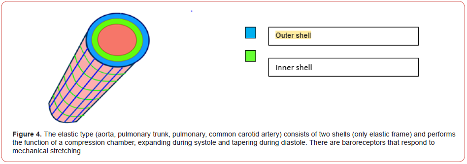
The elastic type (aorta, pulmonary trunk, pulmonary, common carotid artery) consists of two shells (only elastic frame) and performs the function of a compression chamber, expanding during systole and tapering during diastole. There are baroreceptors that respond to mechanical stretching (Figure 4).
The elastic and muscular type (subclavian, external and internal iliac, femoral, mesenteric artery, celiac trunk) joins the muscular layer (a spiral arrangement of fibers). This arrangement of fibers while reducing the flow makes the blood spiral. The baroreceptors disappear and sympathetic adrenergic vasoconstrictor fibers appear (Figure 5).

The muscle type (vertebral, brachial, radial, popliteal, arteries of the brain, etc.) has the greatest ability to tighten the blood in the spiral and push it forward. This is achieved by the spiral arrangement of fibers not only in the muscular layer, but in the external elastic membrane too (Figure 6).


The muscle-elastic type (small artery) disappears in the external elastic membrane. The muscular layer of the spiral arrangement of fibers. In the inner shell the thickness of the connective tissue and internal elastic membrane decreases (Figure 7).
Precapillary type – a thinner muscular layer with the reduced thickness of the connective tissue and internal elastic membrane which becomes fenestrated. This leads to the contact of endothelial cells with smooth muscle cells, which have the opportunity to respond to hormone-active substances that get into the blood. (Figure 8).

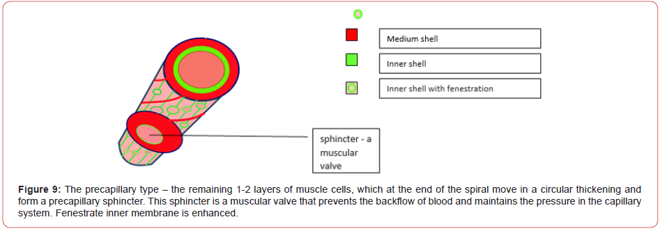
The precapillary type – the remaining 1-2 layers of muscle cells, which at the end of the spiral move in a circular thickening and form a precapillary sphincter. This sphincter is a muscular valve that prevents the backflow of blood and maintains the pressure in the capillary system. Fenestrate inner membrane is enhanced (Figure 9).

We presented the features of blood flow in different types of
arteries in the form of diagrams [6].
Diagram of blood flow in various types of arteries:
The direction of blood flow in Elastic type arteries (Figure 10).
The direction of blood flow in Elastic and muscular type arteries
(Figure 11).
The direction of blood flow in Muscle, Muscle-elastic,
Precapillary types arteries (Figure12).
The obtained data clearly confirm the main role of the vessels
in the movement of blood in the bloodstream considering that the
length of all arteries ( L) is 27,91 to 504,59 km.


Cardiac output gives the blood a spiral movement. This is the trigger for contraction of the arteries, which have a very important anatomical feature - a spiral orientation of muscle fibers. This allows a spiral support of blood flow throughout their length until it gets to the beginning of the capillary bed.
Nervous Regulation of the Cardiovascular System
I tried to take a fresh look at the existing data on the neurohumoral
regulation of the work of arteries and veins with the help
of the Hyper-cycle modern theory - the principle of the natural
self-organization causing integration and coordinated evolution
of the system of functionally connected self-replicated units [44].
This theory allows to understand and systematize the principles
of regulation of various functional systems of an organism and the
development of various pathological processes. The regulation and
synchronization of activity of all systems in an organism happen
due to neuro- and humoral regulation. The nervous system is
autonomous and its regulating influence is carried out through a
complex of nervous cells, but humoral regulation can’t be carried
out without the existence of organs producing hormones and the
vascular system that delivers hormones and other biologically
active agents to target organs.
Afferent impulses from the baroreceptors reach the cardio
inhibitory center and the vasomotor center located in medulla
oblongata, as well as in other departments of the Central nervous
system. Depending on stretching of the artery wall the frequency
of these pulses varies which results in wave-like inhibition of the
sympathetic centers and the stimulation of the parasympathetic
centers. Thus, the Central nervous system regulates the tone of
the sympathetic vasoconstrictor fibers, and the frequency and the
strength of contractions, the heart and blood vessels [45-49].
Innervations of blood vessels are carried out through a
superficial plexus in the outer layer, admissions or edge, the plexus
on the border of the outer and middle shells. From there fibers go
to the average shell wall and form an intramuscular plexus, which
is especially pronounced in the arterial wall. Individual nerve fibers
penetrate to the inner layer of the wall. 50) These plexuses’ afferent
impulses from the baroreceptors directly affect all the contractile
elements of the wall around the blood vessels. Given that the heart
muscle works offline, these plexuses synchronize the contractions
of the heart and blood vessels. All these make a complex and very
stable self-regulating system. An important feature is that the
velocity of propagation of pulse wave through the vessels is much
higher than the flow velocity. The pulse wave propagates to the
arterioles stops in 0.2 s, whereas the particles of blood ejected by
the heart during this time are only reaching the descending aorta
[51,52,53].
This means that the pulse wave after its passing creates a zone
of lower pressure than the pressure in the aorta. The blood is just
absorbed into the bloodstream after a systolic ejection.
Thus, the vascular pump of a recession is rigidly synchronized
with the work of the heart. This allows to pump blood with minimal
cost across the vascular bed. The Central nervous system and
hormonal factors regulate the redistribution of blood at a lower
level, depending on the needs of various parts of the body.
Clinical Results of Using IFLT for Treating the Diseases of the Nervous System and Brain Encephalopathy
Fedin A.I. et al. [54] showed that ILBI has a positive influence on
the brain’s functional condition and the general emotions of patients
with atherosclerotic dysfunctional circulatory encephalopathy.
Mikhailov V.A: [55] IFLT (wavelength, 640 nm.) was performed
in patients with Hypertensive encephalopathy (13 patients) - I
group and Progressive vascular encephalopathy (14 patients) – II
group. IFLT was performed when medication was not effective. In
24 cases positive dynamics was noted – a reduction of headache,
dizziness, reduction of noise in the head, improvement of memory
and performance. 3 patients did not notice significant changes in
their health. Gonchar-Zaykin A.P. et al [2] reported on the efficacy
of ILBI (630 nm; exposure time - 60 min over 3-6 sessions) for
29 patients with I – III st. of encephalopathy (group I), with 34
patients forming the control group II. The longest interval between ILBI sessions was 3 months. As a result of ILBI treatment all
patients began to feel much better, their physical and psychoemotional
activity increased, and their complaints regarding
headaches, vertigo, nausea and tinnitus became considerably less.
In group I the arterial pressure significantly decreased in the I
st.- by 60%, at stage II, 49 %, and at stage III, 20% (р < 0.05). The
rheoencephalographic index improved: at the I st - 90%, at the II st.-
56% and at the III st.-10%; the rheography index increased from
0.5 to 1.3 W; asymmetrical pulse filling decreased, especially for
the patients with dysfunctional circulatory encephalopathy (DE) I
st. and II st. The vessels tonus normalized and the venous outflow
improved considerably. In addition, the extent of neurological
symptoms such as oral automatism, muscle hyper-tonus, and
autonomic-regulated vascular dysfunction decreased. The health of
the II group also improved with a rheoencephalographic index of
60% in I st. patients and of 33% in stages II and III. The results of
this study showed that ILBI increases the functional condition of
the central nervous system of DE patients, reduces the resistance
of an organism to medication. The greatest effect for ILBI was
observed in patients with I st and II st. of dysfunctional circulatory
encephalopathy (DE).
Diseases of the Peripheral Nervous System
Mikhailov V.A: [55] using IFLT (wavelength, 640 nm) in addition to transcutaneous laser therapy (wavelength 890 and 640 nm) was aimed to relieve local inflammatory and allergic reactions. Laser treatment started when medication was not effective. The number of patients was s follows: Radiculopathy of various parts of the spinal cord - 21 patients..Traumatic neuritis of the upper extremities - 5 patients, Traumatic neuritis of the lower extremities -7 patients, Trigeminal and facial neuralgia - 14patients, Neuritis of the facial nerve - 16 patients. In 88,9 % of patients there was a positive reaction to the use of laser therapy.
Acute disorders of cerebral circulation
Mikhailov V.A.: [55] IFLT (wavelength 640 nm) performed for treating patients with transient ischemic attacks (25 patients) and ischemic cerebral infarction (9 patients). As a rule, laser therapy was started after discharge from the hospital, when appropriate therapy was carried out, but various disorders persisted (motor, speech, sensory, visual and coordination disorders). In patients with ischemic cerebral infarction, additional transcutaneous laser therapy (890 nm) on the projection of the lesion was prescribed to reduce edema and improve microcirculation. All patients with transient ischemic attack showed positive dynamics after the use of IFLT. In 3 patients with ischemic cerebral infarction after the use of IFLT there were no changes.
Multiple sclerosis
Mikhailov V.A: [2] IFLT (wavelength, 640 nm; dose- 0.9 J) used in 6 patients. ILBI (wavelength, 630 nm; dose -0.9 J) performed in 10 patients. All patients in both groups had the cerebrospinal form of multiple sclerosis. In IFLT group remission of diseases was 2.9 months on average, in ILBI group – 2.1 months (period of observation -1 year). Mikhailov V.A.: [55] Combination of IFLT (wavelength, 640 nm; dose- 1.6 J) with transcutaneous laser therapy (890 nm.), massage of the head and medication therapy (15 patients). All patients had the cerebrospinal form of multiple sclerosis. Period of observation - 1-15 years. The period of remission for patients with cerebrospinal form receiving combination therapy with IFLT is presented in Table 2.
Table 2: The period of remission for patients with the cerebrospinal form of Multiple sclerosis receiving combination therapy with IFLT.

Parkinson’s Disease
Mikhailov V.A.: [2] 9 patients have been treated with ILBI: 6 patients with IFLT and combination therapy (kryolaser massage, transcutaneous laser therapy (890 nm), massage of the head and medications), and 3 patients were receiving ILBI (630 nm). Reduction of tremor, reduction of gait unsteadiness and improvement of the general state of health were achieved in all patients. The period of remission in patients with IFLT was 3.4 months and in patients with ILBI - 2.1 months (period of observation -1 year). Mikhailov V.A.: [55] In 6 patients IFLT (with combination therapy) was maintained for 5 years (2 times per year). Long-term remission was in 4 patients (1 patient -15 years, 2 patients - 10 years, 1 patient - 7 years). The patient with 15-years remission was my mother. She has Parkinsonism for 35 years. She had IFLT for the last 15 years. Until the last moment, she could serve herself on her own.
Alzheimer’s Disease
Mikhailov V.A.: [55] The results of using IFLT on patients with Alzheimer’s disease (4 patients) were unsatisfactory.
Other Diseases of the nervous system
Ester Yi Liu and Shin-Tsu Chang 56) used the ILBI for treating Transverse myelitis, Guillain-Barré and Sjögren’s syndrome combined with two other diseases. After 2 courses of laser therapy better motor and sensory recovery was obtained. A close relationship between motor/sensory function and the dorsolateral prefrontal cortex was observed from the brain function images.
Discussion
The regulation and synchronization of all systems’ activity
is the essential basis of any organism’s activity, and it is not
important how these goals can be reached. The more complicated
the system is and the longer it functions, the more system failures
will occur and mistakes will be made and accumulated. Living
organisms are influenced during their entire lifetime by a set
of negative factors - both external and internal. These negative
impacts gradually accumulate and this consistently leads to the
emergence of pathological changes at first at the molecular, then
at the cellular, and finally at the tissue level. This inevitably leads
to the disturbance of activity of a variety of the affected organism’s
systems. As a matter of fact, any illness represents a failure in one of the organism’s systems concomitantly with a disturbance of its
function. The outcome of the pathology and further activity of an
affected organism depends on the organism’s ability to restore the
function of out-of-order systems [4].
Any defect in the nervous system is one of the most severe
defects any organism can suffer. This is confirmed by the fact
that mortality rates associated with vascular diseases have now
achieved the top score worldwide [1]. Therefore, the maintenance
of functional activity of the blood vascular system is the most
important problem of contemporary medicine. Irregularities in
other systems can possibly be corrected by means of medicaments,
but to use only them in the treatment of cardiovascular diseases is
simply not enough. The advent of hypertension, malnutrition of the
myocardium, the elasticity dis¬orders in vessel walls and stenosis
all arise after pathological dysfunctions have occurred in the vessel
walls, which impair or destroy the ability of these vessels to contract
and. to pump the ever-essential blood to the recipient organs and
tissues. The disturbance of the transport function of vessels can
severely interrupt the delivery of oxygen, various cells, hormones
and nutrients to their targets. In addition, the drainage function of
blood and lymphatic vessels is broken, so that impurities, waste
matter and toxins are not efficiently drained from organs and
tissue, and in this case dysfunction is inevitable.
The studies and literature accumulated in more than 30 years
have proved that the visible red light in the waveband of 630-640
nm have a powerful influence on vascular wall and microcirculation,
[9-20] neomicrovascularization and neoangiogenesis [19,21,22].
These structures and processes play a very important role in the
activity of the nervous system and brain. Pathological changes that
occur in them are the starting point for the development of many
diseases in these systems. The same visible red waveband has
also demonstrated proven effects on the various systems which
contribute to the organism’s survival: the nervous system [23-31]
and the neuro-endocrine system [32-38].
IFLT has a systemic effect on the body. All systems of the body
react to it. The immune system also plays an important role in the
development of diseases of the nervous system and brain. Studies
by many authors have confirmed this fact [30,57,58].
Conclusions
In this study, we determined the variability index of the optic nerve sheath diameter as a reference point that allows the clinician to make decisions quickly, at the bedside, through a simple method of neuromonitoring that can impact treatment, the prognosis, and, probably, the sequelae. The early diagnosis of intracranial hypertension with a non-invasive method favors the neuromonitoring of neurocritical patients. A larger sample of patients is required to be conclusive. Finally, Raffiz M et al in their study determined that an ONSD of 5.205 mm allows the detection of traumatic and non-traumatic neurosurgical patients with an early increase in IH [12]. Therefore, based on our results and taking this ONSD cut-off point, we suggest the following management algorithm (Figure 4):
Acknowledgement
None.
Conflict of Interest
No conflict of interest.
References
- (2017) Lancet Neurology 16.
- Mikhailov V (2007) Intravenous Laser Blood Irradiation. Greece 103.
- Mikhailov V (2009) Intravenous Laser Blood irradiation (ILBI). Laser Therapy 18(4): 310- 311.
- Mikhaylov VA (2015) Use of intravenous laser blood irradiation (ILBI) at 630-640 nm to prevent vascular diseases and to increase life expectancy. Laser Therapy vol 24(1): 15-26.
- Mikhaylov VA (2016) Ming Chien Kao Awards. Laser Therapy vol25(1): 9-10.
- Mikhaylov VA (2017) A newly discovered way of the function of cardio-vascular system and the latest theory of the development of Hypertension and other cardio-vascular diseases. Adstr. “2nd International Conference on Hypertension & Healthcare” Amsterdam, Netherlands 11-13.
- Mikhaylov VA (2008) A newly discovered way of the function of cardio-vascular system and the latest theory of the development of Hypertension and other cardio-vascular diseases. EC Cardiology (ECCY) 5(4): 179-187.
- Mikhaylov VA Mikhaylova TY (2019) Anatomical-Physiological And Mathematical Justification Of The New Principle Of The Function Of The Cardiovascular System And The Development Of Cardiovascular Diseases. International Journal of Hematology and Blood Research Vol1(1): 10-15.
- Badur G I (1993) The dynamics of clinical given and fast changes of phospholipids in patients with a stenocardia under irradiation of the blood by the helium the neon laser. PhD Thesis: 15.
- Dudchenko MA et al. (1992) The clinical-biochemical model of influence of laser therapy on the course of stable forms of ischemic illness of heart, hypertension ill¬ness atherosclerosis and coronary insufficiency. Kiev: 48 53.
- Ionin A P (1992) Influence of low-energy irradiation of helium - neon laser on clinical and functional parameters of cardiovascular system on patients with various forms of a stenocardia. Ph. D. Thesis. Ekaterinburg: 24.
- Kapustina GM (1990) Treating of various forms of ischemic illnesses of heart at radiation of the helium - neon laser. Sci D Moscow 42 p.
- Borisov A Dvorkina MI Schastin NN (1981) Clinical and experimental research of influence of the laser on vessels. Application of methods and means of laser technique in biology and medicine. Kiev 112-114.
- Musienko SM (1983) The change of transcapillary exchange in patients with chronic venous insufficiency of the lower extremities: author. Ph.D., Kuibyshev.
- Zhukov B.N. et al. (1988) The use of low-level laser radiation in the conditions of regional ischemia. Morphological aspects of organs hemocirculation. Kuibyshev: 88-94.
- Zhukov BN Kirichenko DN et al. (1989) Intravenous application of laser radiation in an experiment. Use of lasers and diagnostic techniques in medicine. Kuibyshev: 67-68.
- Zhukov BN Lisov NA (1996) Laser irradiation in experi¬mental and clinical angiology. Samara.
- Shchastnyj SA Volkov VV Kuzovlev VV (1985) Influence of radiation of the helium - neon laser on the functional condition of peripheral blood cir¬culation in the treatment of children with persistent wounds. Clin. Surgery. N(6): 124- 127.
- Васкег J (1988)Biostimulation laser Reality and Perspectives. 4941.
- Gausman BJ et al. (1989) Intravascular irradiation of blood by helium-neon laser in the treatment of diabetic patients with purulent-necrotic lesions of the lower extrem¬ities. The effect of low-energy laser irradiation on blood. Kiev 70-72.
- Kozlov VI et al. (1989) Stimulation of microcirculation with low-level laser radiation. Moscow part 1 90-93
- Kozlov VI Terman OA Lozhkevitch AA (1992) Laser diagnostics and therapy of microcirculatory infringe-ments. Lasers in medical practice. Thesis 2 Conf. of the Moscow Region 127-128.
- Konovalov EP et al. (1989) Impact of intravascular laser irradiation of blood on the functional activity of a number of physiological systems with purulent-septic complications. Kiev 102-103.
- Rakhishev AP et al. (1973) Laser light is a stimulator of regeneration of peripheral nerve. Some aspects of the study of the peripheral nervous system. Alma-Ata 23-124.
- Serov VN et al. (1987) Laser therapy in endocrinology gynecology. Rostov University, 1988, 120 p.
- Walkner J.B. et al: Laser Therapy for pain of trigeminal neuralgia. Pain (29):585.
- Rochkind S (1986) He-Ne low energy laser - is it com¬pletely harmless (letter). J Biomed Eng (8): 1- 77.
- Rochkind S Nissan M Alon M et al. (2001) Effects of laser irradiation on the spinal cord for the regener¬ation of crushed peripheral nerve in rats. Lasers in Surgery and Medicine 28(3): 216-219.
- Kositsyn RS et al. (1989) Biostimulation laser regeneration of damaged nerve. Lasers and Medicine part I 93-94.
- Ohshiro Т Calderhead R (1989) The role of low reactive level laser therapy in revitalising failing grafts and flaps // Laser Surg. Med 1 -31.
- Wang Yu Zhu Jing et al. (1999) Vascular Low Level Laser Irradiation Therapy in Treatment of Brain Injury. Acta Laser Biology Sinica Vol8(2):
- InychinVM(1967) On some reasons of biological efficiency of laser light of a monochromatic red part of a spectrum. On the biological action of monochromatic red light. Alma-Ata 5-15.
- Serov VN et al. (1988) Laser therapy in endocrinology gynecology. Rostov University 120.
- Grishchenko LV et al. (1997) Laser Metrology in the applica¬tion of lasers in medicine and biology. Application of lasers in medicine and biology. Yalta 173-174.
- Peshev DP et al.(1993) Laser treatment in obstetric-gyne¬cologic practice. Saransk 152.
- Koshelev VN Semina EA Kamaljan AB (1995) Comparative evaluation of the efficiency of transcutaneous and intravascular laser irradiation in diabetic angiopathias. Materials of the international con¬gress “Clinical and experimental applications of new laser technology.” Moscow-Kazan, Russia 391-393.
- Zhukov BN Kirichenko DN et al. (1989) Intravenous application of laser radiation in an experiment. Use of lasers and diagnostic techniques in medicine. Kuibyshev 67-68.
- Filgus GA (1984) Influence of a pituitary gland in the reaction of endocrine systems on the irritation of cervix uterus various physical factors. Laser and magneto-laser therapy in medicine. Tyumen 21-22.
- Parashin VB Itkin GP (2005) Biomechanical circulation. Bauman Moscow State University.
- Shepherd JT Abboud FM (1983) Handbook of Physiology, Section2 The Cardiovascular System. Vol.III. Peripheral Circulation and Organ Blood Flow. Bethesda, Maryland, American Physiological Society.
- McDonald DA (1974) Blood Flow in Arteries. 2nd Ed. London, Arnold.
- Guyton AC Young DB (1979) Cardiovascular Physiology Baltimore. University Park III Vol(18):
- Prives MG Lysenkov NK I Bushkovich VI (1985) Human Anatomy, Moscow, "Medicine".
- Schuster P (1982) The Hyper-cycle principles of macromolecules self-organization. M Mir 270.
- Gorbatenkova EA et al. (1990) The effect of endovascular laser irradiation on the LPO parameters and endo-toxication in experimental peritonitis. New in laser medicine and surgery Moscow, part 2 33- 34.
- Latifullin IA et al. (1989) Products of lipid peroxidation, blood enzymes and kinins in myocardial infarction following endovascular laser therapy, Lasers and Medicine. Moscow part 1100-101.
- Smotrin SM (1982) Influence of laser radiation on an exchange of serotoninum in patients with trophic ulcers of the bottom finitenesses. Application of laser radiation and a magnetic field in biology and medicine Minsk 18-19.
- Baynozarova BJ (1977) The influence of monochromatic polar¬ized red light on activity NAD-depending dehydrogenase of cycle Krebs. Biological action of laser radiation. Alma-Ata 70-74.
- Djugurian MA (1986) Activity of dehydrogenase and citochromokinase of the brain and the cardiac muscle of rats under the influence of laser radiation of various wavelengths. Ph.D. Thesis in Biology Kiev.
- Alekseeva AV Ivanov AV Minaev P et al. (1971) A study of action of laser radiation on blood cells. In: Mathematical models of biological systems. Moscow 102-103.
- Moroz AA (1976) ATF activity and ATF maintenance in some organs of the rats after exposure to monochromatic red color. Hygiene and sanitary (11): 110-111.
- Adghimolaev ТА et al. (1976) On the mechanism of laser radiation action on structure and function of a nervous cell. Problems of bioenergetics and stimulation of an organism by laser radiation. Alma- Ata.
- Zubkova SM (1978) On the mechanism of biological action of radiation of the helium-neon laser. Biological sciences M (7): 30-37.
- Fedin AI at al. (1991) The intravascular laser therapy influence on the psychological condition of patients with atherosclerotic dyscirculatory encephalopathy. Proceedings of International Conference “New Developments in Laser Medicine” Moscow 122.
- Mikhaylov VA (2019) Use of the intravenous frequency laser therapy (IFLT) of 640 nm in the complex treatment of the nervous system and brain diseases. International Conference on Clinical and Medical Case Reports Zurich,Switzerland Aug19-20.
- Ester Yi Liu Shin-Tsu Chang (2019) Journal Scientific Csi & Tech Res BJSTR, MS ID 002572, Publ Feb14.
- Mikhailov VA Skobelkin OK (1989) The application of low-level laser irradiation in the preoperative period in patients with cancer. Book of Abstracts, International Congress “Use of lasers in surgery and medicine,” Moscow part III 40-41.
- Longo L Terapia (1986) laser Italy, Firenze.
-
Vladimir A Mikhaylov. Intravenous Laser Therapy in the Complex Treatment of Nervous System and Brain Diseases and the New Development Mechanism of the Diseases. Arch Neurol & Neurosci. 11(4): 2021. ANN.MS.ID.000767.
-
Nervous System, Brain Diseases, vascular system, nervous tissue, multiple sclerosis, parkinsonism, Alzheimer's disease, Parkinson's disease, Encephalopathy, nerve, blood plasma.
-

This work is licensed under a Creative Commons Attribution-NonCommercial 4.0 International License.
- Abstract
- Introduction
- Material and Methods
- Experimental Studies of the Red Waveband Efficacy
- Formation of New Capillary Tubes
- The Anatomical-Physiological and Mathematical Justification of the Development of Cardiovascular and Nervous Diseases
- Clinical Results of Using IFLT for Treating the Diseases of the Nervous System and Brain Encephalopathy
- Discussion
- Conclusion
- Acknowledgement
- Conflict of Interest
- References


