




 Case Report
Surgical Management of Fully Embedded IUD Using Ultrasound Guidance: A Case Report
Received Date: January 26, 2026; Published Date: February 23, 2026
Abstract
We present a case of a levonorgestrel IUD embedded completely within the myometrium diagnosed two years after placement six weeks after a Cesarean delivery. The patient underwent two in-office removal attempts, an in-office hysteroscopy, and ultimately a laparoscopic ultrasoundguided IUD removal. She plans for future repeat Cesarean delivery.
Keywords: Embedded intrauterine device; Postpartum; Hysteroscopy; Laparoscopy
Abbreviations: IUD : Intrauterine device
Introduction
The practice of placing IUDs six weeks after delivery is recommended by the Centers for Disease Control Medical Eligibility Criteria for Contraceptive Use [1]. In comparison to immediate postpartum IUD insertion, delayed insertion is associated with a higher risk of uterine perforation in breastfeeding individuals with a relative risk of 6.1 [2]. In comparison to non-postpartum insertion, perforation risk is nearly seven times higher during the postpartum period [3]. At six weeks postpartum, most patients have resumed intercourse, but the likelihood of pregnancy in those who are exclusively breastfeeding is 1-2% [4,5]. Historically, IUD string checks were recommended after insertion; however, current guidelines do not recommend this, regardless of timing of insertion [1,6]. We present a unique case of a fully embedded IUD discovered two years after a delayed postpartum levonorgestrel IUD insertion in a breastfeeding patient.
Case Report
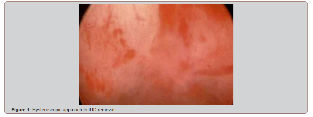
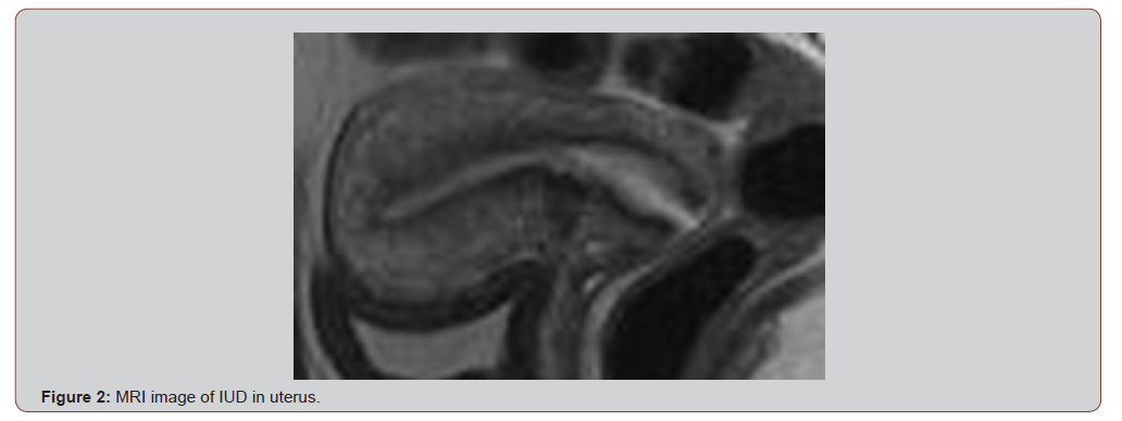
Written, informed consent was obtained for this case report. A 31-year-old G1P1001 requested a levonorgestrel IUD at her six-week postpartum visit. At the time of insertion, she was breastfeeding. Two years later she requested removal due to desired fertility and had been amenorrheic since insertion. During the in-clinic removal attempt, one of the IUD strings popped off after grasping with ring forceps and thus alligator forceps were used to attempt to grasp IUD, but it was unable to be dislodged. Formal ultrasound showed IUD embedded in anterior myometrium. Patient was then seen in complex family planning clinic and opted for repeat in office attempt, at which point strings were not visible, and alligator forceps were able to remove portion of string but unable to grasp IUD itself. Patient was then referred to minimally invasive gynecology. Office hysteroscopy was performed and was unable to visualize any part of IUD (Figure 1). MRI pelvis (Figure 2) showed IUD in lower uterine segment 2mm deep to the mucosa and 5mm from the serosa.
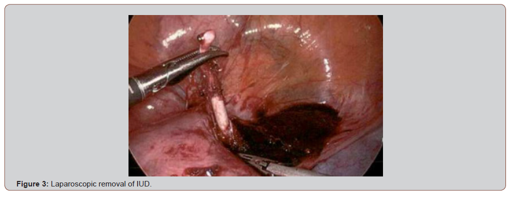
Patient was consented for ultrasound guided diagnostic hysteroscopy with possible laparoscopy. Hysteroscopy under transabdominal ultrasound guidance revealed the stem of IUD close to the endometrium and a bipolar hook was used to make an incision over this area but IUD was not encountered. Decision was made to convert to laparoscopic approach with transvaginal ultrasound guidance. The right broad ligament was opened and IUD was palpable. Uterine incision was made at the right lateral myometrium. Dissection of this area revealed one arm extending posterior and caudad to the round ligament. This was grasped and broke free from the IUD. Uterine incision was extended inferior and lateral. Stem of IUD was grasped and remainder of IUD successfully removed from uterus (Figure 3).
Discussion
The case presented describes a levonorgestrel IUD that was placed at the six-week postpartum visit after a Cesarean delivery for a breastfeeding patient, which was ultimately diagnosed as fully embedded within the myometrium two years after insertion. Diagnosis and treatment of embedded IUDs depend on the location and level of embedment, with hysteroscopy and laparoscopy recommended based on where the majority of the IUD lies [7-9]. Ultrasound is the recommended imaging modality for locating IUDs; however, when there remains uncertainty about the level of embedment, which impacts approach to removal, CT or MRI imaging may be helpful [10]. When IUDs are completely embedded within the myometrium, some describe using a tissue shaver device with fluoroscopy to remove the IUD while others describe laparoscopic removal using preoperative imaging to guide removal [11,12]. Given the rarity of fully embedded IUDs, it is debatable whether routine follow up is warranted for all IUD insertions. However, if embedment is not recognized and IUD strings are removed, surgical removal can become highly complex.
Conclusions
We submit that when an ultrasound is unable to identify any part of the IUD in the endometrial cavity, confirmatory formal imaging should be obtained before further removal attempts especially in those desiring future pregnancies. We recommend tailored counseling when placing IUDs in the postpartum period, particularly regarding perforation and expulsion, and routine follow up with ultrasound for those with multiple risk factors or unusual symptoms. We recommend investigation as to the best imaging modality to determine level of embedment as a future area of research.
Author Contributions
Conceptualization, JC, SC, writing – original draft, JC, writing – reviewing and editing, SC, supervision, S.C. All authors have read and agreed to the published version of the manuscript.
Funding
The research received no external funding.
Institutional Review Board Statement
Ethical review and approval were waived for this study due to the type of study (case report).
Conflicts of Interest
The authors declare no conflicts of interest.
References
- Nguyen AT, Curtis KM, Tepper NK, Kortsmit K, Brittain AW, et al. (2024) U.S. Medical Eligibility Criteria for Contraceptive Use, 2024. MMWR Recomm Rep 73(4): 1-126.
- Committee on Practice Bulletins-Gynecology, Long-Acting Reversible Contraception Work Group. (2017) Practice Bulletin No. 186: Long-Acting Reversible Contraception: Implants and Intrauterine Devices. Obstet Gynecol 130(5): e251-e269.
- Reed SD, Zhou X, Ichikawa L, Gatz JL, Peipert JF, et al. (2022) Intrauterine device-related uterine perforation incidence and risk (APEX-IUD): a large multisite cohort study. Lancet 399(10341): 2103-2112.
- Averbach S, Kully G, Hinz E, Dey A, Berkley H, et al. (2023) Early vs Interval Postpartum Intrauterine Device Placement: A Randomized Clinical Trial. JAMA 329(11): 910-917.
- Grandi G, Del Savio MC, Tassi A, Facchinetti F (2023) Postpartum contraception: A matter of guidelines. Int J Gynaecol Obstet 164(1): 56-65.
- American College of Obstetricians and Gynecologists’ Committee on Obstetric Practice (2016) Committee Opinion No. 670: Immediate Postpartum Long-Acting Reversible Contraception. Obstet Gynecol 128(2): e32-e37.
- Asto MRD, Habana MAE (2018) Hysteroscopic-guided Removal of Retained Intrauterine Device: Experience at an Academic Tertiary Hospital. Gynecol Minim Invasive Ther 7(2): 56-60.
- Vitale SG, Di Spiezio Sardo A, Riemma G, De Franciscis P, Alonso Pacheco L, et al. (2022) In-office hysteroscopic removal of retained or fragmented intrauterine device without anesthesia: a cross-sectional analysis of an international survey. Updates Surg 74(3): 1079-1085.
- Rahnemai-Azar AA, Apfel T, Naghshizadian R, Cosgrove JM, Farkas DT (2014) Laparoscopic removal of migrated intrauterine device embedded in intestine. JSLS 18(3): e2014.00122.
- Lee M, Melamud K, Petrocelli R, Slywotzky C, Prabhu V (2024) Comprehensive multimodality imaging review of reproductive interventions and their complications. Clin Imaging 116: 110312.
- Kwon CS, Bender LS, Schiff LD (2024) Fully embedded IUD: hysteroscopic management using x-ray. Ann Case Rep 9: 101979.
- Hur C, Goldberg J (2021) The laparoscopic removal of an embedded copper IUD fragment. Fert Steri 116(3): e468.
-

This work is licensed under a Creative Commons Attribution-NonCommercial 4.0 International License.




