 Research Article
Femoral Length Estimation from its Fragmentary State in Nigerian Population
Bienonwu Emmanuel Osemeke1 and Benwoke Woroma Ibiwari2*
1Department of Anatomy, School of Basic Medical Sciences, Igbinedion University Okada
2Department of Anatomy, Rivers State University of Science and Technology, Port Harcourt
Bienonwu Emmanuel Osemeke, Department of Anatomy, Rivers State University of Science and Technology, Port Harcourt, Nigeria.
Received Date: June 02, 2023; Published Date: June 21, 2023
Abstract
In forensic science, the identification of a deceased person is of great medico legal importance. To establish the identity of a person, stature is one of the criteria. To know stature of individual, length of long bone(s) (i.e., humerus, tibia, femur, and ulna) is/are needed. In Crime scene investigations, most times depending on the nature of the crime a complete body may not be found and instead fragments of bones or dismembered body parts maybe found. It then becomes necessary to estimate the physical characteristics (i.e., stature, height, race) of the victim with the available resources. The present study was aimed at estimating the total length of femur from fragments in Nigerian population. Maximum femoral length and measures of five (5) fragments of 200 femora (97 rights and 103 left) were obtained by means of a measuring tape and a vernier caliper from museums of various Anatomy Departments in Nigerian Universities. The mean length of femur bone on the left side was 44.92 ±3.28cm while on the right side was 44.71±3.32cm. The mean length of segments on left side were segment-1=8.00±1.19, segment-2= 10.19±1.35, segment-3= 10.35±1.48, segment-4= 12.52±1.10 and segment-5= 3.86±0.42, while mean length of segments on right side were segment-1= 8.07±1.13, segment-2= 10.37±1.34, segment-3= 10.02±1.63, segment- 4= 12.43±1.22 and segment-5= 3.82±0.43 A relationship was formulated to calculate length of femur in respect to each segment and this would be used ultimately to determine stature. The knowledge of Morphometric values of different femoral segments has important forensic and clinical implications.
Keywords: Femur, Fragments, Stature, Forensic, Nigerian population
Introduction
Establishing the biological profile of deceased persons is of great medico legal importance. To establish the identity of a person, stature is one of the criteria and to achieve this, the length of long bones is required [1,2]. In cases of natural disasters such as plane crashes, earthquakes or crush injuries, long bones are usually fractured or crushed making it difficult to estimate stature of body. In such a scenery, the ratio between fragment of a long bone and length of that bone would help in estimating height of that individual. Therefore, the length of the fragment of the long bone and the total length of that long bone were calculated and the ratio between each fragment and the total length of the bone was found out. Then, by applying the regression formula, the stature of the individual can be estimated [3]. Which is one of the big Four parameters in establishing identity (age, sex, race, and stature) [4]. The femur is the longest and strongest bone in the human body. Found in the lower limb(thigh) It has shaft, proximal end, and distal end. The shaft is slightly convex anteriorly [5].
Femur is established to be one of the long bones which helps in assessing the stature of the individual more accurately compared to the other long bones [6]. Being the largest and heaviest bone in the skeleton and because of its strength and density it is frequently recovered in forensic and archaeological settings and therefore provides a good lead in the process of identification [3,7,8]. Grossly mutilated skeletal remains are a big challenge in establishing identity of the deceased. Osteometry plays an important role in medico legal investigation for estimating height which is key to establishing identity. Skeletal remains could also show details of individualizing characteristics such as amputation, fractures, ankylosis, deformities and bone pathologies and to some extent the cause of death which is also essential in establishing identity of the individual [6]. The aim is to enable forensic experts to achieve the goal of personal identification. Pearson derived regression formulae for calculation of stature of an individual by the length of long bones in dry bones [9]. There have been recorded racial differences between stature and the length of long bone in Caucasoid, Negro and Mongoloid [10].
Therefore, there is a need to derive separate, specific formulae for different races [1]. and for different population groups such as Nigeria. Measurement of fragmented bones such as the femur becomes necessary where bodies are presented in such state in order to estimate stature [8,11]. This also has clinical implication in the treatment of femoral fractures where fractured bone needs to be replaced with prosthesis and sizing becomes of importance [9,12]. Morphometric values of femoral segments therefore have important implications in forensic, anatomical, and archaeological cases in establishing identity of unknown bodies and stature. It is also applied in clinical settings in the treatment of femoral fractures.
Materials and Methods
Two hundred (200) dried Femora bones consisting of 97 right and 103 left were used. The bones were obtained from Museums in Anatomy departments of selected Universities in Nigeria. Bones which have undergone degenerative changes were exempted from this study on gross inspection. All measurements were taken with a vernier caliper. A pair of dividers was also used to confirm the measurements at its point and measured with a calibrated ruler. Each measurement was repeated twice, and the mean value was recorded. Measurements were taken in centimeters. The Femur was divided into 5 segments by taking the following landmarks on the basis of the morphological characters of femur from the top of head to distal point on medial condyle, i.e., a-b; b-c; c-d; d-e; e-f [6] (Figure 1).
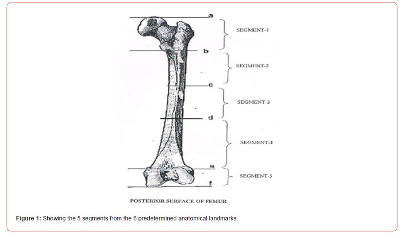
a) The most proximal point on head.
b) Lower border of lesser trochanter.
c) Point where gluteal and spiral lines join to form Linea aspera.
d) Point where Linea aspera divides into medial and lateral supracondylar lines.
e) Most proximal point on the intercondylar fossa.
f) Most distal point of the medial epicondyle.
The following measurements were carried out:
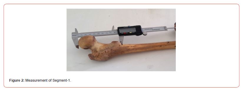
1. Segment-1(a-b) (Figure 2): from the most proximal point (a) to the lowest border of the lesser trochanter (b) as shown below.
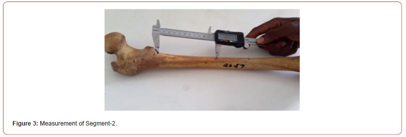
2. Segment-2(b-c) (Figure 3): from the lower border of lesser trochanter (b) to the point where the gluteal line and the spiral line joins to form the linea aspera(c).
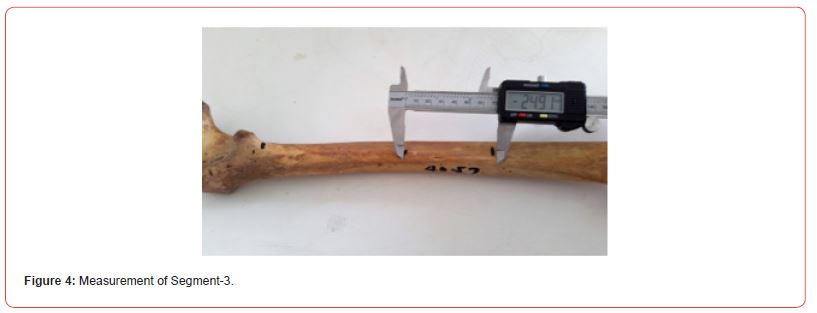
3. Segment-3(c-d) (Figure 4): from point c to the point where the Linea aspera form the lateral and medial supracondylar lines (d).
4. Segment-4(d-e) (Figure 5): from point ‘d’ to the most proximal point on the intercondylar fossa (e).
5. Segment-5 (e-f) (Figure 6): from the most proximal point of the intercondylar fossa (e) to the most distal point of the medial epicondyle (f).
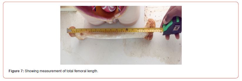
6. Total length of Femur: measures from the most proximal point of the head to the most distal point of the medial epicondyle (Figure 7).
Figures 2-7: Pictures showing measurement of the total length of femur and the measurement of 5 segments using their 6 predetermined anatomical landmarks.


Statistical Analysis
1. Descriptive statistics like mean and standard deviation of segement-1, segment-2, segment-3, segment-4, and segment-5 was calculated.
2. The independent ‘t’ test was used to compare the segment-1, segment-2, segment-3, segment-4 and segment-5 lengths of left and right femur. Data analysis was carried out using the Statistical Package for Social Science (SPSS) package.
3. Linear regression analysis was employed to estimate the stature of an individual from length of femur.
Method of Statistical Analysis
The methods of statistical analysis used in this study include:
1. The student ‘t’ test was used to determine whether there was a statistical difference between two groups in the parameters measured.
2. One way Analysis of Variance (Anova) was used to test the difference between groups. Analysis of Variance is a technique by which the total variation is split into two parts, one between groups and the other within the group. If ‘F’ value is significant, there is a significant difference between group means. Y6 The formula used:

Where MS= Mean of Squares.
3. A simple relationship was applied. This relationship was made considering the right and left, separately for each fragmentary bone measurement to estimate the total length.

Where; 100= the total percentage (%)
Z= Total length (cm)
C= Percentile length of given segment based on this study (%).
X= Length of found segment (cm)
This regression was made considering the right and left, separately for each fragmentary bone measurement. Multiple regressions were used to estimate the total length using fragmentary bone measurement. In this method, the incorporation of variables was made through stepwise regression.
In the entire above test, the “p” value of less than 0.05 was accepted as indicating statistical significance. Data analysis was carried out using Statistical Package for Social Science (SPSS) package18.
Results
The Results are presented in Tables 1-4.
Table 1: Mean lengths of corresponding left segments and right segments of femora along with the standard deviation, minimum, maximum, ‘t’ and ‘p’ values of the segments of femur bone.
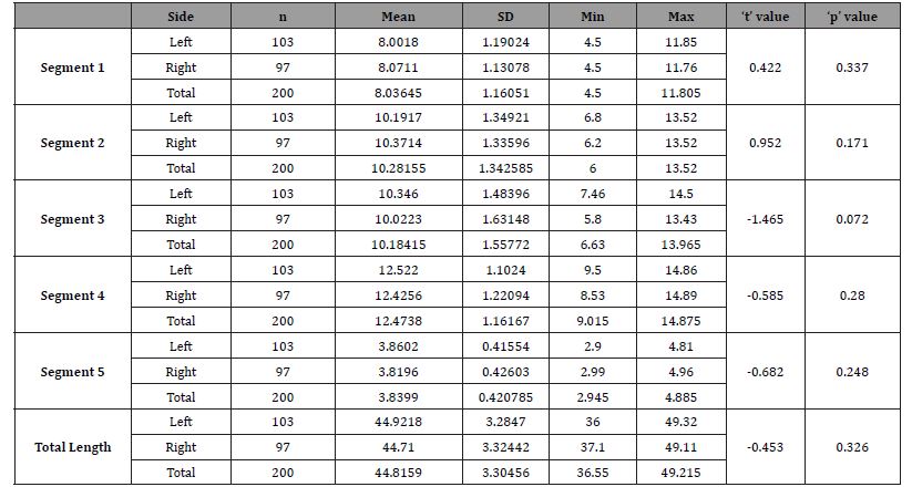
Table 2: Showing the mean lengths of five segments of the left and right femora along with the standard deviation, ‘F’ and ‘p’ values.

Table 3: Showing length of femoral segments in percent along with the standard deviation, ‘f’ and ‘p’ values of the segments of femur.

Table 4: Comparison of Total Femoral Length of this study with other authors.

A linear regression was created for each segment to derive the total length of femur considering the side of femur (left or right).
Y = a + bx
Where Y (dependent variable) = total length of femur
a= intercept
b= slope
x (independent variable) = given length of segment
Substitution:
For right:Segment-1: Y= 27.085 + (2.068 × segment-1)
Segment-2: Y= 24.406 + (1.846 × segment-2)
Segment-3: Y= 34.788 + (0.987 × segment-3)
Segment-4: Y= 27.986 + (1.289 × segment-4)
Segment-5: Y= 33.980 + (3.096 × segment-5)
For Left:Segment-1: Y= 27.236 + (2.070 × segment-1)
Segment-2: Y= 23.865 + (1.943 × segment-2)
Segment-3: Y= 38.683 + (0.485 × segment-3)
Segment-4: Y= 28.634 + (1.244 × segment-4)
Segment-5: Y= 29.575 + (4.598 × segment-5)
1. The mean length of femur bone on left side was 44.92±3.28cm while on right side was 44.71±3.32cm with ‘t’ value= -0.453 and ‘p’ value= 0.326.
2. The mean length of segments on left side were segment-1=8.00±1.19, segment-2= 10.19±1.35, segment-3= 10.35±1.48, segment-4= 12.52±1.10 and segment-5= 3.86±0.42, along with ‘F’ value= 775.59 and ‘p’ value=<0.005. While mean length of segments on right side were segment-1= 8.07±1.13, segment-2= 10.37±1.34, segment-3= 10.02±1.63, segment- 4= 12.43±1.22 and segment-5= 3.82±0.43, along with ‘F’ value = 671.74 and ‘p’ value= <0.005.
3. On the left side, mean percentage of segments to total femoral length were segment-1= 18.08±2.28, segment-2= 23.73±2.32, segment-3= 21.55±3.73, segment-4= 29.25±2.40 and segment-5= 7.39±0.88, along with ‘F’ value= 1131.77 and ‘p’ value= <0.005. While on the right side, mean percentage of segments to total femoral length were segment-1= 18.27±2.16, segment-2= 24.21±2.39, segment-3= 20.72±3.79, segment-4= 29.52±2.99 and segment-5= 7.28±1.07, along with ‘F’ value= 956.64 and ‘p’ value= <0.005.
4. Femoral length comparison with that of previous studies.
Discussion
Stature Estimation from skeletal remains has great medicolegal implication in forensics. This relies on the length of long bones. The femur is of key interest because it is frequently recovered in most forensic and archeological sites and therefore provides a good lead in the process of identification [3,7]. This is due to its strength and density. Where the femur is damaged (found in fragments), length can be calculated from the fragments of femur found. This is achieved by assessing the ratio between various segments of femur with the length of that bone. Once the length of the femur is obtained, the height of the individual can be calculated using various standard regression formulae. In this study, five (5) segments of the femur were obtained using six anatomical landmarks which varied from previous works but aligned with landmarks used by Shweta et al [6]. This will help enable femur length to be calculated if any of the segmented parts are recovered.
The right and left femur was used in this study which differs from previous studies but agrees with the study by Shweta et al [6]. The mean values of total femur length were identified as 44.92±3.28cm and 44.71±3.32cm for left and right respectively. This agrees with the study carried out by Steels et al [13] on Northeastern Arkansans population but disagrees with that of Shroff [14] and Shweta et al [6]. where lower values were reported. This could be as a result of the height of the people in those populations. The mean values gotten for Segment-1 in this study was 8.00±1.19cm for the left side and 8.07±1.13cm for the right side this was in consonance with study reported by Shweta et al 6 with similar values (8.08±0.70cm) but differs from that of Steels et al [13] on Northeastern Arkansans population (7.35± 0.61 cm). This may be due to different anatomical landmarks used. Segment-1 in the present study was taken from highest point on the head to the lowest point of lesser trochanter which is like the study conducted by Shroff [14] and Shweta et al6 whereas the points for segment-1, taken by Steels et al [13], was highest point of head and the midpoint of the lesser trochanter.
In this study, the proportion of segment-1 to total length of femur was 18.07± 2.29% and 18.26± 2.15% for the left and right femur respectively this is in line with the study done by Shweta et al.9 which reported 18.58± 0.62% but it varied from the study done by Steels et al [13] and Shroff [14]. This can be used for estimation of stature of individual by using Steele’s regression formulae. Segment-2 was measured as the distance between the lower border of lesser trochanter and point where gluteal and spiral lines join to form linea aspera. Its proportion to femoral length was 23.73±2.32% for the left and 24.21±2.39% for the right. Although the landmarks used in this study were same with that of Shweta et al [6] it gave higher proportion when compared to Shweta et al [6]. which recorded 19.05± 0.34%. this may be due to the higher femoral length recorded for the Nigerian population. The landmarks used differs from that of Steels et al [13] who measured segment-2 as distance between mid-points of lesser trochanter to most proximal extension of popliteal surface where medial and lateral supracondylar lines become parallel below the Linea aspera. They reported a proportion of 56.1% for segment -2. The segments taken in the study have enough scope to derive a regression equation that can be derived from this segment to find the precise length of femur which will help in determining the height of that individual. Segment-3 was measured as the distance between point c (Point where gluteal and spiral lines join to form Linea aspera) to the point where the Linea aspera form the lateral and medial supracondylar lines (d). these landmarks were same as that of Shweta et al [6] but differs from that of Steels et al [13]. Its proportion to femoral length was 21.55±3.73% for the left and 20.72±3.79% for the right. segment-4 is between lines where Linea aspera divides into medial and lateral supracondylar lines, to the most proximal point on intercondylar fossa and it measured 12.52±1.10cm for left and 12.43±1.22cm for right this was lower than that reported by Shweta et al [6] who reported 13.71±1.74cm even though same anatomical landmarks were used. Proportion of segment-4 to the total length was 29.25±2.40% and 29.52±2.99% for the left and right femur respectively as against 31.56± 0.39% reported by Shweta et al [6] reported for South Indian Population. The difference may be due to race, growth pattern as well as nutrition.
Segment-5 was measured from the most proximal point of the intercondylar fossa (e) to the most distal point of the medial epicondyle (f). same anatomical points as with Shweta et al [6] it measured 3.86±0.42cm and 3.82±0.43cm for the left and right respectively this was higher than that reported by Shweta et al [6] which gave 2.93± 0.50 cm with a proportion of 7.39±0.88% and 7.28±1.07% for the left and right respectively. Which was higher than that of Shweta et al.6 which was 6.75± 0.27%. This also may be due to differences in race, growth pattern as well as environment. The ratio and proportion between the fragmentary length of femur and the total length of femur, for estimating the total length of femur, has been derived in this study and can be used to calculate or estimate stature [15,16].
Conclusion
From the result of the study, it can be concluded that femur length can be derived from fragments, which could help further in the estimation of stature. This was achieved by dividing the femur bone into five (5) segments using six (6) anatomical landmarks and creating a regression formula for each segment16. Femur length can then be calculated using any of the available segments. This is important in forensics where identification of unknown bodies and stature is key. to solve most mystery cases in order to identify unknown bodies and stature. Morphometric values of femoral segments are also useful to clinicians in the treatment of femoral fractures. This study therefore provides a data base of mean values of different morphometric measurements from the femur for the Nigerian population.
Acknowledgment
None.
Conflict of Interest
No conflict of interest.
References
- Ajay MP, Kanan PS, Jatin G, Brijesh A, Agarwal GC (2015) Reconstruction of total length of femur from its proximal and distal fragments. International Journal of Anatomical Research 3(4): 1665-1668.
- Dayal Manisha R, Maryna Steyn, Kuykendall Kevin L (2008) Stature estimation from bones of south African whites, south. African journal of science 104: 3-4.
- Rother P, Jahn W, Hunger H, Kurp K (1980) Determination of body height from fragment of the femur. Gegenbaurs Morphol Jahrb 126(6): 873–83.
- Shende MR, Parekh NAJ (2009) The study of reconstruction of total length of ulna from its fragments and its medicolegal perspectives. J. Anat. Soc. India 58(2): 130-134.
- Standring Susan (2008) Gray’s Anatomy, The Anatomical basis of clinical practice. 40th edition. London: Elsevier Churchill Livingstone pp. 1360–13
- Shweta S, Roopa K (2013) Estimation of total length of femur from its fragments in South Indian population. Journal of Clinical and Diagnostic Research 7(10): 2111-2115.
- Bienonwu EO, Fawehimi HR, Olotu EJ, Ihunwo IW (2017) Sex ans Bilateral Differences of Femoral Condyle Morphology of Nigerians: An Osteometric Study. Journal of Anatomical Sciences 8(1): 159-165.
- Bidmos MA (2008) Estimation of stature using fragmentary femora in indigenous south Africans. Int J Legal Med 122(4): 293-299.
- Pearson K (1899) Mathematical contribution to the theory of evolution. On the reconstruction of the stature of prehistoric races. Philosphical Transanctions of the Royal Society 192: 169-244.
- Pan N (1924) Length of long bones and their proportion to body height in Hindus. J Anat 58: 374-378.
- Stevenson PH (1929) On racial differences in stature long regression formulae, with special reference to stature reconstruction formulae for Chinese. Biom 21(1-4): 303-3.
- Prasad Rajendra (1996) Selvakert, Reconstruction of femur length from markers of its proximal end. Clinical Anatomy 9(1): 28-33.
- Steele DG, McKern TW (1969) A method for assessment of maximum long bone length and living stature from fragmentary long bones. American Journal of Physical Anthropology 31: 215-228.
- Shroff AG, Panse AA, Diwan CV (1999) Estimation of length of femur from its fragments. Journal of the Anatomical Society of India 48(1): 1-5.
- Singh S, Nair SK, Anjankar V, Bankwar V, Satpathy DK et al. (2013) Regression equation for estimation of femur length in central Indians from intertrochanteric crest. Journal of Indian Academy of Forensic Medicine 3(3): 223-226.
- Bienonwu EO, Benwoke WI, Okwuonu UC, Omotoso DR (2020) Osteometric Morphometry of the proximal Tibia end in Nigerian Population. International. Journal of Applied Research 6(7): 117-123.
-
Bienonwu Emmanuel Osemeke and Benwoke Woroma Ibiwari*. Femoral Length Estimation from its Fragmentary State in Nigerian Population. Acad J Health Sci & Res. 1(1): 2023. AJHSR.MS.ID.000502.
Research, Beginners, Medicine, Hypothesis, Blinding, Lung cancer, Clinical trials
-

This work is licensed under a Creative Commons Attribution-NonCommercial 4.0 International License.


