


 Case Report
Removal of an Intragastric Foreign Body: A Case Report
1,2 Department of Gastroenterology, People’s Hospital of Ningxia Hui Autonomous Region, 301 Zhengyuan North Street Yinchuan, China
Received Date: October 09, 2026; Published Date: October 15, 2026
Abstract
Herein, we present the case of a 31-year-old male patient admitted to the emergency department due to “chest pain for 1 day” after swallowing a foreign object. Chest computed tomography (CT) revealed a “long metal foreign body shadow” approximately 17 cm in length in the stomach. This object was subsequently removed using an endoscope. When removing sharp foreign bodies, it is very important to use protective devices (such as transparent caps, outer tubes, and protective covers) at the front end of the endoscope to avoid damage to the digestive tract mucosa.
Case Report
Herein, we present the case of a 31-year-old male patient (Gao) admitted to the emergency department at 19:41 on October 13, 2025 due to “chest pain for 1 day” after swallowing a foreign object. Due to a mental illness, the patient was represented by a family member. The patient developed chest pain and pharyngeal discomfort after swallowing a metal foreign object (bracelet) one day prior. The family initially sought medical attention at Ning’an Hospital in Yinchuan, Ningxia, where relevant tests and examinations were performed.
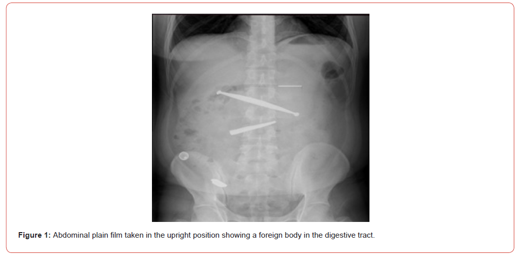
The results of examination were as follows: Brain computed tomography revealed no obvious abnormalities. Chest CT revealed a “long metal foreign body shadow” approximately 17 cm in length in the stomach. Doctors at the first hospital recommended that the patient seek medical attention from a superior hospital, resulting in his admission to our center. At our hospital, improved standing abdominal plain film revealed a foreign object in the digestive tract (Figure 1). Further, chest CT revealed the following: plain chest CT revealed no abnormalities, while metal-density shadows were observed in the gastric cavity, duodenum, and proximal jejunum, indicating a foreign body in the digestive tract (approximately 17 cm in length).
After consultation with the gastroenterology department, this digestive tract foreign body was considered to be extremely narrow and metallic and potentially corrosive, with estimates indicating it had been swallowed more than 24 hours prior. Immediate removal was deemed necessary to prevent bleeding and perforation risks. However, due to the patient’s mental illness, it was difficult to ensure cooperation with ordinary endoscopes. The risk of pain during endoscopic surgery in the gastroscopy room is high. Therefore, after communicating with the anesthesia department, we decided to perform foreign body removal in the operating room.
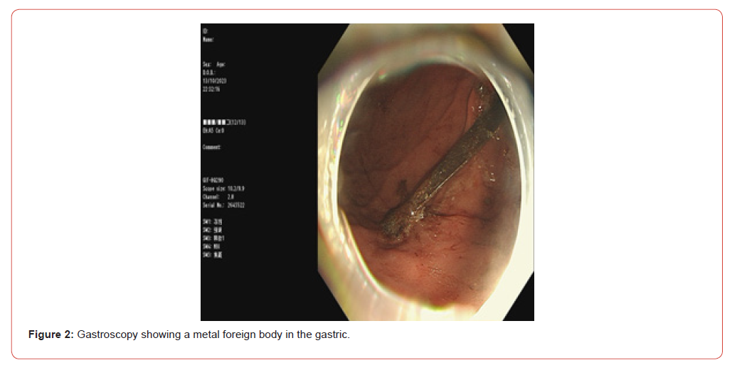
Discussions were conducted with the family, who ultimately agreed for us to remove the foreign object in the operating room. At 21:30 on October 13th, the patient entered the operating room. After achieving satisfactory anesthesia, a physician advanced a gastroscope, equipped with a transparent cap at the front end, into the cavity, Under the gastroscope, a metal foreign body spanning ~17 cm in length across the gastric body was visible (Figure 2), with one end embedded in the pyloric entrance, accompanied by the formation of a huge ulcer at the pyloric entrance. A large amount of old blood was visible in the fundus and gastric body. During the surgery, the foreign body was repeatedly grabbed with foreign body forceps and snares, and the head end was smoothly grasped. However, as the foreign body was too narrow and long, it was not possible to collect it into the transparent cap. However, it was considered that if the foreign body could not be collected into the transparent cap, it may scratch the esophagus and mediastinum when pulled from the gastric cavity, posing a significant risk to the patient.
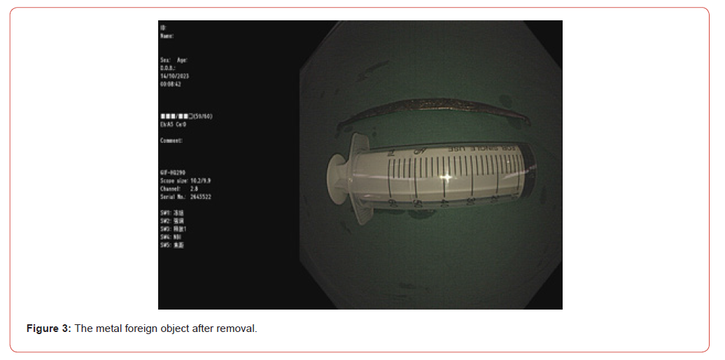
During surgery, communication with the gastroenterology department was consistently performed. After clinicians in this department became aware of the patient’s condition, they considered the surgical trauma to be significant and did not recommend surgical treatment. Our physician and assistant worked closely together again, collecting the large and narrow foreign object into the transparent cap, and carefully, quickly, and cautiously removing the large foreign object (Figure 3). After removing the foreign body, another endoscopic observation was performed along the cavity. Scanning of the oral cavity revealed that the mucosa was intact without obvious scratches, and only one ulcerative surface was formed at the pyloric opening. After the surgery, the patient’s vital signs were stable according to electrocardiogram monitoring.
Discussion
Upper gastrointestinal foreign bodies may include a variety of objects in the upper gastrointestinal tract that cannot be digested and are not discharged in a timely manner. Ingestion of a foreign body which then becomes lodged in the upper gastrointestinal tract is a common clinical emergency, accounting for 4% of all emergency endoscopic diagnoses and treatment [1]. If not handled in a timely manner, this condition may cause serious complications, and even lead to death. Common foreign bodies in the upper gastrointestinal tract include short and blunt, long, sharp, metallic, corrosive, or magnetic foreign bodies, in addition to food clumps in the esophagus, drug bags, etc. The endoscopic treatment methods vary between different cases [2].
Generally, corrosive sharp foreign bodies need to be removed under emergency gastroscopy. The foreign object swallowed by our patient was a sharp and corrosive foreign object, with a length of up to 17 cm, thus, we performed endoscopy using a gastroscope equipped with a transparent cap on the end of the stomach lens during intubation performed under general anesthesia. Initially, we attempted to use foreign body forceps to grab the object; however, these attempts were unsuccessful. Later, we used a snare to secure one end and remove the foreign body from the body, which was difficult. This experience suggest that it is very important to us protective devices (such as transparent caps, outer tubes, and protective covers) at the front of the endoscope to avoid damage to the digestive tract mucosa when removing sharp foreign bodies.
References
- S Mosca, Manes G, Martino R, Amitrano L, Bottino V, et al. (2001) Endoscopic management of foreign bodies in the upper gastrointestinal tract:report on a series of 414 adult patients Endoscopy 33(8): 692-696.
- Digestive Disease Branch of the Chinese Medical Association (2016) Consensus Opinion of Chinese Experts on endoscopic Treatment of Foreign Bodies in the Upper Gastrointestinal Tract [J]Chinese Journal of Digestive Endoscopy 33: 19-22.
-

This work is licensed under a Creative Commons Attribution-NonCommercial 4.0 International License.