



 Research Article
A New Way of Reconstruction in Pancreaticoduodenectomy. “The Anatomical Sequence”. Preliminary Report
1Servicio de Cirugía General del Hospital Tránsito Cáceres de Allende
2Cátedra de Cirugía II – UHC 4. Universidad Nacional de Córdoba, Córdoba, Argentina
Received Date:November 25, 2025; Published Date:December 04, 2025
Abstract
Introduction: performing the biliary anastomosis first and then the gastric anastomosis is usually the norm in the reconstruction of
pancreatoduodenectomy. In this way, episodes of cholangitis and bile reflux to the stomach would be avoided than if it were done anatomically. We
have used an anatomical reconstruction sequence in order to assess whether there are more episodes of cholangitis or reflux.
Material and Method: 17 patients who underwent pancreticoduodenectomy. Gastric and biliary anastomoses were constructed in that order
(anatomical). Cholangitis and duodeno-gastric bile reflux were evaluated with clinics, radiology and tomography.
Results: There was one death. In the remaining 16, there were no episodes of cholangitis or duodenal gastric bile reflux that caused symptoms.
Conclusion: The anatomical sequence is safe and original. Not increase episodes of cholangitis or alkaline gastritis due to reflux. This type of
reconstruction favors endoscopic access to the bile duct in easy way.
Keywords:Pancreatoduodenectomy; Complications; Cholangitis; Endoscopy
Introduction
Multiple techniques for reconstruction of continuity after a pancreatoduodenectomy (PD) have been proposed. All of them with the aim of reducing the number of postoperative pancreatic fistula (POPF), delayed gastric evacuation, biliary complications or hemorrhage [1-5]. There are few works that focus on biliary complications (Stenosis, reflux, postoperative colangitis) [6-10]. In all the techniques there is a common point, the sequence of the biliary and gastric anastomoses are inverted to the anatomical or normal sequence with the reason that in this way the reflux within the biliary tract and cholangitis would be reduced as a consequence of the absence of the papilla of Vater and in the same way there would be a decrease in biliary reflux towards the stomach and esophagus (Alkaline reflux). In all mentinoed techniquesits recomended tnat the distance between biliary and gastric anastomosis must be major tan 40 cm [6]. The experience with hepatic-duodenostomy in the treatment of choledochal cysts has not shown a higher percentage of cholangitis [11] and is the only technique that is similar to one proposed by us. The aim of the present study is to present a novel technique where we perform gastric and biliary continuity with anatomical sequence in the PD and to evaluate whether there is a higher number of cholangitis or esophageal-gastric biliary reflux attributable to this reconstruction.
Material and Method
Technique
After resection the duodenum-pancreas was completed, the continuity of the different elements was reconstructed in two ways:
a. Type 1: Reconstruction on a Roux-en-Y loop: in one
of the loops, a pancreatico-jejunal-end-to-end anastomosis
was performed with Hunt stitches, in the other loop, first a
gastro-entero-end-to-end anastomosis was performed and
approximately 15 cm from this, a hepatic-jejunal-end-to-side
anastomosis.
b. Type 2: Reconstruction through a single loop: an end-toend
anastomosis was performed between the jejunal loop and
the stomach and approximately 15 cm away an end-to-side
biliary anastomosis. Pancreatic continuity was established
through a pancreatico-gastro-anastomosis. Pyloric preservation
was performed in all of them (Figure 1).

The anastomosis between the stomach and the jejunum was performed using continuous sutures in two layers with absorbable thread. The biliary-enteric anastomosis in the posterior plane was performed with interrupted stitches that were knotted in a delayed manner, and the anterior surface was performed using a continuous suture with absorbable thread (Vicryl 3/0). In no patient was the biliary anastomosis intubated. In all patients, a catheter was placed in the Wirsung duct. In type 2 reconstruction, the jejunal loop was raised behind the mesenteric vessels, reconstructing the duodenal “C” (Figure 2 & 3), while in the type 2 reconstruction, the loop in which the pancreas was anastomosed was raised in this manner, while the alimentary loop was raised in a trans-meso-colonic manner. A nasogastric tube was placed in all patients and was advanced past the biliary anastomosis. Two multi-lumen drains were placed in the abdominal cavity, one below the pancreatic anastomosis and the other in the Winslow hiatus.


Inclusion criteria were: patients over 18 years of age, with albumin greater than 3 g/dl, in tumors resectable by imaging. Borderline patients were excluded. All patients signed an informed consent form, knowing not only the procedure but also the possible complications of the procedure. Data collected prospectively from patients undergoing PD in which the anatomic sequence of reconstruction of the gastric and biliary anastomoses was used were retrospectively analyzed. Patient demographic data and data regarding the surgery were analyzed. Bile duct dilatation was considered when the diameter of the bile duct was greater than 8 mm measured by ultrasound, and Wirsung duct dilatation was considered when it measured more than 3 mm measured by threephase computed tomography (CT). For postoperative complications (POPF, delayed gastric emptying and hemorrhage) the classification proposed by the ISGPS [12-14] was used. For biliary leak, the proposed ISGLS classification was used [15]. Cholangitis was defined as proposed by the 2013 Tokyo Guidelines [16]. Early cholangitis was considered to be that which appeared in the first 30 days postoperatively, after that time it was considered late, and refractory cholangitis was considered to be that which recurred 3 or more times. Complications were classified as proposed by Dindo-Clavien [17]. Amylase determination of the drain leading to the pancreatic anastomosis was performed on days 1, 3 and 5 postoperatively, and in some cases also on day 7. Patients were studied postoperatively with triphasic CT and serial esophagealgastroduodenal radiographs to try to detect reflux of iodinated contrast into the bile duct (Figures 2 & 3). Bile reflux into the stomach and esophagus was assessed clinically or by endoscopy if the patient consented to the procedure. Quantitative variables were assessed with range and mean and while qualitative variables were assessed with average and percentage.
Results
Seventeen patients were operated on with this technique. One patient died within the first 90 days. Of the remaining 16 patients, 9 patients presented some degree of pneumobilia observed by CT scan, in 1 of them there was a slight but persistent elevation of alkaline phosphatase that did not reach double the normal value. None of the patients presented clinical or laboratory signs suggestive of cholangitis or transient elevation of bilirubin, nor symptoms or signs of gastro-oesophageal reflux. 5 patients agreed to undergo an upper digestive endoscopy, but no bile was observed in the stomach. The rest of the data can be seen in Tables 1 & 2.
Table 1:Demographics and pre-operatives’ data.
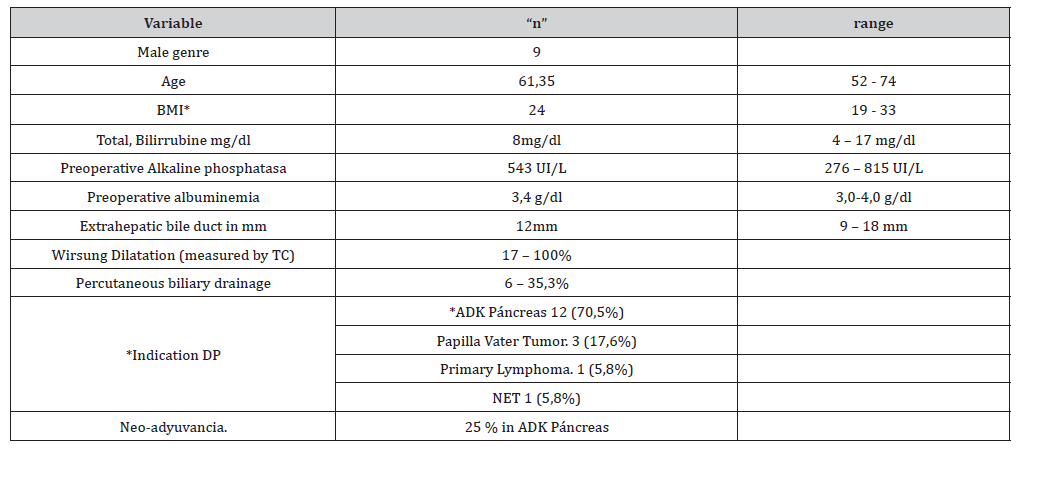
BMI: Body mass Index; ADK: Adenocarcinoma; PD: Pancreatoduodenectomy
Table 2:Intra and post-operative data.
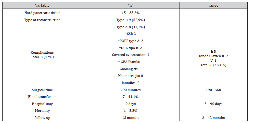
SSI: Surgical Site Infection; POPF: Postoperatice pancreatic fistula; DGE: Delayed Gatsric Emptying; GEA: Gastro-entero-anastomosis
Discussion
The number of previously reported cholangitis episodes in choleco-duodenostomy (CD)is according to some authors around 10% [6]. This technique (CD) has been the probable origin of the theory that the anatomical reconstruction favors the reflux of food into the biliary tract, the presence of the blind sac between the anastomosis and the papilla would justify stagnation and subsequent ascending infection. Some authors in 2146 patients undergoing choledoco-duodenostomy report an incidence of 0.73% [18]. Patil et al. [11] used hepatic-duodenostomy as a biliarydigestive anastomosis in the treatment of choledochal cysts in 56 patients and report only one cholangitis secondary to stenosis of the anastomosis 18 years after its creation (0.56%), which would cast doubt on reflux as the cause of cholangitis. These authors performed the anastomosis with the duodenum, 2 cm from the pylorus and always at the level of the biliary crossroads, although they do not report the diameter of the same. On the other hand, the experience with Billroth 1 type reconstructions in gastrectomies has been abandoned because the biliary reflux from the duodenum to the gastric remnant could become disabling and this would also justify the conduct of placing the biliary anastomosis before the gastric one. There is little literature referring to biliary complications of PD. Cholangitis is reported by different authors between 1 and 18.6%. (2-6, 8, 9, 19, 20), most of the episodes appear within the first 30 days [4,9].
Ueda et al. (19) among 18 patients with refractory cholangitis, report that in 17 of them the same appeared in the first year after surgery. End-to-side hepatic-duodenostomy is the procedure that most closely resembles the anatomic sequence reconstruction we have used. In a comparison between hepatic-jejunal anastomosis and hepatic-duodenostomy in the treatment of choledochal cysts, the authors report that patients with hepatic-jejunal anastomosis had more cholangitis than those who underwent hepaticduodenostomy (15% vs 3%) [21]. In the same sense, but comparing both anastomoses in liver transplantation, the percentage of cholangitis was the same between both anastomoses (HD 14% vs HJ 12%) [22]. It is likely that pyloric preservation in our patients plays a fundamental role both in avoiding cholangitis as well as in avoiding bile reflux into the stomach and the consequent alkaline gastritis or even esophagitis. Among the causes of cholangitis in PD, anastomotic stenosis, stones, intestinal obstruction, afferent loop syndrome and jejunal peristalsis disorders are the most frequently reported [6,9]. The cause of early episodes of cholangitis, which are the most frequent, [4] could be attributed to minimal biliary stenosis due to acute inflammation, ileus, peristalsis disorders and contamination by resistant germs, [9] while late episodes would be associated to a greater extent with anastomotic stenosis. Some authors [8] have found that the main predictive factor of stenosis is a thin bile duct. Other authors [20] reaffirm this concept by stating that a bile duct less than 15 mm in diameter is a risk factor for stenosis and based on this they propose performing a hepaticoplasty to increase the diameter of the anastomotic mouth and promote a greater flow of bile into the intestine. Our patients had a mean diameter of 12 mm and there were no episodes of cholangitis in the mean follow-up of 13 months.
Other factors associated with postoperative cholangitis are resection for benign pathology, prolonged surgery time and persistent elevation of alkaline phosphatase [19], the latter with a value greater than 440 should lead to expect a stenosis [6]. The use of preoperative percutaneous biliary drainage or stents would favor the appearance of cholangitis by causing micro-traumas in the biliary tract [6,10]. Because we agree with this last observation, we leave the percutaneous drains preferably before the biliary crossroads functioning as external biliary drainage, we have noticed that in those patients with drains that run through the common bile duct they produce traumatic choledocytitis that, depending on the time it is left in place, makes its dissection difficult in surgery and forces an anastomosis to be made over an inflamed biliary edge. We have used the anatomical sequence in 16 patients with no episodes of cholangitis or bile reflux into the stomach to date, and this new arrangement seemed more harmonious to us. It facilitates endoscopic access to the biliary tract in a simple manner, a very important factor since, as far as we know, endoscopic access in other types of reconstructions is extremely cumbersome, [23] expanding therapeutic procedures. In the imaging studies we were able to observe a small amount of reflux with complete filling of the stomach and a significant portion of the jejunum in the patients in whom we performed serial esophageal-gastro-duodenal radiographs.
In conclusion, the proposed technique is novel. [24] There are still a few cases for a definitive assertion, this should be increased and will probably require multi-center participation for this objective. In future communications we will try to make up for the lack of more in-depth complementary methods to confirm the absence of cholangitis and gastric reflux, as well as the performance of an ERCP to determine one of the strong points of this reconstruction, which is biliary access by endoscopy.
Authors Participation
E. De elias, F. López, D. Mariot y L. Viotto were in charge of the bibliographic search, its selection and the preparation of the tables and figures. J. Layún carried out the data loading and the results. G. Nari wrote the paper. All authors reviewed the finished work.
References
- Nari G, Granero L, Silva J, Layún J, Mariot D et al. (2019) Pancreatic Fistula after pancreaticoduodenectomy. Pancreaticogastrostomy vs pancreaticojejunostomy. Rev Argent Cirug 111: 79-89.
- Yeo Ch, Cameron J, Sohn T, Lillemoe K, Pitt H et al. (1997) Six hundred fifty consecutive pancreaticoduodenctomies in the 1990s. Ann Surg 228: 248-257.
- Braga M, Capretti G, Pecorelli N, Balzano G, Doglioni C et al. (2011) A prognostico score to predict major complications after pancreaticoduodenectomy. Ann Surg 254: 702-708.
- Ven Fong Z, Ferrone C, Thayer S, Wargo J, SahoraK et al. (2013) Understanding hospital readmissions after pancreaticoduodenectomy: can we prevent them? J Gastrointest Surg 18(1): 137-144.
- Yamaguchi K, Tanaka M, Chijiiwa K, Nagakawa T, Imamura M et al. (1999) Early and late complications of pylorus-preserving pancreaticoduodenectomy in Japan 1998. J Hepatobiliary Pancreat Surg 6: 303-311.
- Ito Y, Abe Y, Kitago M, Itano O Kitagawa Y (2018) Predictive factors of late cholangutis in patients undergoing pancreaticoduodenectomy. World J Surg Oncol 16: 19-27.
- Samada Y, Yamada N, Taguchi M, Morishima K, Kasahara N et al. (2014) Recurrent Cholangitis by biliarystasis due to non-obstructive afferent loop síndrome after pylorus preserving pancreaticoduodenectomy: repor of a case. Int Surg 99: 426-431.
- Duconseil P, Turrini O, Ewald J, Berdah S, Mourtadier V et al. (2014) Biliary complications after pancreaticoduodenectomy: Skinny bile ducts are surgeon`s enemies. World J Surg 38: 2946-2951.
- Malgras B, Duron S, Gaujoux S, Dokmak S, Aissilhou B et al. (2016) Early biliary complications following pancreaticoduodenectomy: prevalence and risk factors. HPB 18(4): 367-374.
- House M, Cameron J, Schulick R, Campbell K, Sauter P et al. (2006) Incidence and outcome of biliary strictures after pancreaticoduodenectomy. Ann Surg 243: 571-578.
- Patil V, Kanetkar V, Talpallikar M (2015) Hepaticoduodenostomyfor biliary reconstruction after surgical resection of choledochal cyst: a 25-yearexperience. Indian J Surg 72: 240-244.
- Bassi C, Dervenis C, Butturini G, Fingerhut A, Yeo C et al. (2005) Postoperative pancreatic fistula:An international study group (ISPGF) definition. Surgery 138: 8-13.
- Wente M, Bassi C, Dervenis C, Fingerhut A, Gouma D et al. (2007) Delayed gastric emptying (DGE) after pancreatic surgery. a suggested definition by the International Study Group of Pancreatic Surgery (ISPGS). Surgery 142: 761-768.
- Wente M, Veit J, Bassi C, Dervenis C, Fingerhut A et al. (2007) Postpancreatectomy Hemorrhage (PPH): An International Study Group of Pancreatic Surgery definition. Surgery 142: 20-25.
- Brook Smith M, Figueras J, Ullah S, Rees M, Vauthey J et al. (2015) Prospective evaluation of The International Study Group for Liver Surgery definitionof bile leak after liver resection and the roleof routine operative drainage: an international multicentre study. HPB 17: 46-51.
- Kimura Y, Takada T, Strasber S, Pitt H, Gouma D et al. (2013) TG13current terminology, etiology and epidemiology of acute colangitis and cholecystitis.J Hepatobiliary Pancreat Sci 20: 8-23.
- Dindo D, Demartines N, Clavien P (2004) Classification of surgical complications: a new proposal with evaluation in a cohort of 6336 patients and results of a survey. Ann Surg 240: 205-213.
- Suiffet W, Ituño C (1974) Coledocoduodenostomia: Indicaciones, Tecnica y Resultados. Cir del Uruguay 44: 71-77.
- Ueda H, Ban D, Kudo A, Ochiai T, Tanaka S et al. (2017) Refractory long term colangitis after pancreaticoduodenectomy: a retrospective study. World J Surg.
- Hiyoshi M, Wada T, Tsuchimochi Y, Hamada T, Yano K et al. (2016) Hepaticoplasty prevents colangitis after pancreaticoudodenectomy in patients with small bile ducts. Int J Surg 35: 7-12.
- Santore T, Behar B, Blinman T, Doolin E, Hedrick H et al. (2011) Hepaticoduodenostomy vs hepaticojejunostomy for reconstruction after resection of choledochal cyst. J Pediatric Surg 46: 209-213.
- Campsen J, Zimmerman M, Mandell M, Wachs M, Bak T et al. (2009) Hepaticoduodenostomy is an alternative to Roux-en-Y hepaticojejunostomy for biliary reconstruction in live donor liver transplantation. Transplantation 87: 1842-1845.
- Krutsri Ch, Kida M, Yamauchi H, Iwai T, Imaizumi H et al. (2019) Current status of endoscopic retrograde cholangiopancreatographyin patients with surgically altered anatomy. World J Gastroenterol 25: 3313-3333.
- Nari G, Lopez A, Layun J, Mariot D, Lopez F et al. (2022) Is the anatomical sequece of gastric and biliary anastomosis in the pancreatoduodenectomy reconstruction the cause in the incidence of colangitis? A technical variant presentation and initial results. Arq Bras Cir Digest 35: e1688.
-

This work is licensed under a Creative Commons Attribution-NonCommercial 4.0 International License.


 a Creative Commons Attribution 4.0 International License. Based on a work at www.irispublishers.com.
Best viewed in

 | Above IE 9.0 version | Privacy Policy