 Research Article
Biological and Therapeutic Role of Dendritic Cells in Lymphomas
Laura Galvez-Carvajal1, Elísabeth Pérez-Ruiz1, Silvia Sequero2, Eduardo Zamorano1, Andrés Mesas1, Sofía Ruíz1, Inmaculada Ramos1, Emilio Alba1, Isabel Barragan1 and Antonio Rueda-Domínguez1*
1Medical Oncology Intercenter Unit, Regional and Virgen de la Victoria University Hospitals, IBIMA, Málaga, Spain
2Medical Oncology Unit. San Cecilio Hospital, Granada, Spain
Antonio Rueda-Domínguez, Medical Oncology Intercenter Unit, Regional and Virgen de la Victoria, University Hospitals, IBIMA, Málaga, Spain.
Received Date: April 04, 2022; Published Date: April 27, 2022
Abstract
Dendritic cells, via their antigen-presenting function, play a fundamental role in the antitumour immune response. In patients with lymphoma, dendritic cells activity has been associated with tumour progression and prognosis, and hence cause-specific survival. Moreover, some gene expression signatures with prognostic value for follicular lymphoma patients are dendritic cell specific. Vaccines based on the specific stimulation of dendritic cells have recently been developed, with very promising results in animal models and in early clinical trials, with an objective response rate of over 30%. Therefore, immunotherapy based on dendritic cells may become an integral part of advanced treatment for patients with lymphoproliferative syndrome.
Keywords:Tumour microenvironment; Dendritic cells; Lymphoma; Dendritic cell vaccines
Introduction
The immune system plays a key role in the control and survival of tumour cells, and the investigation of various immunotherapy strategies is currently revolutionizing cancer treatment. In this respect, one of the most promising strategies is cellular immunotherapy to generate specific antitumour T cells and dendritic cell-based vaccines [1-4]. Although dendritic cells (DCs) are the main antigen-presenting cells, their function and biology are still largely unknown. These cells are activated after phagocytosis and antigen processing, and are capable of stimulating naive T cells, thus playing a crucial role in promoting the host’s immune response by means of appropriate tumour antigens [5-9]. The clinicalbiological and prognostic relevance of several non-neoplastic cell types within the tumour microenvironment has been extensively studied in lymphomas. However, much less is known about the role of DCs as possible mediators of immune escape and/or tumour progression [10,11].
DCs and natural killer (NK) cells are the main effector cells of the innate immune system. Dendritic cells stimulate the activation of NK cells, improving interferon-γ (IFN-γ) secretion and increasing NK cell proliferation and antitumour activity. This interaction fosters the interconnection of innate and adaptive immunity, thus enhancing the quality of the immune response, enabling the recognition and elimination of microbial pathogens and transformed cells and inducing the initiation and regulation of the adaptive immune response [12,13]. This phenomenon has been observed, among other cases, in patients with localised (stage II/IIIA) breast cancer, for whom treatment with vaccines based on dendritic cells has facilitated an increase in the number of NK cells in peripheral blood, an expansion of tumour-specific CD8+ T cells and their differentiation into IFN-γ-producing effector cells [14-18].
The high toxicity of standard cancer treatment has driven the search for increasingly specific targeted antitumour therapies. DCs that are genetically modified to express specific proteins offer a promising line of treatment against infections and neoplastic processes [19], heightening the capacity to stimulate the production of CD4+ and CD8+ T cells against specific antigens [12, 20-24]. This outcome is corroborated by the correlation between the immune response induced by vaccines based on dendritic cells and increased patient survival. In recent years, dendritic cell-based immunotherapy has been widely investigated in patients with different types of haematological malignancies [25] and has obtained very promising results with respect to follicular lymphoma [26,27]. Furthermore, high expectations have been raised by the combination of immunomodulatory drugs and vaccines against tumour antigens [28,29]. In the following sections, we review the role of DCs in the progression of lymphoproliferative syndromes and consider their potential use in the treatment of these neoplasms.
Dendritic Cells: Characteristics, Function and Biology
Dendritic cells, derived from CD34+ pluripotent stem cells, are large, irregularly shaped cells with numerous membrane protrusions, prominent mitochondria, endosomes and lysosomes, all of which are involved in the identification, uptake and processing of antigens [9]. These cells present a number of surface antigens that distinguish them from macrophages and monocytes, such as CMRF-44, CD83, adhesion molecules (CD11a, CD11c), co-stimulatory antigens (CD80, CD40 and CD86) and molecules of the major histocompatibility complexes (MHC) I and II [1]. Following antigen absorption and processing, DCs mature and migrate through the tissues to the secondary lymphoid organs. This maturation occurs with the recognition of pathogen associated molecular patterns (PAMPS), binding to Toll-like receptors on the cell surface. Recognition of these receptors triggers a cascade of intracellular signals that activates nuclear factor jD, resulting in dendritic cell maturation with an increased expression of costimulatory molecules, adhesion molecules and cytokines. In the secondary lymphoid organs, these mature DCs with the processed antigen activate the T cells in association with the molecules of the MHC. This activation also requires a series of co-stimulatory molecules, consisting of the CD8 (B7.1) and CD86 (B7.2) expressed by the antigen-presenting cell, which bind to the CD28 and CD40 ligands present on the surface of the T cells [9]. These costimulatory molecules determine whether the T cells induce a type 1 (inflammatory) or type 2 (humoural) immune response, or one derived from the Th17 cells producing Granulocyte Colony- Stimulating Factor (G-CSF) and Granulocyte Macrophage Colony- Stimulating Factor (GM-CSF) [1,2,30,31].
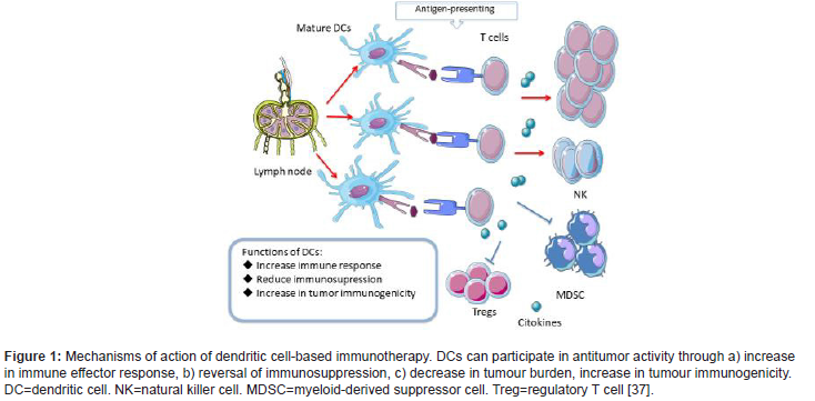
DCs overexpress these co-stimulatory molecules only if they recognise signals that indicate that the cell is under stress, such as the presence of inflammatory cytokines, high levels of pathogens, and prostaglandins. The inflammatory cytokines that play a critical role in antitumour activity are tumor necrosis factor α (TNF-α) and IFN-γ [32]. In contrast, TGF-β favours tumour progression and the development of metastases [32,33]. IL-15 is the most important inflammatory cytokine in cell activation, facilitating the survival of memory CD8-α T cells. IL-15 activated dendritic cells present antitumour potential, which has been observed in various breast cancer cell lines in vitro [34,35]. If the antigen is recognised without binding to a Toll-like receptor, the DCs will remain immature. In this case, the T cells will not be activated, and there may be a proliferation of regulatory T cells (Tregs) [9]. DCs, thus, may induce a lack of response (or anergy), through the expansion of Tregs, negatively regulating the immune response [31,36]. Figure 1 reflects the mechanisms by which dendritic cells participate in antitumor activity (Figure 1).
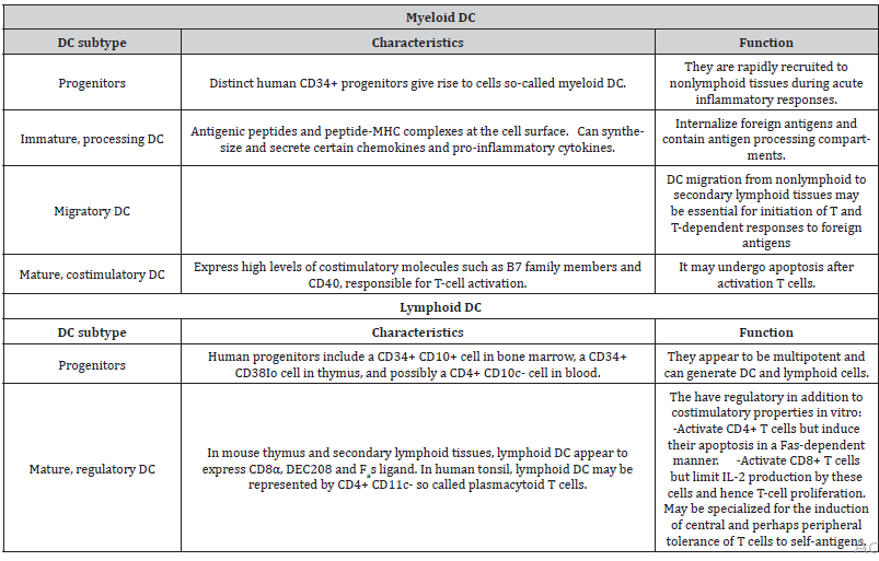
Various subtypes of DCs can be found in peripheral blood, including conventional dendritic cells (cDC) CD11C+, also termed myeloid dendritic cells (mDC), consisting of CD1C+ (mDC1) or CD141+ (mDC2) cells, and plasmacytoid dendritic cells (pDC) CD11C-, composed of CD123+ or CD303+ cells [38-41]. Table 1 shows the different subtypes of dendritic cells. mDC play a fundamental role in pathogen detection, in the presentation of antigens to T lymphocytes and in the specific stimulation of CD4 + and CD8+ T cells. pDC, on the other hand, exert strong antiviral activity through the production of IFN-1 and contribute to the antitumour response through a synergistic interaction with the mDC [42]. One type of partially mature dendritic cells is formed from human CD14+ monocytes in the presence of GM-CSF and IFN-α (IFN-DC). These cells favour the Th1-type cellular response and stimulate the immune response of CD8+ T cells [43-46] (Table 1).
Table 1: Dendritic cell subsets [47].
Role of Dendritic Cells in Lymphomas
Many studies have corroborated the potential of DCs for use as prognostic biomarkers in lymphoproliferative syndromes, or in some cases even for supporting the diagnosis. For example, Fiore et al. (2006) analysed DCs in 55 patients with non-Hodgkin lymphoma (NHL) and in 33 reactive lymph nodes, observing that the presence of these cells in the reactive lymph nodes was greater than in the cases of NHL, while the pDC/mDC ratio was similar. The conclusion drawn was that the presence of DCs crucially induces a specific antitumour immune response. The lower presence of DCs in patients with NHL explains the decreased expression of CD62L and CCR7 (dendritic cell receptors). With NHL, phenotypic alteration and a decreased presence of DCs in the tumour infiltrate may contribute to the loss of antitumour immune control in these patients [48]; this outcome has been documented in a variety of haematologic malignancies, including chronic lymphocytic leukaemia, hairy cell leukaemia [49], acute lymphoblastic leukaemia [50], multiple myeloma [51,52], chronic myeloid leukaemia [53-56], classical Hodgkin lymphoma (cHL) [56] and other cancers [57], as well as inflammatory disorders such as systemic lupus erythematosus [58] and uveitis [59]. This immunosuppressive state of the tumour microenvironment could be secondary to the metabolic stress, hipoxia and cytokine secretion that can negatively affect DCs [57]. cHL is a monoclonal lymphoid neoplasm composed of mononuclear Hodgkin cells and multinucleated Reed-Sternberg cells (HRS) embedded in an overwhelming mix of inflammatory and immune cells that contribute to tumour expansion by providing survival signals and promoting immune escape [60,61]. The significant impact of the inflammatory microenvironment in cHL motivated a study of the role of DCs in 52 patients diagnosed with de novo cHL, of whom 50 were treated with the ABVD polychemotherapy regimen (doxorubicin, bleomycin, vinblastine, dacarbazine). The count of all DCs subtypes was lower in patients with cHL compared to healthy controls. Moreover, the mDC count was lower in patients with advanced stage disease compared to those with an earlier stage, both for mDC1 and for mDC2. In addition, the mDC2 count decreased in cases with bulky disease and extranodal disease. These data suggest that a lower presence of mDC2 in the tumor microenvironment is related to a more advanced stage. The preeminent role of mDC2 in antigen presentation and in the induction of antitumour immunity, with respect to other DCs subtypes [62–64] could explain these facts [62–64]. Low levels of mDC1, mDC2 and pDC have also been reported in patients with B symptoms. It is not clear why cHL is characterized by a lower presence of DCs in the tumor microenvironment and why an increase is observed during chemotherapy treatment [56].
In the case of follicular lymphoma (FL), a better understanding of the molecular events responsible for differences in patients’ survival could facilitate a more accurate method for stratifying risk, guiding treatment and suggesting new therapeutic approaches. This question is particularly important because prognostic models based on the clinical variables of routine clinical practice are not useful in determining the best initial treatment. From a cohort of 191 biopsies of treatment-naive FL patients, gene expression profiles were studied in a training cohort of 95 samples and validated in an independent cohort of 96 samples [65]. Two gene expression signatures were obtained (immune-response 1 and 2) capable of predicting survival at the start of treatment. These signatures were composed of genes related to the expression of non-malignant immune cells present in the tumour. The binary model obtained from the joint consideration of these two signatures was a better predictor of survival than one obtained from either signature alone. The survival results were grouped into four quartiles, with the following median periods of survival: quartile 1, 13.6 years; quartile 2, 11.1 years; quartile 3, 10.8 years; quartile 4, 3.9 years. The “immune response 1” signature included genes encoding T cell markers (CD7, CD8B1, ITK, LEF1 and STAT4) and also genes that are highly expressed in macrophages (ACTN1 and TNFSF13B). The “immune-response 2” signature, on the other hand, included genes expressed mainly in macrophages, DCs or both (TLR5, FCGR1A, SEPT10, LGMN and C3AR1) [65-71].
These findings suggest that the survival of patients with FL is correlated with the molecular profile of non-malignant immune cells present in the tumour at diagnosis (including DCs). This understanding provides us with a tool to investigate different aspects of the immune response in FL that could positively or negatively influence the evolution of the disease. The identification of genetic signatures related to the immune response might constitute a valuable basis for studying biomarkers related to different subpopulations of immune cells that could promote or hinder the survival of malignant lymphoid cell clones [65]. Primary cutaneous marginal zone B-cell lymphoma (PCMZL) is an indolent lymphoma with a five-year survival of 95-98% [72,73]. Histologically, it is composed of a proliferation of small, centrocyte-like lymphocytes, monoclonal plasma cells, reactive germ centres and small T cells [74]. It presents multiple clinical pathological characteristics that are common to cutaneous Bcell pseudolymphomas (CBCL). However, PCMZL can present high levels of CD123+ pDC in the tumour (100% of cases), unlike CBCL, where these infiltrates are much less extensive (64%). They are even less frequent in cutaneous centre follicular lymphomas (13%) and are totally absent from primary cutaneous diffuse large B-cell lymphoma, leg type. The presence of high intratumoural levels of pDC supports the diagnosis of PCMZL. Furthermore, the pDC in PCMZL lack the expression of CD68 whereas in the absence of cutaneous lymphomas the pDC express both CD123 and CD68 [75]. The presence of high levels of pDC in this lymphoma may explain the large numbers of T cells generally found there, which can account for up to 60% of the tumour cell component [76]. These data support the hypothesis that pDC and T and B cells are necessary for the development of PCMZL, especially pDC (which is present from very early lesions). Therefore, the use of treatment strategies targeting antigen-driven T-cells or even the pDC responsible for the stimulation of B cells could be very effective in the treatment of PCMZL [77].
The Role of Dendritic Cells in Treating Lymphoproliferative Syndromes
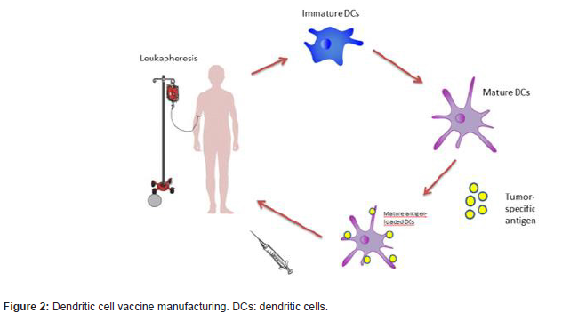
Recent studies have highlighted the potential role of DCs in the treatment of patients with malignant tumours. These cells can be loaded with specific tumour antigens that trigger an antitumour response by specific T cells (Figure 2). To achieve this, the dendritic cells are cultured with tumour peptides or proteins, or the tumour antigen is expressed through the inoculation of adenoviruses that transfer genetic material to encode these specific tumour antigens [9]. Dendritics cells are obtained from peripheral blood. To mobilize dendritic cells to the peripheral blood it is recommended to use GM-CSF. When the regimen includes GM-CSF, mobilization of DCs to peripheral blood is 3-5 times more effective [78]; this contrasts with NK cell mobilisation, which is just as efficient regardless of whether G-CSF, GM-CSF or GM-CSF followed by G-CSF is used [79]. In this respect, Avigan et al. (1999) [78] obtained a greater quantity of DCs when the peripheral blood DCs precursors in patients were mobilised with GM-CSF + G-CSF, compared with the use of G-CSF alone (Figure 2).
However, there are some limitations to the use of DCs as therapeutics agents, especially due to the scarcity of certain subgroups of DCs in peripheral blood. In consequence, most dendritic cell-based vaccines employ mDC differentiated from circulating monocytes in peripheral blood, due to the relative ease with which these monocytes can be obtained [80]. DCs differentiation can be induced from monocytes cultured with IL4 and GM-CSF [81] or from CD34 cells [82] cultured with a wide variety of growth factors such as GM-CSF + TNF-α, GM-CSF + SCF and IL4, TNF-α and ligand flt3 with TGF-β [51,83,84].
Most studies of dendritic cell-based vaccines in lymphoproliferative syndromes have been performed in patients with FL. This is the most common form of indolent lymphoma (accounting for 70% of the total) and is usually diagnosed at advanced stages of the disease [85]. The standard treatment is a combination of rituximab with chemotherapy followed by rituximab maintenance. Although most patients will relapse, over 80% of patients obtain long-lasting remission, this approach is complex, expensive and heightens both acute and long-term toxicity [86,87]. n general, subsequent lines of treatment are progressively less effective; thus, toxicity increases, and the patient’s quality of life deteriorates [88,89]. When relapses occur, most patients experience a greater risk of transformation and treatment-induced toxicity [85,88,90]. Therefore, effective long-term management is necessary. Recent studies have shown that dendritic cell-based treatment enables a specific immune response associated with sustained tumour regression in some patients with low tumour burden FL. The objective response rate with sustained remission (33-50%), as shown in Table 2, is clearly greater than that obtained in studies of solid tumours, which corroborates the sensitivity of FL to this treatment strategy [80] (Table 2).
Table 2:Main clinical trials with dendritic cell-based vaccines in follicular lymphoma.
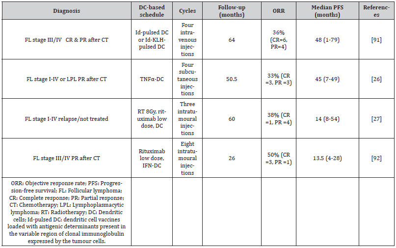
Timmerman et al. (2002) [91] studied 35 patients with relapsed or refractory FL, treated after chemotherapy with dendritic cell vaccines loaded with antigenic determinants present in the variable region of clonal immunoglobulin expressed by the tumour cells (Id-pulsed DC vaccination). Of the 18 patients with residual disease at the time of the vaccine, four had tumour regression and 16 remained progression-free at a median 43 months after chemotherapy. Six patients with progression after this vaccine received another with the Id- KLH protein (idiotype protein bound to the highly immunogenic transporter protein keyhole limpet hemocyanin). Tumour regression took place in three patients (two with complete response and one with partial response). These data suggest that Id-pulsed DC vaccination can induce a humoral and cellular immune response with lasting tumour regression, and that subsequent vaccination with ID-KLH can induce tumour regression despite apparent resistance to the initial Id-pulsed DC vaccination [91]. In another study, 25 patients in initial remission after cytoreductive chemotherapy received three monthly infusions of Idpulsed DC followed by a fourth, 2-6 months later. After vaccination, 23 of the 25 patients developed a T cell response and 36% of those with an evaluable tumour had an objective response to the vaccine [91]. Bendandi et al. (1999) [93], too, obtained excellent diseasefree survival, in 18 of 20 patients with FL treated with Id-KLH DC + GM-CSF, although this study was limited to patients who achieved a complete response after chemotherapy. These findings suggest that the response to Id-pulsed DC is more frequently obtained in patients presenting a complete response when the vaccine is received.
A second generation of vaccines combines Id-pulsed DC with chemokines, enabling the DCs to capture the antigen. Sakamaki et al. (2014) associated this vaccination strategy with lenalidomide, an immunomodulatory drug that reduces the number of systemic myeloid derived suppressor cells (MDSCs), and Tregs, thus improving tumour immunosuppression [94]. This approach is consistent with previous studies showing that lenalidomide inhibits the proliferation and function of Tregs [95,96] and increases the quantity of NK cells [97]. In a study by Di Nicola et al. (2020), 33% (6/18) of heavily pretreated and/or refractory patients with follicular and lymphoplasmacytic lymphoma obtained lasting clinical remission after repeated four-dose dendritic cell vaccination [26]. Another 33% remained stable, during a median follow-up of 50 months. Among the responders, NK cells were activated, which correlated with a reduction in the frequency of Tregs. Combining anti-tumour vaccines with immunomodulatory drugs is considered a very promising approach to boost the immunity of tumour-specific T cells and eradicate residual malignant cells. Dendritic cell-based strategies can safely be combined with radiation therapy, immune checkpoint inhibitors, immunomodulators and metronomic chemotherapy. However, clinical trials are still at a very early stage.
The optimal conditions for the culture of effective mDC is unknown, but a quick and easy method is to perform the culture in the presence of IFN-α and GM-CSF, which obtains partially mature and highly active mDC from peripheral blood monocytes (IFNDC) [46,98]. IFN, which presents as a signal of damage in cases of infection/inflammation, can initiate a cascade of differentiation of monocytes into dendritic cells, resulting in the activation of NK cells, the generation of a Th1 helper T lymphocyte immune response and the induction of a CD8+ T cell response towards both pathogens and neoplastic cells. The use of IFN-DC enables antitumour vaccines to be created endogenously [27,99,100]. In the phase I clinical trial of Cox et al. (2019), eight patients with relapsed or refractory FL were sequentially treated with the intranodal administration of low-dose rituximab followed by IFN-DC not loaded with tumour antigens. In this process, the first four cycles were administered every two weeks and the remaining four cycles, at four-week intervals. GMCSF and IFN-α facilitated the differentiation of blood monocytes to DCs, thus incorporating the tumour antigen and activating the CD8+ T cells faster than with conventional DCs and enhancing the antitumour immune response [45,101-103]. Among the eight patients, the overall response rate was 50% (4/8). Three patients obtained a complete metabolic response, and two maintained a remission (median follow-up 26 months, range 11-47). These results demonstrate the induction of a systemic response based on an abscopal effect and highlight the effectiveness of IFN-DC treatment in inducing a long-lasting clinical response, together with the ability to promote the stimulation of peripheral tumourspecific T cells, through the endogenous formation of an effective antitumour vaccine [92].
As concerns the association of radiotherapy and dendritic cell-based vaccination, radiotherapy promotes the cellular and molecular reprogramming of the tumour microenvironment, which in turn favours the maturation of DCs into effective antigenpresenting cells [104]. Kolstad et al. (2015) [27] studied fourteen patients with stage III/IV FL, in first-line treatment or in relapse, who were treated with 8 Gy radiotherapy in a specific lymph node región followed by the intranodal injection of rituximab at low doses (5mg) and cDC + GM-CSF. The treatment was repeated three times (weeks 1, 3 and 5), in a different node each time. A strong correlation was observed between the reduction in tumour area and the magnitude of the antitumour response of the CD8+ T cells generated. Five of the 14 patients (36%) obtained a clinical response, regardless of the tumour load, and the abscopal effect associated with a specific T-cell response in peripheral blood was present in 50% of the patients. However, the median time to progression was similar for all patients, irrespective of the antitumour immune response [27]. Although this is a controversial aspect of the study, it is unlikely that the systemic effect observed was exclusively due to the administration of intranodal rituximab, since the dose used was 100 times lower than that normally supplied intravenously when rituximab is used in monotherapy.
Conclusions
Although it is difficult to draw firm conclusions from the limited number of clinical trials conducted (which are heterogeneous and at very early stages of research), it is apparent that dendritic cell vaccines have achieved effective T-cell responses against tumour antigens, with promising clinical results, with an objective response rate exceeding 30% in patients with lymphoproliferative syndromes, a rate significantly higher than that obtained with solid tumours (0-16%). In addition, expectations have been raised by the association of these vaccines with existing immunomodular ones in the treatment of low-grade lymphomas. Future research efforts should be aimed at extending our understanding of the complex role played by the immune response in tumour development and progression, and at developing new strategies to overcome the inhibition of the immune response by tumour cells.
Acknowledgement
None.
Disclosures
Lectures and advisory fees from Takeda and BMS. Investigational fee from Celgene.
Conflict of Interest
No conflict of interest.
References
- Hart DNJ (1997) Dendritic Cells: Unique Leukocyte Populations Which Control the Primary Immune Response. Blood, 90(9): 3245-3287.
- Mittal S, Marshall NA, Barker RN, Vickers MA (2008) Immunomodulation against leukemias and lymphomas: A realistic future treatment? Critical Reviews in Oncology/Hematology. febrero de 65(2): 101-108.
- Rooney CM, Smith CA, Ng CYC, Loftin SK, Sixbey JW, et al. (1998) Infusion of Cytotoxic T Cells for the Prevention and Treatment of Epstein-Barr Virus–Induced Lymphoma in Allogeneic Transplant Recipients. Blood 92(5): 1549-55.
- Santos PM, Butterfield LH (2018) Dendritic Cell–Based Cancer Vaccines. JI 200(2): 443-9.
- Steinman RM (2012) Decisions About Dendritic Cells: Past, Present, and Future. Annu Rev Immunol 30(1): 1-22.
- Steinman RM, Cohn ZA (1973) Identification of a Novel Cell Type in Peripheral Lymphoid Organs of Mice. Journal of Experimental Medicine 137(5): 1142-62.
- Banchereau J, Briere F, Caux C, Davoust J, Lebecque S, et al. (2000) Immunobiology of Dendritic Cells. Annu Rev Immunol 18(1): 767-811.
- Chan CW, Housseau F (2008) The ‘kiss of death’ by dendritic cells to cancer cells. Cell Death Differ 15(1): 58-69.
- Davison GM (2010) Dendritic cells, T-cells and their possible role in the treatment of leukaemia and lymphoma. Transfusion and Apheresis Science 42(2): 189-192.
- Scott DW, Gascoyne RD (2014) The tumour microenvironment in B cell lymphomas. Nat Rev Cancer 14(8): 517-534.
- Aldinucci D, Gloghini A, Pinto A, De Filippi R, Carbone A (2010) The classical Hodgkin’s lymphoma microenvironment and its role in promoting tumour growth and immune escape: Hodgkin’s lymphoma microenvironment and promotion of tumour growth and immune escape. J Pathol 221(3): 248-263.
- Xu J, Chakrabarti AK, Tan JL, Ge L, Gambotto A, et al. (2007) Essential role of the TNF-TNFR2 cognate interaction in mouse dendritic cell–natural killer cell crosstalk. Blood, 109(8): 3333-3341.
- Goodier MR, Londei M (2000) Lipopolysaccharide Stimulates the Proliferation of Human CD56 + CD3 − NK Cells: A Regulatory Role of Monocytes and IL-10. J Immunol 165(1): 139-147.
- Moretta A (2002) Natural killer cells and dendritic cells: rendezvous in abused tissues. Nat Rev Immunol 2(12): 957-965.
- Ruggeri L (2002) Effectiveness of Donor Natural Killer Cell Alloreactivity in Mismatched Hematopoietic Transplants. Science 295(5562): 2097-2100.
- Farag SS, Fehniger TA, Ruggeri L, Velardi A, Caligiuri MA (2002) Natural killer cell receptors: new biology and insights into the graft-versus-leukemia effect. Blood. 100(6): 1935-1947.
- Kapsenberg ML (2003) Dendritic-cell control of pathogen-driven T-cell polarization. Nat Rev Immunol 3(12): 984-993.
- Qi C-J, Ning Y-L, Han Y-S, Min H-Y, Ye H, et al. (2012) Autologous dendritic cell vaccine for estrogen receptor (ER)/progestin receptor (PR) double-negative breast cancer. Cancer Immunol Immunother. 61(9): 1415-1424.
- Butterfield LH, Comin-Anduix B, Vujanovic L, Lee Y, Dissette VB, et al. (2008) Adenovirus MART-1– engineered Autologous Dendritic Cell Vaccine for Metastatic Melanoma. Journal of Immunotherapy 31(3): 294-309.
- Vujanovic L, Whiteside TL, Potter DM, Chu J, Ferrone S, et al. (2009) Regulation of antigen presentation machinery in human dendritic cells by recombinant adenovirus. Cancer Immunol Immunother 58(1): 121-133.
- Evdokimova VN, Liu Y, Potter DM, Butterfield LH (2007) AFP-specific CD4+ Helper T-cell Responses in Healthy Donors and HCC Patients. Journal of Immunotherapy 30(4): 425-437.
- Fernandez NC, Lozier A, Flament C, Ricciardi-Castagnoli P, Bellet D, et al. (1999) Dendritic cells directly trigger NK cell functions: Cross-talk relevant in innate anti-tumor immune responses in vivo. Nat Med 5(4): 405-411.
- Ferlazzo G, Tsang ML, Moretta L, Melioli G, Steinman RM, et al. (2002) Human Dendritic Cells Activate Resting Natural Killer (NK) Cells and Are Recognized via the NKp30 Receptor by Activated NK Cells. Journal of Experimental Medicine 195(3): 343-351.
- Piccioli D, Sbrana S, Melandri E, Valiante NM (2002) Contact-dependent Stimulation and Inhibition of Dendritic Cells by Natural Killer Cells. Journal of Experimental Medicine 2002 195(3): 335-341.
- Weinstock M, Rosenblatt J, Avigan D (2017) Dendritic Cell Therapies for Hematologic Malignancies. Molecular Therapy - Methods & Clinical Development 5: 66-75.
- Di Nicola M, Zappasodi R, Carlo-Stella C, Mortarini R, Pupa SM, et al. (2009) Vaccination with autologous tumor-loaded dendritic cells induces clinical and immunologic responses in indolent B-cell lymphoma patients with relapsed and measurable disease: a pilot study. Blood 113(1): 18-27.
- Kolstad A, Kumari S, Walczak M, Madsbu U, Hagtvedt T, et al. (2015) Sequential intranodal immunotherapy induces antitumor immunity and correlated regression of disseminated follicular lymphoma. Blood 125(1): 82-89.
- Garg AD, More S, Rufo N, Mece O, Sassano ML, et al. (2017) Trial watch: Immunogenic cell death induction by anticancer chemotherapeutics. OncoImmunology 6(12): e1386829.
- Garg AD, Coulie PG, Van den Eynde BJ, Agostinis P (2017) Integrating Next-Generation Dendritic Cell Vaccines into the Current Cancer Immunotherapy Landscape. Trends in Immunology 38(8): 577-593.
- van Kooyk Y, Geijtenbeek TBH (2003) DC-SIGN: escape mechanism for pathogens. Nat Rev Immunol 3(9): 697-709.
- Nencioni A, Grünebach F, Schmidt SM, Müller MR, Boy D, et al. (2008) The use of dendritic cells in cancer immunotherapy. Critical Reviews in Oncology/Hematology. 65(3): 191-199.
- Salomon BL, Leclerc M, Tosello J, Ronin E, Piaggio E, et al. (2018) Tumor Necrosis Factor α and Regulatory T Cells in Oncoimmunology. Front Immunol 9: 444.
- Szondy Z, Sarang Z, Kiss B, Garabuczi É, Köröskényi K (2017) Anti-inflammatory Mechanisms Triggered by Apoptotic Cells during Their Clearance. Front Immunol 8: 909.
- Waldmann TA (2006) The biology of interleukin-2 and interleukin-15: implications for cancer therapy and vaccine design. Nat Rev Immunol 6(8): 595-601.
- Zhang X, Sun S, Hwang I, Tough DF, Sprent J (1998) Potent and Selective Stimulation of Memory-Phenotype CD8+ T Cells In Vivo by IL-15. Immunity 8(5): 591-599.
- Coquerelle C, Moser M (2008) Are dendritic cells central to regulatory T cell function? Immunology Letters 119(1-2): 12-16.
- Anguille S, Smits EL, Lion E, van Tendeloo VF, Berneman ZN (2014) Clinical use of dendritic cells for cancer therapy. The Lancet Oncology 15(7): e257-67.
- Rovati B, Mariucci S, Manzoni M, Bencardino K, Danova M (2009) Flow cytometric detection of circulating dendritic cells in healthy subjects. Eur J Histochem 52(1): 45.
- Ziegler-Heitbrock L, Ancuta P, Crowe S, Dalod M, Grau V, et al. (2010) Nomenclature of monocytes and dendritic cells in blood. Blood 116(16): e74-80.
- Reizis B, Bunin A, Ghosh HS, Lewis KL, Sisirak V (2011) Plasmacytoid Dendritic Cells: Recent Progress and Open Questions. Annu Rev Immunol 29(1): 163-183.
- Tel J, Smits EL, Anguille S, Joshi RN, Figdor CG, et al. (2012) Human plasmacytoid dendritic cells are equipped with antigen-presenting and tumoricidal capacities. Blood 120(19): 3936-3944.
- Segura E, Villadangos JA (2009) Antigen presentation by dendritic cells in vivo. Current Opinion in Immunology 21(1): 105-110.
- Lapenta C, Santini SM, Spada M, Donati S, Urbani F, et al. (2006) IFN-α-conditioned dendritic cells are highly efficient in inducing cross-priming CD8+ T cells against exogenous viral antigens. Eur J Immunol 36(8): 2046-2060.
- Parlato S, Santini SM, Lapenta C, Di Pucchio T, Logozzi M, et al. (2001) Expression of CCR-7, MIP-3β, and Th-1 chemokines in type I IFN-induced monocyte-derived dendritic cells: importance for the rapid acquisition of potent migratory and functional activities. Blood 98(10): 3022-3029.
- Lapenta C, Santini SM, Logozzi M, Spada M, Andreotti M, et al. (2003) Potent Immune Response against HIV-1 and Protection from Virus Challenge in hu-PBL-SCID Mice Immunized with Inactivated Virus-pulsed Dendritic Cells Generated in the Presence of IFN-α. Journal of Experimental Medicine 198(2): 361-367.
- Santini SM, Lapenta C, Belardelli F (2005) Type I Interferons as Regulators of the Differentiation/Activation of Human Dendritic Cells. En: Carr DJJ, editor. Interferon Methods and Protocols [Internet]. Totowa, NJ: Humana Press p.167-181.
- Austyn JM (1998) Dendritic cells: Current Opinion in Hematology. 5(1): 3-15.
- Fiore F, Von Bergwelt-Baildon MS, Drebber U, Beyer M, Popov A, et al. (2006) Dendritic cells are significantly reduced in non-Hodgkin’s lymphoma and express less CCR7 and CD62L. Leukemia & Lymphoma 47(4): 613-622.
- Bourguin-Plonquet A, Rouard H, Roudot-Thoraval F, Bellanger C, Marquet J, et al. (2002) Severe decrease in peripheral blood dendritic cells in hairy cell leukaemia: Short Report. British Journal of Haematology 116(3): 595-597.
- Mami NB, Mohty M, Chambost H, Gaugler B, Olive D (2004) Blood dendritic cells in patients with acute lymphoblastic leukaemia: Short Report. British Journal of Haematology 126(1): 77-80.
- Ratta M, Fagnoni F, Curti A, Vescovini R, Sansoni P, et al. (2002) Dendritic cells are functionally defective in multiple myeloma: the role of interleukin-6. Blood 100(1): 230-237.
- Brimnes MK, Svane IM, Johnsen HE (2006) Impaired functionality and phenotypic profile of dendritic cells from patients with multiple myeloma. Clin Exp Immunol 144(1): 76-84.
- Mohty M, Isnardon D, Vey N, Brière F, Blaise D, et al. (2002) Low blood dendritic cells in chronic myeloid leukaemia patients correlates with loss of CD34 + /CD38 - primitive haematopoietic progenitors: Short Report. British Journal of Haematology 119(1): 115-118.
- Dong R, Cwynarski K, Entwistle A, Marelli-Berg F, Dazzi F, et al. (2003) Dendritic cells from CML patients have altered actin organization, reduced antigen processing, and impaired migration. Blood 101(9): 3560-3567.
- Boissel N, Rousselot P, Raffoux E, Cayuela J-M, Maarek O, et al. (2004) Defective blood dendritic cells in chronic myeloid leukemia correlate with high plasmatic VEGF and are not normalized by imatinib mesylate. Leukemia 18(10): 1656-1661.
- Galati D, Zanotta S, Corazzelli G, Bruzzese D, Capobianco G, et al. (2019) Circulating dendritic cells deficiencies as a new biomarker in classical Hodgkin lymphoma. Br J Haematol 184(4): 594-604.
- Saxena M, Bhardwaj N (2018) Re-Emergence of Dendritic Cell Vaccines for Cancer Treatment. Trends in Cancer 4(2): 119-137.
- Klarquist J, Zhou Z, Shen N, Janssen EM (2016) Dendritic Cells in Systemic Lupus Erythematosus: From Pathogenic Players to Therapeutic Tools. Mediators of Inflammation 2016: 1-12.
- Verhagen FH, Hiddingh S, Rijken R, Pandit A, Leijten E, et al. (2018) High-Dimensional Profiling Reveals Heterogeneity of the Th17 Subset and Its Association With Systemic Immunomodulatory Treatment in Non-infectious Uveitis. Front Immunol 9: 2519.
- Siegel RL, Miller KD, Jemal A (2015) Cancer statistics, 2015: Cancer Statistics, 2015. CA: A Cancer Journal for Clinicians 65(1): 5-29.
- Steidl C, Lee T, Shah SP, Farinha P, Han G, et al. (2010) Tumor-Associated Macrophages and Survival in Classic Hodgkin’s Lymphoma. N Engl J Med 362(10): 875-885.
- Bachem A, Güttler S, Hartung E, Ebstein F, Schaefer M, et al. (2010) Superior antigen cross-presentation and XCR1 expression define human CD11c+CD141+ cells as homologues of mouse CD8+ dendritic cells. Journal of Experimental Medicine 207(6): 1273-1281.
- Jongbloed SL, Kassianos AJ, McDonald KJ, Clark GJ, Ju X, et al. (2010) Human CD141+ (BDCA-3)+ dendritic cells (DCs) represent a unique myeloid DC subset that cross-presents necrotic cell antigens. Journal of Experimental Medicine 207(6): 1247-1260.
- Roberts EW, Broz ML, Binnewies M, Headley MB, Nelson AE, et al. (2016) Critical Role for CD103 + /CD141 + Dendritic Cells Bearing CCR7 for Tumor Antigen Trafficking and Priming of T Cell Immunity in Melanoma. Cancer Cell 30(2): 324-336.
- Dave SS, Wright G, Tan B, Rosenwald A, Gascoyne RD, et al. (2004) Prediction of Survival in Follicular Lymphoma Based on Molecular Features of Tumor-Infiltrating Immune Cells. N Engl J Med 351(21): 2159-2169.
- Li DN, Matthews SP, Antoniou AN, Mazzeo D, Watts C (2003) Multistep Autoactivation of Asparaginyl Endopeptidase in Vitro and in Vivo. Journal of Biological Chemistry 278(40): 38980-38990.
- Sui L, Zhang W, Liu Q, Chen T, Li N, et al. (2003) Cloning and functional characterization of human septin 10, a novel member of septin family cloned from dendritic cells. Biochemical and Biophysical Research Communications 304(2): 393-398.
- Hayashi F, Smith KD, Ozinsky A, Hawn TR, Yi EC, et al. (2001) The innate immune response to bacterial flagellin is mediated by Toll-like receptor 5. Nature 410(6832): 1099-1103.
- Muzio M, Bosisio D, Polentarutti N, D’amico G, Stoppacciaro A, et al. (2000) Differential Expression and Regulation of Toll-Like Receptors (TLR) in Human Leukocytes: Selective Expression of TLR3 in Dendritic Cells. J Immunol 164(11): 5998-6004.
- Roglić A, Prossnitz ER, Cavanagh SL, Pan Z, Zou A, et al. (1996) cDNA cloning of a novel G protein-coupled receptor with a large extracellular loop structure. Biochimica et Biophysica Acta (BBA) - Gene Structure and Expression 1305(1-2): 39-43.
- Ames RS, Li Y, Sarau HM, Nuthulaganti P, Foley JJ, et al. (1996) Molecular Cloning and Characterization of the Human Anaphylatoxin C3a Receptor. Journal of Biological Chemistry 271(34): 20231-20234.
- Hoefnagel JJ (2005) Primary Cutaneous Marginal Zone B-Cell Lymphoma: Clinical and Therapeutic Features in 50 Cases. Arch Dermatol. 141(9): 1139.
- Golling P, Cozzio A, Dummer R, French L, Kempf W (2008) Primary cutaneous B-cell lymphomas – Clinicopathological, prognostic and therapeutic characterisation of 54 cases according to the WHO-EORTC classification and the ISCL/EORTC TNM classification system for primary cutaneous lymphomas other than mycosis fungoides and Sézary syndrome. Leukemia & Lymphoma 49(6): 1094-1103.
- Takino H, Li C, Hu S, Kuo T-T, Geissinger E, et al. (2008) Primary cutaneous marginal zone B-cell lymphoma: a molecular and clinicopathological study of cases from Asia, Germany, and the United States. Mod Pathol 21(12): 1517-1526.
- Liu YJ (2005) IPC: Professional Type 1 Interferon-Producing Cells and Plasmacytoid Dendritic Cell Precursors. Annu Rev Immunol 23(1): 275-306.
- Servitje O, Gallardo F, Estrach T, Pujol RM, Blanco A, et al. (2002) Primary cutaneous marginal zone B-cell lymphoma: a clinical, histopathological, immunophenotypic and molecular genetic study of 22 cases. Br J Dermatol 147(6): 1147-1158.
- Albini A, Sporn MB (2007) The tumour microenvironment as a target for chemoprevention. Nat Rev Cancer 7(2): 139-147.
- Avigan D, Wu Z, Gong J, Joyce R, Levine J, et al. (1999) Selective in vivo mobilization with granulocyte macrophage colony-stimulating factor (GM-CSF)/granulocyte-CSF as compared to G-CSF alone of dendritic cell progenitors from peripheral blood progenitor cells in patients with advanced breast cancer undergoing autologous transplantation. Clin Cancer Res 5(10): 2735-2741.
- Gazitt Y, Shaughnessy P, Devore P (2001) Mobilization of Dendritic Cells and NK Cells in Non-Hodgkin’s Lymphoma Patients Mobilized with Different Growth Factors. Journal of Hematotherapy & Stem Cell Research 10(1): 177-186.
- Cox MC, Lapenta C, Santini SM (2020) Advances and perspectives of dendritic cell-based active immunotherapies in follicular lymphoma. Cancer Immunol Immunother 69(6): 913-25.
- Kiertscher SM, Roth MD (1996) Human CD14 + leukocytes acquire the phenotype and function of antigen-presenting dendritic cells when cultured in GM-CSF and IL-4. J Leukoc Biol 59(2): 208-218.
- Steinman RM (1991) The Dendritic Cell System and its Role in Immunogenicity. Annu Rev Immunol 9(1): 271-296.
- Strunk D, Rappersberger K, Egger C, Strobl H, Kromer E, et al. (1996) Generation of human dendritic cells/Langerhans cells from circulating CD34+ hematopoietic progenitor cells. Blood 87(4): 1292-1302.
- Caux C, Dezutter-Dambuyant C, Schmitt D, Banchereau J (1992) GM-CSF and TNF-α cooperate in the generation of dendritic Langerhans cells. Nature. 360(6401): 258-261.
- Freedman A (2018) Follicular lymphoma: 2018 update on diagnosis and management. Am J Hematol 93(2): 296-305.
- Bachy E, Seymour JF, Feugier P, Offner F, López-Guillermo A, et al. (2019) Sustained Progression-Free Survival Benefit of Rituximab Maintenance in Patients With Follicular Lymphoma: Long-Term Results of the PRIMA Study. JCO 37(31): 2815-2824.
- Hiddemann W, Barbui AM, Canales MA, Cannell PK, Collins GP, et al. (2018) Immunochemotherapy With Obinutuzumab or Rituximab for Previously Untreated Follicular Lymphoma in the GALLIUM Study: Influence of Chemotherapy on Efficacy and Safety. JCO 36(23): 2395-2404.
- Casulo C, Nastoupil L, Fowler NH, Friedberg JW, Flowers CR (2017) Unmet needs in the first-line treatment of follicular lymphoma. Annals of Oncology 28(9): 2094-106.
- Maddocks K, Barr PM, Cheson BD, Little RF, Baizer L, et al. (2017) Recommendations for Clinical Trial Development in Follicular Lymphoma. JNCI J Natl Cancer Inst. 109(3): djw255.
- Danese MD, Reyes CM, Gleeson ML, Halperin M, Skettino SL, et al. (2016) Estimating the Population Benefits and Costs of Rituximab Therapy in the United States from 1998 to 2013 Using Real-World Data. Medical Care 54(4): 343-9.
- Timmerman JM, Czerwinski DK, Davis TA, Hsu FJ, Benike C, et al. (2002) Idiotype-pulsed dendritic cell vaccination for B-cell lymphoma: clinical and immune responses in 35 patients. Blood 99(5): 1517-26.
- Cox MC, Castiello L, Mattei M, Santodonato L, D’Agostino G, et al. (2019) Clinical and Antitumor Immune Responses in Relapsed/Refractory Follicular Lymphoma Patients after Intranodal Injections of IFNα-Dendritic Cells and Rituximab: a Phase I Clinical Trial. Clin Cancer Res 25(17): 5231-41.
- Bendandi M, Gocke CD, Kobrin CB, Benko FA, Sternas LA, et al. (1999) Complete molecular remissions induced by patient-specific vaccination plus granulocyte–monocyte colony-stimulating factor against lymphoma. Nat Med 5(10): 1171-7.
- Sakamaki I, Kwak LW, Cha S, Yi Q, Lerman B, et al. (2014) Lenalidomide enhances the protective effect of a therapeutic vaccine and reverses immune suppression in mice bearing established lymphomas. Leukemia 28(2): 329-37.
- Galustian C, Meyer B, Labarthe M-C, Dredge K, Klaschka D, et al. (2009) The anti-cancer agents lenalidomide and pomalidomide inhibit the proliferation and function of T regulatory cells. Cancer Immunol Immunother 58(7): 1033-45.
- Luptakova K, Rosenblatt J, Glotzbecker B, Mills H, Stroopinsky D, et al. (2013) Lenalidomide enhances anti-myeloma cellular immunity. Cancer Immunol Immunother 62(1): 39-49.
- Zhu D, Corral LG, Fleming YW, Stein B (2008) Immunomodulatory drugs Revlimid® (lenalidomide) and CC-4047 induce apoptosis of both hematological and solid tumor cells through NK cell activation. Cancer Immunol Immunother 57(12): 1849-59.
- Santini SM, Lapenta C, Santodonato L, D’Agostino G, Belardelli F, et al. (2009) IFN-alpha in the Generation of Dendritic Cells for Cancer Immunotherapy. In: Lombardi G, Riffo-Vasquez Y (Eds.) Dendritic Cells [Internet]. Berlin, Heidelberg: Springer Berlin Heidelberg pp.295-317.
- Subbiah V, Murthy R, Hong DS, Prins RM, Hosing C, et al. (2018) Cytokines Produced by Dendritic Cells Administered Intratumorally Correlate with Clinical Outcome in Patients with Diverse Cancers. Clin Cancer Res 24(16): 3845-3856.
- Rozera C, Cappellini GA, D’Agostino G, Santodonato L, Castiello L, et al. (2015) Intratumoral injection of IFN-alpha dendritic cells after dacarbazine activates anti-tumor immunity: results from a phase I trial in advanced melanoma. J Transl Med 13(1): 139.
- Lapenta C, Donati S, Spadaro F, Castaldo P, Belardelli F, et al. (2016) NK Cell Activation in the Antitumor Response Induced by IFN-α Dendritic Cells Loaded with Apoptotic Cells from Follicular Lymphoma Patients. JI 197(3): 795-806.
- Spadaro F, Lapenta C, Donati S, Abalsamo L, Barnaba V, et al. (2012) IFN-α enhances cross-presentation in human dendritic cells by modulating antigen survival, endocytic routing, and processing. Blood 119(6): 1407-1417.
- Santini SM, Lapenta C, Logozzi M, Parlato S, Spada M, et al. (2000) Type I Interferon as a Powerful Adjuvant for Monocyte-Derived Dendritic Cell Development and Activity in Vitro and in Hu-Pbl-Scid Mice. Journal of Experimental Medicine 191(10): 1777-1788.
- Demaria S, Bhardwaj N, McBride WH, Formenti SC (2005) Combining radiotherapy and immunotherapy: A revived partnership. International Journal of Radiation Oncology*Biology*Physics 63(3): 655-666.
-
Laura Galvez-Carvajal, Elísabeth Pérez-Ruiz, Silvia Sequero, Eduardo Zamorano and Antonio Rueda-Domínguez etc al. Biological and Therapeutic Role of Dendritic Cells in Lymphomas. Adv Can Res & Clinical Imag. 3(4): 2022. ACRCI.MS.ID.000567.
-
Tumour microenvironment, Dendritic cells, Lymphoma, Dendritic cell vaccines
-

This work is licensed under a Creative Commons Attribution-NonCommercial 4.0 International License.


