 Case Study
Lung Adenocarcinoma in Adolescence: A Case Study
Amir Rezaei1, Mahsa Rekabi1, Sharareh Seifi2, Mihan Pourabdollah1, Susan Rahimi3 and Sepideh Darougar4*
1Pediatric Respiratory Diseases Research Center, National Research Institute of Tuberculosis and Lung Diseases (NRITLD), Shahid Beheshti University of Medical Sciences, Tehran, Iran
2Research Center of Thoracic Oncology (RCTO), National Research Institute of Tuberculosis and Lung Diseases (NRITLD), Shahid Beheshti University of Medical Sciences, Tehran, Iran
3National Research Institute of Tuberculosis and Lung Diseases (NRITLD), Shahid Beheshti University of Medical Sciences, Tehran, Iran
*4Department of Pediatrics, Faculty of Medicine, Tehran Medical Sciences, Islamic Azad University, Tehran, Iran
Sepideh Darougar, Department of Pediatrics, Faculty of Medicine, Tehran Medical Sciences, Islamic Azad University, Tehran, Iran
Received Date: August 21, 2024; Published Date: September 03, 2024
Abstract
Lung cancer is the leading cause of cancer-related mortality among adults in the world, yet primary lung cancers are rare in children and adolescents. Childhood lung adenocarcinoma comprises approximately 15% of pulmonary tumors with an incidence of 0.2% of all childhood malignancies. This study is the report of a 16-year-old female with a fulminant course who was admitted to hospital with persistent non-resolving respiratory symptoms, initially mimicking influenza infection without a favorable response to outpatient treatments. On the chest x-ray, pleural effusion on both sides as well as pericardial effusion were seen and the high-resolution computed tomography (HR-CT) revealed diffuse parenchymal involvement, and lymphadenopathies along with necrotic changes. A left supraclavicular lymph node biopsy was performed, which was indicative of metastatic adenocarcinoma. After the initiation of chemotherapy, she developed respiratory distress and unconsciousness later and was transferred to ICU for further treatment. However, her situation did not improve, and unfortunately the pulmonary dysfunction progressed to a sudden arrest without a response to cardiopulmonary resuscitation. This patient is presented to conclude that since early detection may affect the outcome and survival rate, a high index of suspicion, as well as appropriate imaging studies, are important to diagnose the condition earlier and in lower stages, the results of which could be improving the quality of life.
Keywords: Lung adenocarcinoma; malignancy; adolescence; childhood
Introduction
Lung cancer is the leading cause of cancer-related mortality among adults in the world [1], yet primary lung cancers are rare in children and adolescents. According to a classification by WHO, malignant lung tumors of epithelial origin include non-small cell lung cancer, which is subdivided into adenocarcinoma, squamous cell carcinoma, and large-cell carcinoma. Other pulmonary carcinomas include small cell lung cancers or neuroendocrine tumors. Childhood lung adenocarcinoma comprises approximately 15% of all pulmonary tumors [2], with an incidence of 0.2% of all childhood malignancies. Common risk factors of adenocarcinoma in adults include active or passive smoking, underlying infections, air pollution, and pulmonary cysts, which are not found in children. However, molecular mutations may be a more prominent cause in children [3]. This study is the report of a 16-year-old female with a fulminant course who was admitted to hospital with persistent non-resolving respiratory symptoms, initially mimicking influenza infection without a favorable complete response to outpatient treatments.
Case Study


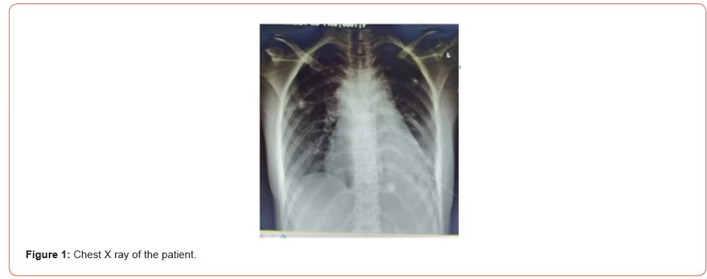
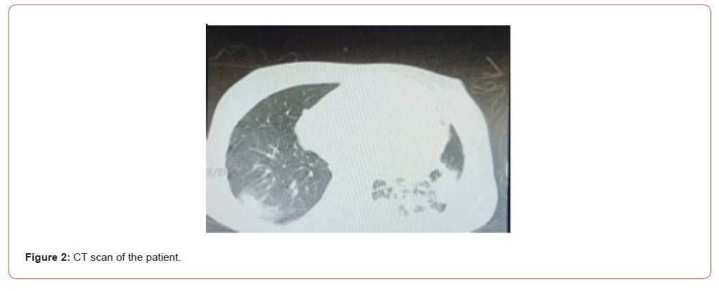
A 16-year-old girl was admitted with the symptoms of a severe common cold, including fever, dry cough, and dyspnea, which were initially attributed to influenza infection. On the chest x-ray, pleural effusion on both sides as well as pericardial effusion were seen as shown in Figure 1. The high-resolution computed tomography (HR-CT) revealed diffuse parenchymal involvement, and lymphadenopathies along with necrotic changes (Figures 1&2). The drained pericardial fluid appeared to be red, bloody, and exudative. The collected samples of a diagnostic pleural tap were sent to the laboratory for gram stain, culture, and TB specific evaluations, the results of which were all negative. Following that, a chest tube was inserted. Meanwhile, the patient’s blood test revealed increased acute phase reactants (leukocytosis with increased ESR and CRP levels). Vasculitis lab tests were also conduced, showing negative results as well (Table 1). Later in the course of the disease, she complained of pain in her right hip, making her unable to walk. A color Doppler ultrasonography indicated deep vein thrombosis in the common iliac vein.
Thereafter, computed tomography/pulmonary angiography (CTPA) was considered to detect the source of deep vein thrombosis as well as respiratory symptoms, revealing massive pulmonary embolism. Since the risk of venous thromboembolism and pulmonary embolism are four to seven times higher in patients with malignancies than those free from cancer [4], exploration for lung cancer as the leading cause of pulmonary emboli was initiated. Therefore, the patient underwent CT guided lung biopsy, the result of which was pulmonary adenocarcinoma. Meanwhile, left supraclavicular lymph node biopsy was also performed, which showed metastatic adenocarcinoma as well. The tumor was classified as stage IV according to the TNM classification. No risk factors could be considered in her case as her past history showed no impact of such risk factors on pulmonary adenocarcinoma. In echocardiography, a trivial mitral regurgitation, severe tricuspid regurgitation and a left ventricular ejection fraction of 72% were reported.
After the initiation of chemotherapy, she unfortunately developed respiratory distress and later unconsciousness in the course of the therapy and was therefore transferred to ICU for further treatment. However, her situation did not improve, and the pulmonary dysfunction progressed to a sudden arrest. Cardiopulmonary resuscitation was performed and continued for 45 minutes, yet spontaneous circulation was not achieved, and the patient passed away (Table 1).
Table 1: The patient’s lab data.

Discussion
Primary pulmonary carcinoma in children usually has a high mortality rate of more than 90%, with an aggressive growth pattern. The condition is rare in children and therefore the diagnosis may be challenging in this age group. Although genetic factors play a major role in the development of lung adenocarcinoma in children without common environmental risk factors, the patient in this study did not have even a positive family history in this regard. Pulmonary adenocarcinoma can affect children of any age by usually manifesting with respiratory symptoms such as cough, chest pain, shortness of breath, and hemoptysis. In most lung adenocarcinoma cases, there is a 6-to-9-month delay for the final diagnosis. The 5-year survival rate of primary lung carcinoma is approximately 25%. Aggressive growth patterns of lung carcinoma with high mortality rates are reported in different studies. Although the incidence of this disease is rare, making it challenging to make accurate estimations, an average survival of 7 months after diagnosis is reported in previous studies.
In this study, the patient’s failure to respond to antibiotics in the course of the treatment cast doubt on the initial diagnosis. This made the likelihood of a worse-case scenario much more probable. As there is no specific radiographic pattern for pulmonary adenocarcinoma, the patient’s lung CT scan did not show a space occupying lesion indicative of any malignancy. However, the tissue sample obtained from the supraclavicular node revealed the malignant nature of the disease. Once the diagnosis is made and the tumor staging is determined, an appropriate treatment regimen should be started accordingly. However, there are no specific chemotherapy regimens recommended for the pediatric age group. No matter which chemotherapy regimen is used for this disease, no child has unfortunately ever survived longer than 19 months. The patient in this case passed away even earlier. To conclude, although pulmonary malignancies are rare in children, the suspicion about such malignancies should be taken into consideration in patients with persistent or recurring respiratory manifestations. Since early detection may affect the outcome and survival rate, a high index of suspicion, as well as appropriate imaging studies, is important to diagnose the condition earlier and in lower stages, the results of which could be improving the quality of life.
Acknowledgement
None.
Conflict of Interest
No conflict of Interest.
References
- Weipeng Shao, Jie Liu, Bobo Li, Xiaokang Guo, Jian Sun, et al. (2023) Primary lung cancer in children and adolescents: Analysis of a surveillance, epidemiology and end results database. Front Oncol 13: 1053248.
- Lucia De Martino, Maria Elena Errico, Serena Ruotolo, Daniele Cascone, Stefano Chiaravalli, et al. (2018) Pediatric lung adenocarcinoma presenting with brain metastasis: a case report. J Med Case Rep 12(1): 243.
- Lobat Shahkar, Noora Bigdeli, Mahdi Mazandarani, Narges Lashkarbolouk (2023) A Rare Case of Pulmonary Adenocarcinoma in an 8-Year-Old Patient with Persistent Respiratory Manifestation: A Case Report Study. Case Rep in Oncol 16(1): 739-745.
- Yungu Chen, Yiu Sing Tsang, Xiaoxia Chou, Jiong Hu, Qing Xia (2019) A lung cancer patient with deep vein thrombosis: a case report and literature review. BMC cancer 19(1): 285.
-
Amir Rezaei1, Mahsa Rekabi, Sharareh Seifi, Mihan Pourabdollah, Susan Rahimi and Sepideh Darougar*. Lung Adenocarcinoma in Adolescence: A Case Report. Arch Clin Case Stud. 4(2): 2024. ACCS.MS.ID.000583.
-
Lung adenocarcinoma; malignancy; adolescence; childhood; iris publishers; iris publisher’s group
-

This work is licensed under a Creative Commons Attribution-NonCommercial 4.0 International License.


