 Mini Review
Review of Carbon Nanotube in Future Endogenous H2S Measurement
Xiaochu Wu*
Department of Comparative Biomedical Science, Louisiana State University, USA
Xiaochu Wu, Department of Comparative Biomedical Science, Louisiana State University, Baton Rouge, LA, 70820 USA.
Received Date: July 14, 2021; Published Date: August 16, 2021
Abstract
Endogenous H2S in the mammalian body is considered to play the role of a vascular dilator in the cardiovascular system. The physiological role of hydrogen sulfide depends on its in vivo level. As such, the measurement of hydrogen sulfide with nano-quantity vibration becomes an important subject. Current measurement usually needs a large number of samples, and the process is complex. So, it is important to develop a new sensitive method. Carbon nanotubes can sense the tiny amount of H2S in aqueous, as well as in serum, and the proteins in serum seem not to affect this measurement, so it may be possible to use carbon nanotube as a sensor to detect the tiny difference of H2S in mammalian bodies.
Introduction
The patient will be treated from front and back and it does not matter which side is done first. The membrane of the shock head of the CellSonic machine is to be covered in ultrasound gel and the same gel applied to the body where the doctor or nurse will aim at the lungs. This is important because a pressure wave will travel from the shock head, through the gel and through the body to the lungs. The gel behaves like water and bridges the gap between the machine and the body. Set the energy level to 5 and the number of shocks (also called pulses) to 300. Aim at the lungs. Understand that the lungs are encased in the rib cage so the pulses can only penetrate through the gaps between the ribs. Angle the head one way and then another to catch the covid-19 virus in the lungs. The pressure will kill the virus. We know from many years of wound healing that all infections, be they virus, bacteria or parasites are killed by the pressure pulses. No drugs are used so there will be no side effects.
We also know from curing lung cancer that we do not damage the lungs. Nor is the heart damaged providing care is taken. When the heart is in the line of fire, so to speak, give only 50 pulses and pause a minute or two to let the patient’s heart maintain its own rhythm. Very likely the patient will be gasping for breath with their heart beating fast. Immediately the pulses Since carbon nanotubes (CNTs) were first reported in 1991 [1-3], thousands of papers were published every year. Their unique combination of electrical, magnetic, optical, mechanical, and chemical properties, as well as hollow carbon structures, make CNTs great promises for a wide range of applications, including biosensing [4,5].
CNTs are designated single-walled (SWNTs) or multi-walled (MWNTs). SWNTs, with side-well as small as a nanometer or less, can be grown up to 20 cm in length [6]. SWNTs possess the simplest morphology and can be visualized as a single rolled-up graphene sheet. MWNTs can reach diameters of up to 100 nm and the distance between two walls is very close to the distance between two graphene layers in graphite (~3.5 Å) [7]. Increasing the number of layers in MWNT inevitably also increases the number of defects and thus makes them easier to modify and to functionalize. Depending on their electronic structure, SWNTs can behave either as semiconductors or as metals. However, the MWNTs behave more like metals [8,9].
CNTs have a large specific surface area, which can immobilize greater concentrations of bioreceptor units at their surface. This property of CNTs is widely used in biosensing applications [10]. Conjugating with DNA or aptamers, antibodies, peptides, proteins, or enzymes, CNTs can be developed to a wide variety of cancer biomarkers [11-13]. Consisting of modified screen-printed electrodes with cholesterol esterase, peroxidase, oxidase, and MWNTs, Cholesterol biosensors yield highly sensitive means of quantifying total cholesterol in the blood [14].
In mammals, H2S can be found in the blood, brain, lung, heart, liver, spleen, and kidney [15,16]. Recent research has found that hydrogen sulfide, produced in the gastrointestinal tract by different enzymes, is not simply a toxic gas, but can regulate smooth muscle membrane potential and tone, transmit signals from enteric nerves, and can regulate the immune system [17]. Mammalian cells synthesize hydrogen sulfide (H2S), as well as nitric oxide (NO) and carbon monoxide (CO,) which act as a biological switch, relaxing contracted blood vessels and reducing blood pressures [18,19]. However, the physiological importance of hydrogen sulfide depends on its in vivo level. As such, the measurement of hydrogen sulfide in mammalian bodies becomes an important topic. Compare with traditional measurements which need a complex generic procedure and bulky samples, the new paradigm of measurement should be less invasive and more accurate with fewer samples. To realize this new paradigm, carbon nanotubes can be a good candidate.
Material and method
The powder form carbon nanotubes were got from microwave plasma-enhanced chemical vapor deposition (MPECVD) in experiment 1 [20], and purchased from Shenzhen Nanotechnologies Co. Ltd (Shenzhen, China) in experiment 2 (Wu et al., under publication).
Two serum proteins, namely bovine serum albumin (BSA) (Cat. #A7906) and bovine gamma-globulin (γ-Globulin) (Cat. # G5009) were purchased from Sigma-Aldrich [21].
High concentration hydrogen sulfide gas-saturated solution (90 mM at 30°C), was made by bubbling pure hydrogen sulfide into 40 ml distilled water at 30 °C and 10 lbf/in2, or poundsforce per square inch (1 atm=760 mmHg=14.7 lbf/in2). Lower concentrations (μM level) hydrogen sulfide solutions were diluted from high concentration hydrogen sulfide solutions. Especially, their concentrations are, respectively, 10, 20, 30, 40, 50, and 100 μM.
Different concentrations of pure BSA solutions (25mg/mL, 30mg/mL and 35mg/mL) and γ-Globulin solutions (10mg/ mL, 15mg/mL and 20mg/mL) were made by solving proteins in distilled water.
BSA (30 mg/mL) with different H2S concentration solutions were prepared by solving the powder form BSA in 0 μM, 50μM, 100μM, and 200μM H2S solutions, γ-Globulin (15 mg/mL) with 0 μM, 50μM, 100μM, and 200μM H2S were prepared by the same method. Here 0 μM H2S BSA as well as 0 μM H2S γ-Globulin solutions are control samples
To get the fluorescence intensity verse H2S concentration, a laser scanning microscope (ZEISS LSM 510 META) and Raman microscope (Renishaw system 2000) were used. Purified carbon nanotube first immersed in different concentration hydrogen sulfide (H2S) and distilled water, then dry for measurement in experiment 1, and indifferent concentration of BSA and γ-Globulin in experiment 2. The excitation wavelength for confocal is 514nm and emission wavelength ranges from 539nm to 753nm. for Raman, the excitation wavelength is 514nm and collect illuminant at 518nm to 800 nm.
Result
The results of experiment 1 are as (figure 1).
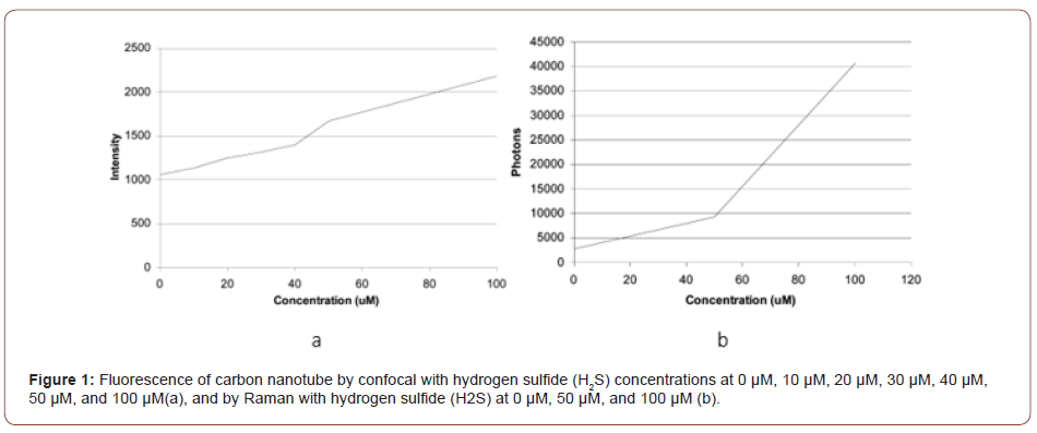
It shows that as H2S concentration increase, the signals increases either. The results show that it is possible to use carbon nanotubes to sensualize the tiny difference of H2S concentrations. The serum contains different proteins. To measure the H2S in blood or serum, the effects of these proteins need to be considered.
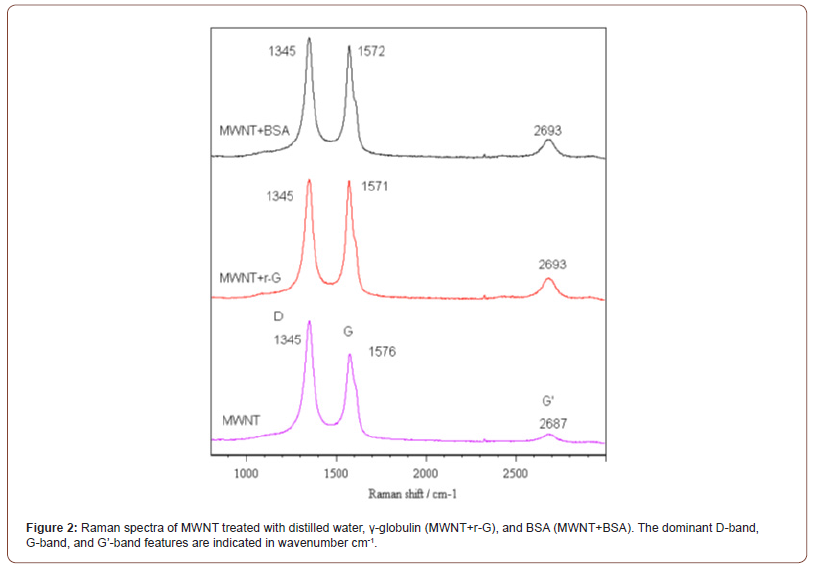
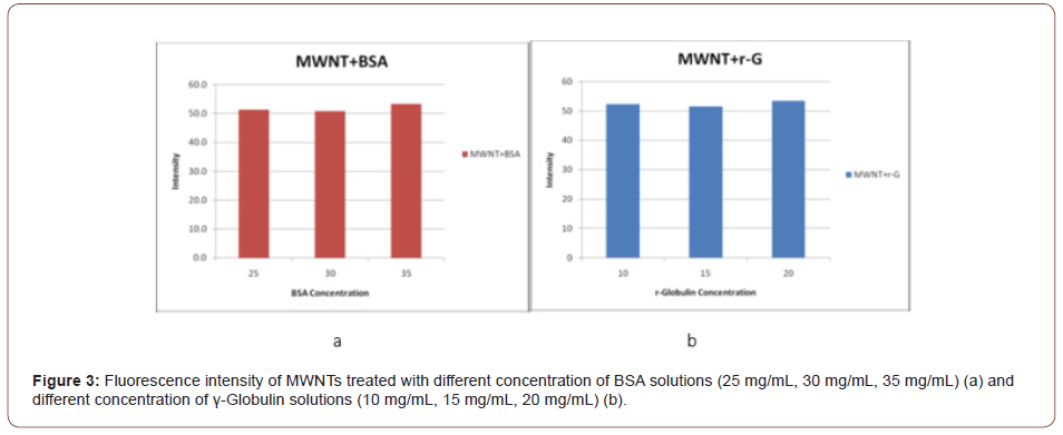
In experiment 2, Wu et. al (manuscript in preparation) used different concentrations of pure BSA solutions (25mg/mL, 30mg/ mL and 35mg/mL) and γ-Globulin solutions (10mg/mL, 15mg/mL and 20mg/mL) to observe the change of correspond intensities. The Reman spectrums show after carbon nanotubes interact with serum or γ-Globulin the G and G’ band shifted. This means that the serum or γ-Globulin is attached to carbon nanotubes (figure 2). However, the intensities keep unchanged with different concentrations of serum or γ-Globulin (Figure 3).
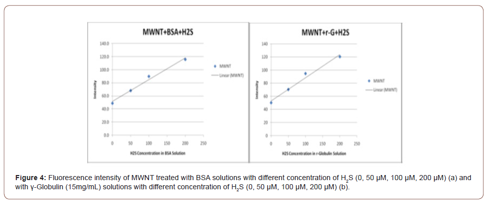
Using confocal or Raman to measure carbon nanotubes treated by serum or γ-Globulin with different concentrations of H2S, it is found that the intensity is different (Figure 4). This result indicates that γ-Globulin or proteins in serum will not affect the sensitivity of using carbon nanotubes as a senor for H2S measurement.
Conclusion
Carbon nanotubes can be used in measuring H2S in the atmosphere as well as in aqueous conditions with high resolution. Although there are a bunch of proteins, it seems these proteins will not affect the sensitivity of the H2S measurement in serum with carbon nanotubes. This opens a future to develop carbon nanotubes as a sensor for H2S measurement in mammalian blood. Of course, there is still a lot of work to do before realizing this objective. For example, the effect of blood cells needs to be considered in feather research.
Acknowledgement
None.
Conflicts of Interest
No conflicts of Interest.
-
Xiaochu Wu. Review of Carbon Nanotube in Future Endogenous H2S Measurement. Arch Biomed Eng & Biotechnol. 5(5): 2021. ABEB.MS.ID.000623.
-
Cardiovascular, Endogenous, Mammalian body, Carbon nanotubes, Electrical, Magnetic, Optical, mechanical, Chemical properties, Morphology, Cholesterol esterase, Peroxidase, Oxidase
-

This work is licensed under a Creative Commons Attribution-NonCommercial 4.0 International License.


