 Research Article
Sternal Force Measurements During Mid-Sternal Retraction in Cadavers: Implications for Chronic Pain?
Dimitri Petsikas1, Joel Couture-Tremblay2, Julia Prince2, Nicole Murrell2, Tim Bryant2, Rachel Phelan3, Debbie DuMerton3, Tarit K Saha3*
1Department of Surgery, Division of Cardiac Surgery, Queen’s University & Kingston Health Sciences Centre, Kingston, Ontario, Canada. K7L 2V7
2Department of Materials & Mechanical Engineering, Queen’s University & Kingston Health Sciences Centre, Kingston, Ontario, Canada. K7L 3N6
3Department of Anesthesiology and Perioperative Medicine, Queen’s University & Kingston Health Sciences Centre, Kingston, Ontario, Canada
Tarit Saha, Department of Anesthesiology and Perioperative Medicine, Queen’s University, Victory 2 Kingston General Hospital site, Kingston Health Sciences Centre, 76 Stuart Street, Kingston, ON, Canada.
Received Date: June 01, 2023; Published Date: June 19, 2023
Summary
Study Objective: Chronic post-sternotomy pain (CPSP) is common following cardiac surgery. We hypothesized that performing sternal retractions more slowly, would require less force and therefore, decrease the nerve/tissue trauma and translate to a reduced incidence/severity of CPSP. The purpose of this investigation was to examine the forces required to reach full retraction when performed slowly versus rapidly (as in cardiac surgery).
Design: Observational
Setting/Participants/Intervention/Outcomes: With ethics and biohazards approvals, a cardiac surgeon (DP) performed sternal retractions on 4 cadavers (2 slow, 2 rapid) with continuous force measurements using a Collins rib retractor fitted with Tekscan FlexiForce sensors and strain gauges for independent measurements from multiple load points.
Result: Measures were relatively stable and reliable. Significant trends were observed with spikes in applied forces with retractor opening, each followed by decay. With rapid retractions, maximal forces were achieved within 30 sec. whereas gradual retractions reached peak forces approaching 15 min. Less average force was required to achieve maximal opening with gradual (289.3N) compared to rapid (315.8N) retraction. Although the temporal profiles appeared similar, measures from force sensors were lower than strain gauges.
Conclusion: Less average force was required when retractions were performed slowly. Force sensor readings were lower than strain gauges, but the similar temporal profile suggests the difference was from sensor location and the forces applied at those points. Future investigations will be required for validation and determination of whether retraction speed may be a modifiable risk factor for reducing nerve/tissue trauma and/or the incidence/severity of CPSP following cardiac surgery.
Keywords: Sternal retraction; Cadavers; Sternal forces; Technological advance; Chronic post-sternotomy pain; Retraction speed
Abbreviations: BMI: Body Mass Index; CPSP: Chronic post-sternotomy pain
Introduction
Sternal retraction during cardiac surgery requires considerable force. Although cardiac surgical practices are variable, sternal retraction for adequate exposure of the heart generally involves opening the sternum to 7-10 cm and given contemporary cardiac surgery practices, it may be reasonable to assume that most are performed expeditiously (i.e., ~30 sec). With the invasiveness of this procedure, it is not surprising that postsurgical pain can be intense and 30-40% go on to develop chronic post-sternotomy pain (CPSP)1-3 which can be severe [1-6]. CPSP is therefore, a major medical problem for cardiac surgery patients, their families, and society in general with associated increases in healthcare costs and lost productivity. Other than a protocol published by our group (available at URL: https://f1000research.com/articles/10-248), [7] to our knowledge, no clinical investigations have examined the forces applied during the sternal retraction maneuver as a possible causal determinant in the subsequent development of CPSP.
We hypothesized that performing the sternal retraction more slowly would require less force and could therefore, potentially translate to a reduction in the amount of nerve/tissue trauma, and a reduced incidence and/or severity of CPSP. The focus of the current investigation was upon the development of a sternal retractor modified to enable continuous force measurements throughout the sternal retraction maneuver and then compare forces required when the sternal retraction was performed gradually versus rapidly (as per standard of care).
Materials and Methods


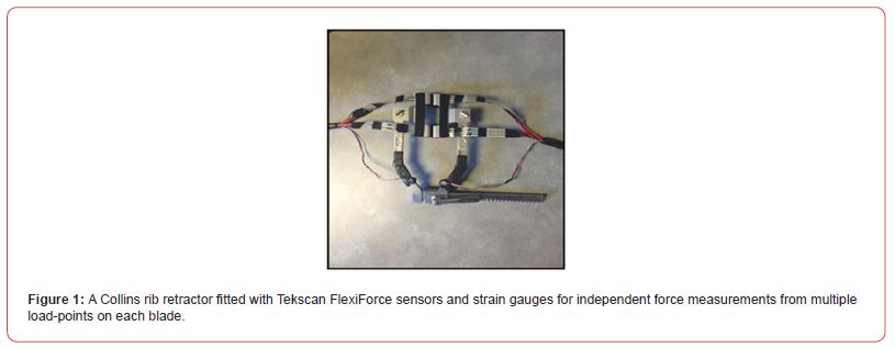
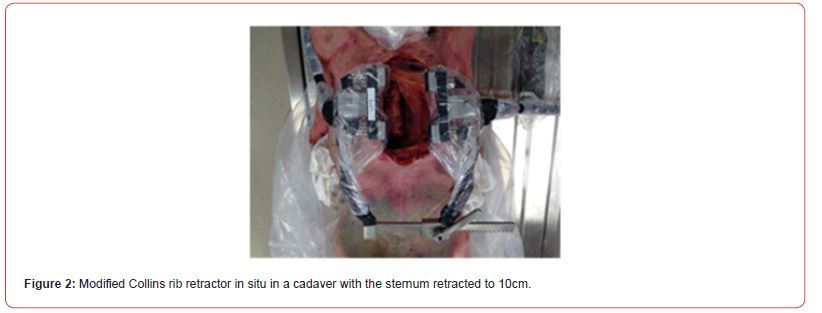
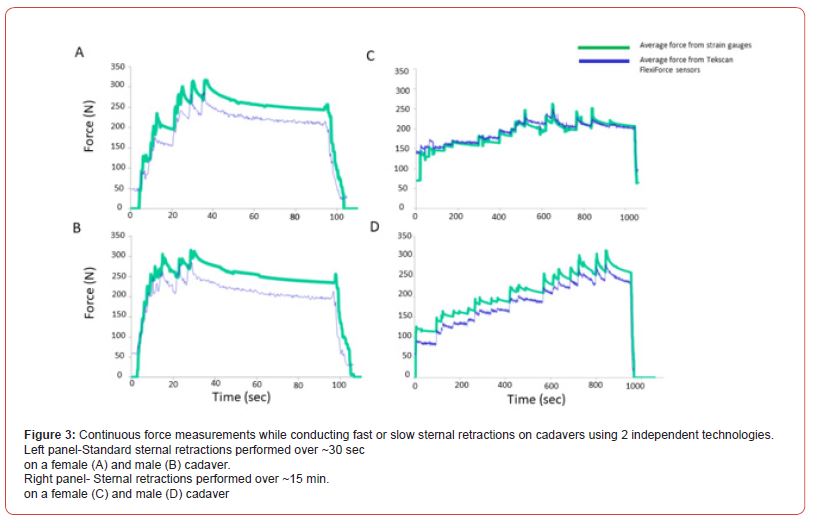
Collins rib retractor was fitted with multiple Tekscan Flex Force sensors and strain gauges for independent force measurements from multiple load points (Fig. 1). Following proof-of-principle trials, institutional ethics board and biohazards approvals were obtained to perform sternal retractions on cadavers donated to the Queen’s University School of Medicine for research purposes. Cadavers were previously frozen, BMI 21- 35, with no thoracic abnormalities, no previous cardiopulmonary resuscitation, or thoracic surgery. All cadavers were over 70 years of age at the time of death and their bodies fresh frozen, but all completely thawed prior to sternal retraction. With the cadaver in the supine position, a cardiac surgeon (DP) performed a standard median sternotomy with an oscillating saw. The modified sternal retractor (encased in a long laparoscopic camera sleeve secured with electrical tape) was placed in position between the sternal edges (Fig. 2). In total, 4 cadavers underwent a sternotomy and sternal retraction in which the sternal edges were opened to a width of 10cm. Two were performed at the standard rate (i.e., time to full retraction ~30sec) and 2 performed slowly (over 15 min), each group included one male and one female cadaver. Average and total applied forces were recorded continuously throughout each retraction (fig.3).
Results and Discussion
Force measurements were relatively stable within the standard vs slow sessions and reproducible across the 2 retractions at each speed. Significant trends were observed with spikes in forces applied to the sternum during retractor opening with each followed by force decay. With the standard (rapid) retractions, maximal forces were achieved within ~30 sec. (Fig. 3A-B) whereas gradual retractions reached peak forces approaching 15 min. (Fig. 3C-D). Less average force was required to achieve maximal opening with gradual (289.3N) versus standard (315.8N) sternal retraction. Although the temporal force profiles appeared similar between force sensors and strain gauges, the forces detected by the former were lower (Fig. 3A-D).

Force measurements were successfully collected continuously throughout standard vs. gradual sternal retractions in 4 cadavers. Less average force was required to achieve full retraction when it was performed gradually. The similar temporal profile between measurement technologies suggests that the measures were valid. The different force measurements provided by the two technologies may be from the strain gauges and force sensors being placed at different locations on the sternal retractor blades at points receiving different force loads during retraction (Fig. 3).
Our results are consistent with those of Bolotin et al (2007) [8] who demonstrated that controlled retraction forces (and the attendant increased time to sternal retraction), resulted in significantly lower average applied forces in anesthetized sheep. They also demonstrated that using controlled force was associated with significantly reduced alterations in heart rate in the anesthetized sheep, suggestive of reduced pain and/or stress compared to those being exposed to the standard retraction forces. Likewise, another study demonstrated that thoracotomies performed with controlled forces (again, over a longer duration), were associated with reduced peak and average applied forces, reduced animal stress, and less tissue damage such as rib fractures [9].
In a more recent human cadaver investigation, Aigner et al. [10] examined the force distribution from curved vs. straight blade retractors in human cadavers and found that the forces applied were localized to different areas depending upon retractor shape. With the straight blade retractor, cranial and caudal sternal forces were greatest whereas mid-sternal forces were greatest with the curved retractor. Although there was no significant difference detected in total forces required with curved vs straight blade retractors (222.8N vs 226.4N respectively), significant differences were observed in males vs. females and in cadavers with and without a state of rigor mortis.
In the current investigation, the Collins retractor blade was straight and none of the cadavers were in a state of rigor mortis. Interestingly, the total average force measures to reach full retraction over 30 sec were higher in the current report than that reported by Aigner et al. (315.8N vs. 290.1N respectively). It is unknown whether this difference is attributable to the speed of the retraction or the status of cadavers (e.g., fresh instead of previously frozen and thawed as in the current study). Alternatively, this difference could be attributable to the difference in retractor design/force-sensing technology. With regards to male vs. female differences, there were too few cadavers in the current study to make any comparison.
A strength of this investigation is in the fact that a single cardiac surgeon performed all sternotomies and retractions thus ensuring consistency with surgical technique. An additional strength is that force measurements were collected concurrently using two different technologies independent of one another. The fact that the results from both technologies were aligned with one another lends credibility to this approach. However, this study has several limitations. A major limitation is the small sample (n=2/ group) which precludes the possibility of drawing any definitive conclusions based on this work. In addition, the cadavers were all >70 years of age at death with a different body habitus and were previously frozen, all factors which may have impacted the results. It is also possible that some patients were taking medications (i.e., steroids) or had comorbidities (osteoporosis, diabetes, chronic kidney disease etc.) prior to death that may have impacted the bone resistance to the applied forces. Finally, it is unknown whether cadaverous tissues (at sub-room temperatures with no vascularity) react to the sternal retraction procedure the same was as living vascularized tissue. However, cadavers were the best option (with at least some clinical relevance) for the current demonstration that provided useful information since the acquired force data for standard vs. gradual sternal retractions were collected relative to one another using two independent measures.
Conclusion
Consistent with previous observations, this study suggests that slower retraction speed requires less average force to achieve the same degree of sternal opening. It also suggests that the currently refitted retractor can provide valid and reliable sternal force measurements throughout the sternal retraction maneuver.
An associated double-blinded randomized controlled trial (NCT02697812) in which coronary artery bypass graft patients are randomly assigned to undergo surgery with standard or gradual sternal retraction is nearing completion. In this study, patients were followed for up to 1 year postoperatively with outcomes including the incidence and severity of CPSP at 6 months postoperatively. Should this study yield significant and\or clinically relevant differences between groups, the next step for our refitted retractor would be to render it sterilizable and wireless for surgical application. With this technological advance, the relationship between the forces applied during sternal retraction, nerve/tissue damage, and the onset and/ or severity of CPSP could be characterized comprehensively.
Funding
This work was supported by the Alison B. Froese Research Fund in Anesthesiology & Perioperative Medicine, Department of Anesthesiology and Perioperative Medicine, Queen’s University. The funding body was not involved in the conception, design, execution or publication of this work.
Disclaimer
The authors report no proprietary or commercial interest in any product mentioned or concept discussed in this article.
Acknowledgement
The authors would like to thank Mr. Richard (Rick) Hunt, and Matt Daalder (Queen’s University, Department of Anatomy) for their assistance.
References
- Steegers M, van de Luijtgaarden A, Noyez, L, Scheffer GJ, Wilder-Smith OH (2007) The role of angina pectoris in chronic pain after coronary artery bypass graft surgery. J Pain 8(8): 667-673.
- Cantero C, Carolina G, Matute, P, Tena B, Rovira I, Carmen G (2011) Prevalence of Postoperative chronic pain after cardiac surgery. Eur J Anaesthes 28:193-194.
- van Gulik L, Janssen LI, Ahlers, SJ, Bruins P, Driessen AH et al. (2011) Risk factors for chronic thoracic pain after cardiac surgery via sternotomy. Eur J Cardiothorac Surg 40(6):1309-1313.
- Myerson J, Thelin S, Gordh, T, Karlsten R (2001) The incidence of chronic post-sternotomy pain after cardiac surgery—a prospective study. Acta Anaesthesiol Scand 45(8): 940-944.
- Bruce J, Drury N, Poobalan, AS, Jeffrey RR, Smith WC et al. (2003) The prevalence of chronic chest and leg pain following cardiac surgery: a historical cohort study. Pain 104(1-2): 265-273.
- Kalso E, Mennander S, Tasmuth, T, Nilsson E (2001) Chronic Post-sternotomy pain. Acta Anaesthesiol Scand 45(8): 935-939.
- Petsikas D, Stewart C, Phelan, R, Allard R, Cummings M, et al (2021) Does the speed of sternal retraction during coronary artery bypass graft surgery affect postoperative pain outcomes? A randomized controlled trial protocol. F1000Res 10: 248-253.
- Bolotin G, Buckner G, Campbell, N, Kocherginsky M, Raman J, et al (2007) Tissue-disruptive forces during median sternotomy. Heart Surg Forum 10(6): E487-E492.
- Bolotin G, Buckner G, Jardine, N, Kiefer AJ, Campbell NB, et al. (2007) A Novel instrumented retractor to monitor tissue disruptive forces during lateral thoracotomy. J Thorac Cardiovasc Surg 133(4): 949-954.
- Aigner P, Eskandary F, Scholglhofer, T, Gottardi R, Aumayr K, et al (2013) Sternal force distribution during median sternotomy retraction. J Thorac Cardiovasc Surg 146(6): 1381-1386.
-
Dimitri Petsikas, Joel Couture-Tremblay, Julia Prince, Nicole Murrell, Tim Bryant, Rachel Phelan, Debbie DuMerton, Tarit K Saha*. Sternal Force Measurements During Mid-Sternal Retraction in Cadavers: Implications for Chronic Pain. Arch Biomed Eng & Biotechnol. 7(3): 2023. ABEB.MS.ID.000662.
-
Sternal retraction, Cadavers, Sternal forces, Technological advance, Chronic post-sternotomy pain, Retraction speed, Body Mass Index; CPSP, Chronic post-sternotomy pain
-

This work is licensed under a Creative Commons Attribution-NonCommercial 4.0 International License.


