

 Research Article
Microscopic appearance and cultivation of objects (Crown Asci-glu-glu cells nema) from agar cultures of antibiotic crown Asci-glu-glu cells stimulated with (Glu-Glu) as a partner
Received Date:December 20, 2025; Published Date:January 20, 2026
Abstract
Synthetic DNA crown cells can be produced using sphingosine (Sph)-DNA-adenosine-monolaurin compounds and egg white. In previous experiments, it was demonstrated that both antibiotic compounds and antibiotic-producing cells could be produced using various combinations of DNA crown cells with partners, such as microorganisms, other cells, extracts and organic compounds. On the other hand, it was unclear whether these antibiotic-producing cells could be cultured. Therefore, in this study, antibiotic-producing cells produced by combining DNA (Ascidian) crown cells with peptides (Glu-Glu) as a partner were cultured on agar plates. The appearance of the resulting cells was then observed under a microscope and objects in the culture were cultivated on agar medium. The findings showed that both cell proliferation and various objects (hereafter referred to as Crown Asci-glu-glu cells nema) were observed in cultures of antibiotic-producing cells (Antibiotic crown Asci-glu-glu cells) produced using egg white powder enclosing DNA (Ascidian, Ascidian sea squirt) crown cells with Glu-Glu as a partner.
Keywords:DNA (Ascidian) crown cells; Sphingosine-DNA; Antibiotic crown Asci-glu-glu cells; Peptide (Glu-Glu); Crown cells nema
Introduction
Self-replicating artificial cells were first reported in 2012 [1] and the principal methods for preparing these artificial cells were reported in 2016 [2]. As their exterior consists of DNA, these cells were referred to as DNA crown cells in 2016 by the present author [3]. Synthetic DNA crown cells were produced using the following four common commercial compounds: Sphingosine (Sph), DNA, adenosine, and monolaurin. The cells developed into fully selfreplicating DNA crown cells when incubated in egg white. In previous studies, it was demonstrated that antibiotic and antibioticproducing cells were produced by combining various DNA crown cells with different partners, such as microorganisms, other cells, extracts, and organic compounds [4-9]. However, the growth characteristics of these antibiotic-producing cells were unclear.
To clarify the characteristics of these cells, antibiotic-producing cells (Antibiotic crown Asci glu-glu cells) were prepared and were cultured on agar plates. Then, Glu-Glu was added. In agar plate cultures after the addition of Glu-Glu, cell proliferation and objects that varied in shape and size were observed. These cells are referred to as Crown Asci glu-glu cells nema. The findings suggested that antibiotic-producing cells produced from DNA crown cells were easy to differentiate, especially with partners. In this study, crown cells nema, which are multicellular or filamentous, are described for the first time. The objects which grown from antibiotic crown Asci-glu-glu cells in the response of the partner (Glu-Glu) are named Crown Asci glu-glu cells nema.
Material and Methods
Antibiotic crown Asci-glu-glu cells were prepared using the following three steps, as described previously [10]. However, the methods are provided here again for reference.
• Step 1 Preparation of DNA crown cells
• Step 2 Preparation of powder
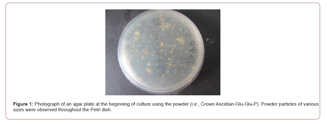
• Step 3 Culture of powder
Materials for these experiments
Sph (Tokyo Kasei, Tokyo, Japan), DNA (from an Ascidian sea squirt), adenosine (Sigma-Aldrich, USA; Wako, Tokyo, Japan), monolaurin (Tokyo Kasei, Tokyo, Japan), and adenosinemonolaurin (A-M), a mixture of adenosine and monolaurin [11,12] were used in this study. Monolaurin solutions were prepared to a final concentration of 0.1 M in distilled water. Agar plates were prepared using standard agar medium (SMA; AS ONE, Japan) and L-glutamyl-L-glutamic acid (Glu-Glu) (PEPTIDE, Institute of Peptides, Japan) was used as a partner and prepared in 1.00 mg/ mL. Edible ascidians were obtained from a local market.
Step 1
Preparation of DNA (Ascidian) crown cells [10-12].
Briefly, 180 mL of Sph (10 mM) and 90 mL of DNA (0.05
μg/mL) were combined, and the mixture was heated and cooled
twice. Then, A-M solution was added and the mixture was incubated
for 15 min at 37°C. Following the addition of monolaurin solution,
the mixture was incubated for 5 min at 37°C to produce synthetic
DNA crown cells.
These cells were then added to egg white and incubated
for 7 days at 37°C. Then, the egg white was recovered and used as
DNA (Ascidian) crown cells.
Step 2
Preparation of powder-enclosed DNA crown cells with Glu-Glu
First, 3 mL of Glu-Glu solution (300 mL Glu-Glu in distilled
water) was mixed with 3 mL of egg white.
The mixture was then incubated for 5 hours at 37°C.
Approximately 20 mL of fresh egg white was then added to the
mixture.
The mixture was then plated onto two Petri dishes and dried
for 1-2 days at 37°C.
The dried material was then collected and ground to a powder
using a mortar and pestle.
The powder, named Crown Ascidian glu-glu-P, was stored at
room temperature and used as necessary.
• Step 3
Cultivation of powder (Preparation of antibiotic producing
cells)
Approximately 50 mg of powder (Crown Ascidian glu-glu -P)
was added to an agar plate and incubated for 2 days at 37°C.
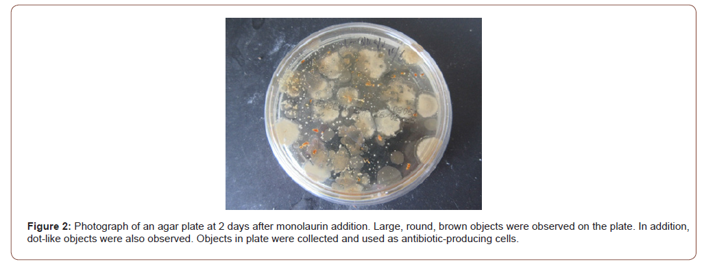
Then, approximately 1.5 mL of 0.1 M monolaurin solution
was poured onto each plate, which was then incubated for 2
days at 37°C. About 6.0 mL of distilled water was then added
to plate and dispersed on the plate surface. Then, the objects
on the plate were recovered. Suspended objects were used as
antibiotic-producing cells.
Cultures of antibiotic producing cells
A total of 200 mL of sample, which had been stored at 4°C for
3 days, was placed onto an agar plate and incubated for 1 day
at 37°C.
Preparation of Crown Asci-glu-glu cells nema
Experiment 1
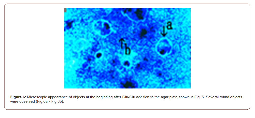
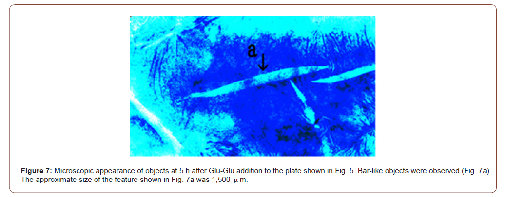
After 7 days of culture of antibiotic producing cells, approximately 1.5 mL distilled water containing 100 mL of Glu-Glu solution (1.0 mg/mL) was added to an agar plate and incubated for 5 hours, 1 day and 2 days at 37°C.
Experiment 2
After 1 day culture of Glu-Glu addition in Experiment 1, objects that grew on the plate were collected and transplanted into a new plate. and incubated for 1 day at 37°C. The objects that then grew on the plate were observed under a microscope.
Results
In previous studies, antibiotic-producing cells (antibiotic crown cells) were produced in combination with various DNA crown cells and partners (e.g., microorganisms such as yeast, Bacillus subtilis, cells such as salmon roe, extracts such as those from bovine meat, and chemical compounds such as peptides) [4-10]. The findings of those studies showed that the antibiotic crown cells could potentially have benefits other than producing antibiotics against Bacillus. However, the characteristics of the antibiotic crown cells were not clarified.
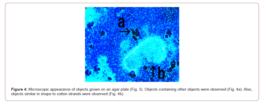
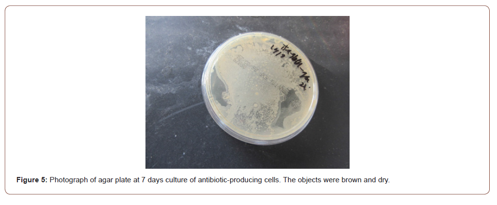
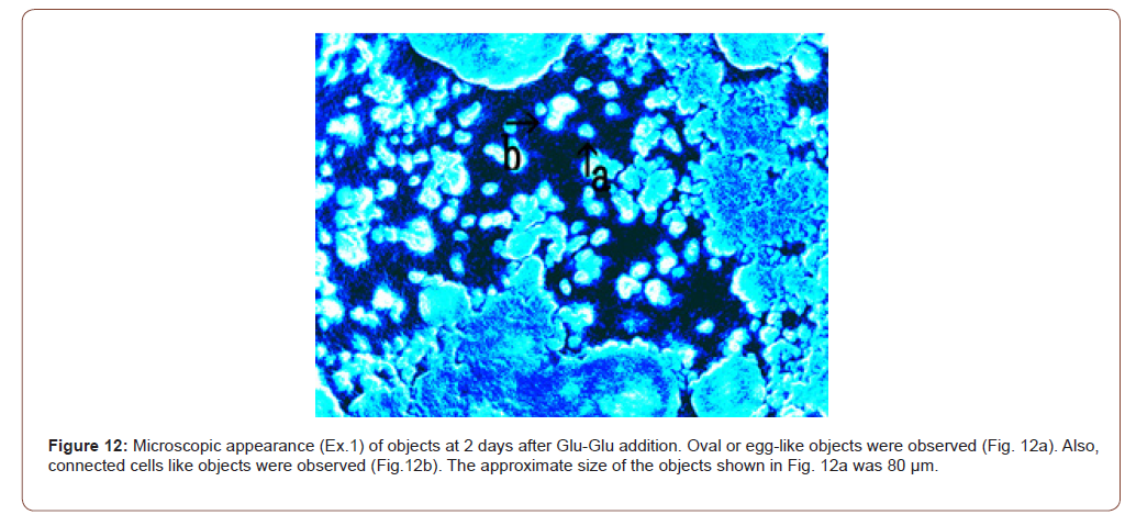
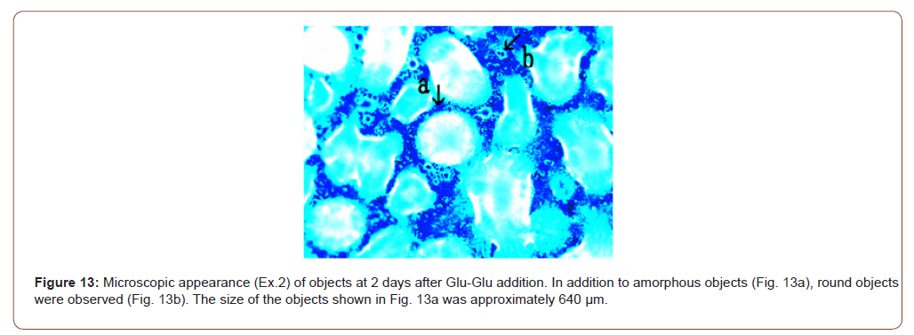
The present experiments were carried out to clarify the growth characteristics of these cells Antibiotic crown cells produced with DNA (Ascidian) crown cells and Glu-Glu as a partner were cultivated on agar plates for 7 days, After 7 days of culture, Glu-Glu was added to agar plates. At 5 hr after the addition, bar-like objects were observed (Figure 7). Interestingly, after 1 day of culture, various uniquely shaped objects were observed with the addition of Glu-Glu (Figures 8-13). These cells were named Crown Asci Glu-Glu cells nema (Crown Asci; Asci: Ascidian used to prepare DNA crown cells; Glu-Glu (Peptides): used as a partner for the crown antibiotic cells; cells nema, as described in the Introduction).
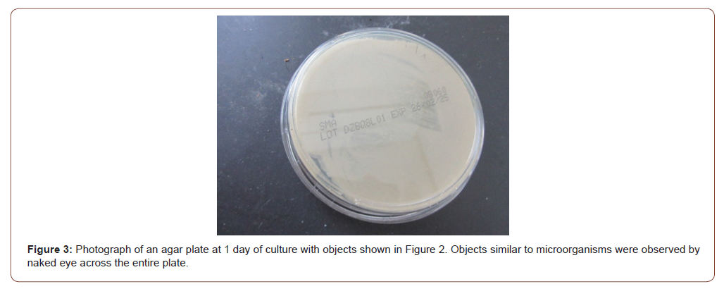
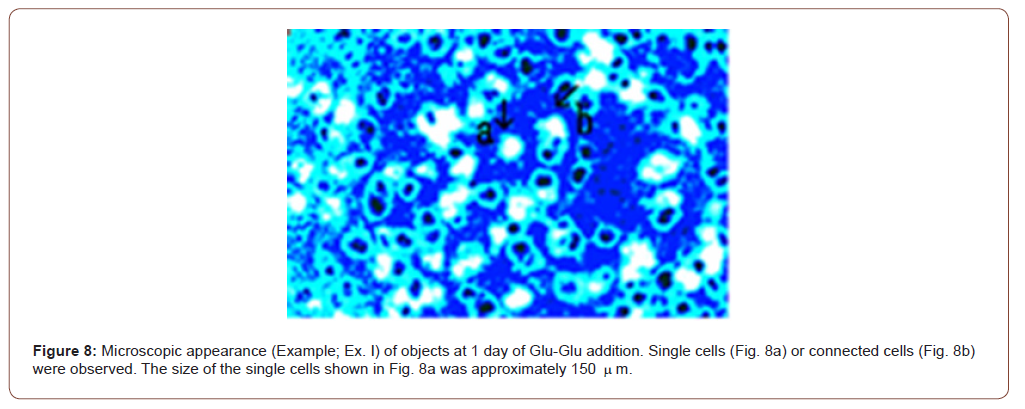
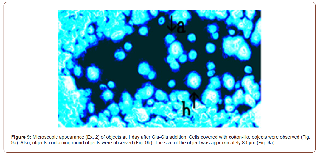
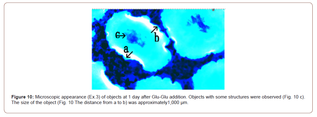
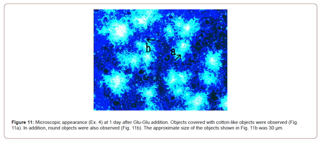
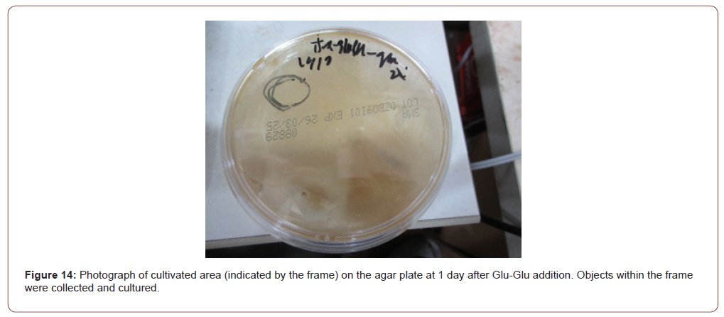
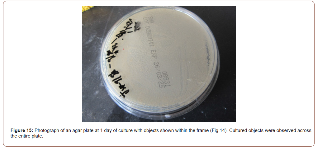
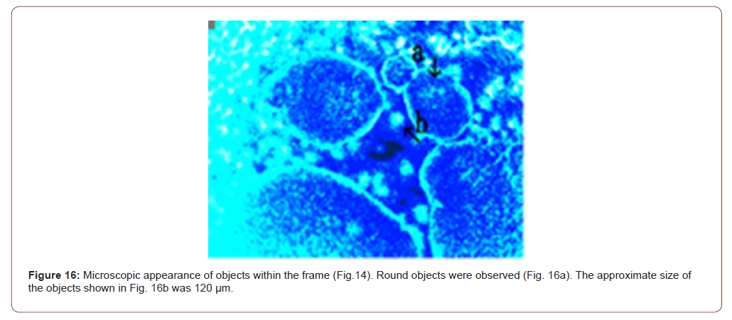
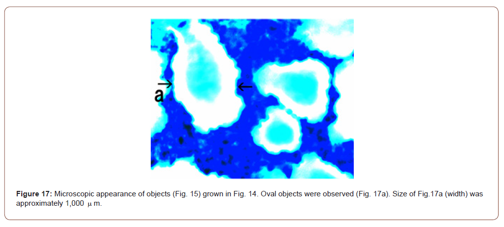
At 1 day after addition, objects within the frame (Figure 14) were collected and cultured. Cultured objects were observed to cover the entire plate (Figure 15), indicating that the Crown Asci glu-glu cells nema were viable. In previous experiments, antibiotic producing cells lost the ability to produce antibiotics and recover with the addition of Glu-Glu, suggesting that changes may occur in these cells [10]. The present results may prove such phenomena, and show that antibiotic crown cells may be easy to stimulate with the partner (Glu-Glu) to form crown cells nema. The purpose of present study was to clarify whether antibiotic crown cells were formed based on the DNA crown cells used, and therefore have added characteristics expected antibiotic (antibacterial to Bacillus subtills).
The present findings demonstrate that antibiotic crown cells tap into hidden abilities of crown cells nema, and were formed from the DNA crown cells that were prepared with Sph, DNA, adenosine, and monolaurin. These findings demonstrate that antibiotic crown cells precede the formation of crown cells nema. Thus, crown cells nema are DNA crown cells. Antibiotic crown cells are not capable of producing antibiotics against Bacillus, but they have the ability to behave as proto-cells for crown cells. In the near future, unlimited kinds of crown cells will be produced. Therefore, although it is very difficult, it may be necessary to develop a taxonomy for these cells in order to better understand crown cells nema. In the first grouping of microorganisms or cells, they were grouped based on their shape or size. Therefore, the grouping of crown cells nema will consider differences in shape and size. However, classifying crown cells nema may be difficult, because crown cells nema were formed with a complex structure that could not be accurately measured for size. In addition, naming them based on their shape and size is difficult.
Animal tissue consists of several cells and crown cells nema may consist of several crown cells. Crown cells nema comprised a mass of crown cells that may form with regenerated DNA crown cells. Based on this idea, crown cells nema may comprise components that have a round or other complex shape. As described previously, DNA crown cells bound easily to each other, microorganisms, and inorganic compounds to form assemblies [13-16]. Moreover, synthetic DNA crown cells formed objects that had various shapes in the stimulation of white and monolaurin, including cell proliferation [17-19]. Crown cells nema may form under these conditions. Conversely, DNA crown cells could be cultivated in liquid medium and cell strains were also established. In general, the growth of these cells was slow in liquid medium [20-23]. On the other hand, crown cells nema could be cultivated on agar plates, and could cover the entire plate the following day [4-10].
However, to clarify the grouping characteristics of crown cells nema, it appears that further experiments on the production of crown cells nema are necessary. The present experiments were conducted using DNA (Ascidian). It was unclear whether the appearance of crown cells nema were observed using DNA crown cells produced using other DNA crown cells, or crown cells nema with partners other than Glu-Glu. Future studies will be conducted to determine whether crown cells nema were produced in combinations other than antibiotic crown Asci-glu-glu cells with Glu-Glu as a partner. The present findings on the proliferation of crown cells nema stimulated with Glu-Glu as a partner have implications for cell proliferation in vivo; for example, lymphocyte proliferation in response to secondary antibody production, allergic reactions, and cancer proliferation. Future developments include various applications in immune system research or cancer development, including the development of therapeutic drugs to treat immune disorder diseases or cancer.
The antibiotic-producing cells identified in this study were designated antibiotic crown Asci glu-glu cells. Additionally, the objects which were derived from antibiotic crown Asci-glu-glu cells after stimulation with a partner (Glu-Glu) are named Crown Asci glu-glu cells nema. The objects consisting of crown cells nema are named crown cells.
References
- Inooka S (2012) Preparation and cultivation of artificial cells. App Cell Biol.
- Inooka S (2016) Preparation of Artificial Cells Using Eggs with Sphingosine-DNA. J, Chem. Eng. Process Technol 17: 277.
- Inooka S (2016) Aggregation of sphingosine-DNA and cell construction using components from egg white. Integrative Molecular Medicine 3(6): 1-5.
- Inooka S (2025) Isolation of Antibiotic Producing Cells from Plate Culture of Egg Powder Enclosing DNA (Bovine Meat) Crown Cells and Beef Extract. Annals of Reviews and Research 12(5).
- Inooka S (2025) Separation of Antibiotic-Producing Cells from Beer Produced in Co-cultures of DNA (Streptomyces) Crown Cells with Yeast. Annals of Reviews and Research 12(3).
- Inooka S (2025) Separation of antibiotic producing cells from culture fluids of egg white powder enclosed DNA(Streptomyces) crown cells and yeast. International Journal of Bioprocess & Biotechnological Advanccments 10(1) 499-504.
- Inooka S (2025) Separation of Antibiotic-Producing Cells from Agar Cultures of Egg Powder-Enclosed DNA (hepato-blastoma cell Line: HepG2) Crown Cell and Yeast. American Journal of Biomedical Science & Research 26(2).
- Inooka S (2025) Isolation of Antibiotic-Producing Cells from Cultures of Egg Cultures of Egg Powder Enclosing DNA (Hepg2) Crown Cells and Salmon Roe. Trends in General Medicine 3(1): 1-5.
- Inooka S (2025) Antibiotic-Producing Cells Produced from A Plate Culture of Egg Powder Enclosing DNA (Nannochloropsis Species) Crown Cells and Yeast. Scientific Journal Biology & Life Sciences 2(2).
- Inooka S (2025) Antibiotic-Producing Cells in Milk Cultures of Egg Powder-Enclosed DNA (Ascidians Sea Squirt) Crown Cells Cultured with Peptide (Glu-Glu) Partners. International Journal of Bioprocess & Biotechnological Advancements 10(1): 505-511.
- Inooka S (2017) Biochemical and Systematic Preparation of Artificial cells. The global Journal of Research in Engineering 17: NOBC.
- Inooka S (2017) Systematic Preparation of Artificial Cells (DNA Crown Cells). J. Chem. Procces Technol 8: 2.
- Inooka S (2021) Microscopic observation of assemblies formed in mixtures of DNA (Human placenta) crown cells with Bacillus subtilis. An archive of organic and inorganic science 2637: 46-09.
- Inooka S (2020) Assembly Formation of Synthetic DNA (E. coli) crown cells with Salt “Reconstruction and Regeneration of Synthetic DNA Crown Cells”. American Journal of Biomedical Science 16: ISSN 2642-1747.
- Inooka S (2022) Cell Proliferation from the Assembly of Synthetic DNA (E. coli) Crown Cells with Stimulation by Monolaurin Twice. Current Trends on Biotechnology & Microbiology.
- Inooka S (2024) Microscopic appearance of Cell Assembly and Proliferation in Agar Cultures of Synthetic DNA (E. coli) Crown Cells with Monolaurin. Chemical & Pharmaceutical Research 6(1): 1-4.
- Inooka S (2024) Synthesis and Microscopic Appearance of Thalus-like Objects tin Synthetic DNA ( coli) Crown Cells Created Using Monolaurin and Egg white. Open Access Journal of Reproductive System and Sexual Disorder 3(2).
- Inooka S (2024) Double Cell-Like Objects Created from synthetic DNA (Human Placenta) Crown Cells using Monolaurin. American. Journal of Medical and Clinical Science 9(2): 1-3.
- Inooka S (2024) Morning glory-and Dandelion-like Objects Created in Agar Cultures of Synthetic DNA (Ascidian) Crown Cells with Monolaurin and Egg white. Annals of Reviews & Research 10(8).
- Inooka S (2022) Preparation of a DNA (E. coli) Crown Cell line in Vitro-Microscopic Appearance of Cells. Annals of Reviews & Research 8(1): ARR.MS.ID.555728.
- Inooka S (2023) Preparation and Microscopic Appearance of a DNA (Human Placenta) Crown Cell Line. Journal of Biotechnology & Bioresearch 5(1): JBB 000603.
- Inooka S (2023) Preparation of a DNA (Akoya pearl oyster) crown cell line. Applied Cell Biology Japan pp. 36.
- Inooka S (2023) Preparation of a DNA (HepG2 is a hepatoblastorma-derived cell line) Crown Cell Line. Journal of Tumor Medicine & Prevention 4(2): ISSN 2575-89OX.
-

This work is licensed under a Creative Commons Attribution-NonCommercial 4.0 International License.