 Review Article
Antibiotic Production in A Milk Culture of Egg White Powder Enclosing DNA (Hepg2) Crown Cells and Salmon Roe
Shoshi Inooka, Japan Association of Science Specialists, Japan
Received Date:May 28, 2025; Published Date:June 16, 2025
Abstract
DNA crown cells can be prepared using sphingosine (Sph)-DNA-adenosine-monolaurin compounds cultured within egg white. Several methods have been established to develop antibiotic-producing cells or antibiotics using DNA crown cells. Antibiotic-producing cells generated from DNA (Lactobacillus) crown cells and Bacillus produced yogurt in the milk cultures and antibiotic could be extracted from the yogurt. These findings suggested that milk could be used in the production of antibiotics using antibiotic-producing cells. In the present experiments, it was examined whether antibiotic-containing yogurt could be produced using a combination of salmon roe with DNA (HepG2) crown cells cultured in milk, and whether the resulting antibiotic could be extracted. The findings showed that this combination of crown cells produced antibiotics and that the antibiotics could be extracted from the yogurt. The resulting antibiotic was named Antibiotic Crown-HepG2-Salmon Roe-yogurt.
Keywords:DNA (HepG2) Crown Cells; Sphingosine-DNA; Antibiotic Crown-HepG2-Salmon Roe-Yogurt
Introduction
Self-replicating artificial cells were first produced in 2012 [1], and the principal methods used to produce them were reported in 2016 [2]. These cells were classified as DNA crown cells in 2016 by the present author [3]. The exterior of these cells consists of DNA. DNA crown cells were produced using four commercially available compounds: Sphingosine (Sph), DNA, adenosine, and monolaurin, incubated in egg white. Numerous kinds of DNA crown cells have been produced to date [4-8], and various strains of these cells have been prepared by the author [9-14]. DNA crown cells have several promising applications in biotechnology and medicine. As a potential application of DNA crown cells in the medical field, methods for producing either antibiotics or antibiotic-producing cells have been described [15-20]. However, the extraction of these antibiotics from culture fluids of antibiotic-producing cells is challenging. In an attempt to use these products, the author used milk to produce antibiotics using antibiotic-producing cells [21]. Using milk as a culture medium is cost effective, straightforward, and antibiotic recovery can be performed quickly. Numerous antibiotic-producing cells can be produced using various combinations of DNA crown cells and other substances. However, it was unclear whether these antibiotic-producing cells could be produced in the antibiotic contained in yogurt The present study examined whether antibiotics to the Bacillus-containing yogurt could be produced in milk cultures of egg white powder used to enclose DNA (HepG2) crown cells in the presence of salmon roe, and whether the antibiotic could be extracted from the yogurt.
Materials and Methods
Materials
DNA (HepG2) crown cells and a powdered cell preparation were prepared as described previously and refrigerated at approximately 4°C [14]. Briefly, the main materials used were the same as those employed in previous studies [14,22,23]: Sph (Tokyo Kasei, Japan), DNA (from HepG2 cells), adenosine (Sigma Aldrich, USA,Wako,Japan) and monolaurin (Tokyo Kasei,Japan). Adenosine-monolaurin (A-M) is produced by mixing adenosine and monolaurin [23]. Agar plates were prepared using standard agar medium (SAM) (AS ONE, Japan). Salmon roe was obtained from a local market. Millipore potato dextrose agar (PDA) was obtained from Kyodo Nyugiou (Tokyo, Japan), Bacillus subtilis from Takahashi (Yamagata, Japan), and milk was obtained from a local market.
Methods
Preparation of DNA (HepG2) crown cells (14, 23)
Step 1: A total of 180 μL of Sph (10 mM) and 90 μL of DNA (0.3
μg/μL) were combined, and the mixture was heated and cooled
twice.
Step 2: A-M solution (100 μL) was added and incubated for 15
min at 37°C.
Step 3: 30 μL of monolaurin was added and the mixture was
incubated for 5 min at 37°C.
Step 4: The mixture was added to egg white and incubated for
7 days at 37°C. The egg white was then recovered and used as
DNA (HepG2) crown cells.
Preparation of powder (18)
a) Distilled water (3 mL) was added to salmon roe (approximate
20 eggs) and mixed with 3 mL of egg white.
b) Mixtures were incubated for 5 h at 37°C.
c) Approximately 25 mL of fresh egg white was added to the mixture
d) The fluid component was poured and spread onto two petri
dishes and dried for 1–2 days at 37°C.
e) The dried materials were collected and ground into a powder
with a mortar and pestle.
f) The powder was stored at room temperature until use.
The powder was referred to as Crown HepG2–Salmon Roe-P.
Preparation of sample to assay antibiotics
A total of 2 g of Crown HepG2–Salmon Roe-P was added to 400 mL of milk in a yogurt maker (Yoguruta, Tanichi, Japan) and the mixture was incubated at 37°C for 3 days.
Extraction of antibiotic from yogurt
The yogurt produced was separated into whey and milk solids using a cloth bag. An equal volume of ethanol was added to the whey and allowed to stand at room temperature for several hours. After collecting the upper layer of fluid, the fluids were concentrated in an incubator at 37°C. The concentrated fluids were used as the sample and stored at room temperature as an antibiotic/sample.
Preparation of agar plates for measuring antibiotic production
The antibody assay was carried out using the agar-well method, as described previously (16). The test bacterium (Bacillus subtilis) was mixed with 200 mL agar medium and poured into petri dishes. A well measuring approximately 2 cm in diameter was then prepared in each plate. The test fluid (approximately 400 μL) was dispensed into each well, and the plates were then incubated for 2 days at 37°C. After incubation, the zone of inhibition was observed.
General observations
Objects on plates and the appearance of the yogurt preparations were observed by the naked eye.
Results
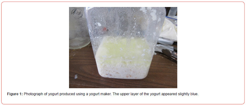
Figure 1 shows a photograph of the yogurt produced in the yogurt maker. The upper layer of the yogurt appeared slightly blue.
Figure 2 shows a photograph of whey after wringing out the cloth bag containing yogurt.
The whey had a slightly blue appearance.
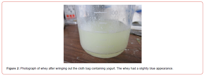
Figure 3 shows a photograph of whey after the addition of ethanol. The whey had a slightly blue appearance.
Figure 4 shows a photograph of a concentrated sample of the upper fluid. The liquid was clear.
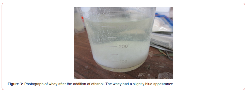

Figure 5 shows a photograph of the yogurt antibiotic assay. A clear zone of inhibition was observed around the well
Figure 6 shows a photograph of the whey antibiotic assay. A clear zone of inhibition was observed around the well.
Figure 7 shows a photograph of a concentrated antibiotic assay. A clear zone of inhibition was observed around the well.
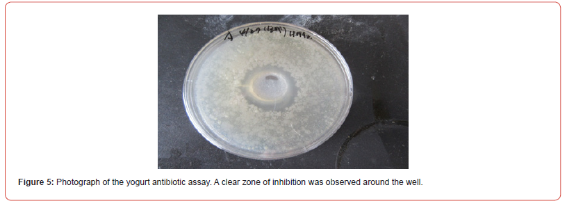

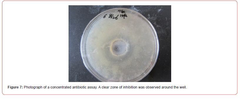
It was previously described that antibiotic-producing cells could be produced following the combination of DNA (HepG2) crown cells and salmon roe [19]. The present experiments examined whether they could be produced antibiotic-containing yogurt in the milk cultures. When the powder was incubated in milk, a similar appearance to yogurt was observed after 3 days of incubation. This yogurt was separated into whey and milk solids, and the antibiotic assay was carried out. Antibiotic activity was observed in whey and in the part obtained via ethanol extraction. Thus, it was demonstrated that antibiotic was produced using milk as a culture medium for powder that was produced from the combination of DNA (HepG2) crown cells with salmon roe. As described previously [21], using milk as a culture medium has several advantages: milk in Japan is sterilized and is relatively cost effective; the composition is indicated and antibiotic can be obtained within about 1 week. However, several challenges remain, particularly whether the present methods are optimal for extracting antibiotics. The extracted antibiotic was completely resolved and the produced antibiotics did not differ from those produced in media other than milk (e.g., malt, agar). These subjects will be clarified in future research on the antibiotic.
On the other hand, antibiotic-producing cells have already been created using several methods [15–20], and it is considered likely that more antibiotic-producing cells will be created in the future. Therefore, it is important to clarify whether these cells produced antibiotic using milk as a medium.
Conclusion
MIn conclusion, the methods used to produce antibiotic in a milk culture of antibiotic-producing cells generated using DNA (HepG2) crown cells and salmon roe have been described. It is expected that these methods will be applied to the production of antibioticproducing cells, with the aim of generating novel and effective antibiotics, including those with activity against Bacillus spp. that could potentially benefit human welfare. The antibiotic developed in this study has been named Antibiotic Crown-HepG2-Salmon-Roeyogurt. This antibiotic was produced using DNA (HepG2) Crown cells with salmon roe and yogurt.
References
- Inooka S (2012) Preparation and cultivation of artificial cells. App Cell Biol 25: 13-18.
- Inooka S (2016) Preparation of Artificial Cells Using Eggs with Sphingosine-DNA. J Chem Eng Process Technol l7: 277.
- Inooka S (2016) Aggregation of sphingosine-DNA and cell construction using components from egg white. Integrative Molecular Medicine 3(6): 1-5.
- Inooka S (2017) Systematic Preparation of Bovine meat DNA Crown Cells. App Cell Biol 30: 13-16.
- Inooka S (2013) Preparation of Artificial Cells for Yogurt Production. App Cell Biol 26: 13-17.
- Inooka S (2014) Preparation of Artificial Placental Cells. App Cell Biol 27: 4-49.
- Inooka S (2019) Preparation of DNA crown cells (artificial cells) using eggs, sphingosine-DNA, and subsequent cell recovery. App Cell Biol 32: 55-64.
- Inooka S (2018) Systematic Preparation of Generated DNA (Akoya pear oyster) Crown Cells. App Cell Biol 31: 21-34.
- Inooka S (2022) Preparation of a DNA (E. coli) Crown Cell line in Vitro-Microscopic Appearance of Cells. Annals of Reviews and Research 8(1).
- Inooka S (2023) Preparation and Microscopic Appearance of a DNA (Human Placenta) Crown Cell Line. Journal of Biotechnology & Bioresearch 5(1).
- Inooka S (2023) Preparation of a DNA (Akoya pearl oyster) crown cell line. Applied Cell Biology Japan 36.
- Inooka S (2022) Microscopic appearance of synthetic DNA (E. coli) crown cells in primary culture. App Cell Biol 35: 71-98.
- Inooka S (2022) Microscopic Appearance of Synthetic DNA (E. coli) Crown Cells in Secondary Cultures. Novel Research in Science 12(3).
- Inooka S, Preparation of a DNA (Hepato-blastoma-Derived Cell line HepG2) Crown Cell line. Journal of Tumor Medicine & Prevence 4(2): 2023.
- Inooka S (2025) Isolation of Antibiotic Producing Cells from Plate Culture of Egg Powder Enclosing DNA (Bovine Meat) Crown Cells and Beef Extract. Annals of Reviews and Research 12(5).
- Inooka S (2025) Separation of Antibiotic-Producing Cells from Beer Produced in Co-cultures of DNA (Streptomyces) Crown Cells with Yeast Annals of Reviews and Research 12(3).
- Inooka S (2025) Separation of antibiotic producing cells from culture fluids of egg white powder enclosed DNA (Streptomyces) crown cells and yeast. International Journal of Bioprocess & Biotechnological Advanccments 10(1) 499-504.
- Inooka S (2025) Separation of Antibiotic-Producing Cells from Agar Cultures of Egg Powder-Enclosed DNA (hepato-blastoma cell Line: HepG2) Crown Cell and Yeast. American Journal of Biomedical Science & Research 26(2).
- Inooka S (2025) Isolation of Antibiotic-Producing Cells from Cultures of Egg Cultures of Egg Powder Enclosing DNA (Hepg2) Crown Cells and Salmon Roe. Trends in General Medicine 3(1): 1-5.
- Inooka S (2025) Antibiotic-Producing Cells Produced from A Plate Culture of Egg Powder Enclosing DNA (Nannochloropsis Species) Crown Cells and Yeast. Scientific Journal Biology & Life Sciences 2: 2.
- Inooka S (2025) The Production of Yogrut Containing Antibiotics from a Milk culture of Egg Powder Enclosing DNA (Lactobacilis species) Crown Cells and Bacillus subtilis and an Attempt to Extract Antibiotics from the Yogurt. Advance Research in Sciences 3(2).
- Inooka S (2017) Biotechnical and Systematic Preparation of Artificial Cells. The global Jornal of Research in Engineering.
- Inooka S (2017) Systematic Preparation of Artificial Cells (DNA Crown Cells). J Chem Eng Procces Technol 8: 327.
-
Shoshi Inooka*. Antibiotic Production in A Milk Culture of Egg White Powder Enclosing DNA (HepG2) Crown Cells and Salmon Roe. Arch Biomed Eng & Biotechnol. 8(2): 2025. ABEB.MS.ID.000682
-
DNA (HepG2) Crown Cells; Sphingosine-DNA; Antibiotic Crown-HepG2-Salmon Roe-Yogurt
-

This work is licensed under a Creative Commons Attribution-NonCommercial 4.0 International License.


