 Opinion Article
Dry Eye in Animals – What Can We Learn from the Human Eye?
Wolfgang G.K. Müller Lierheim, CORONIS Foundation, Munich, Germany.
Received Date: September 02, 2020; Published Date: September 30, 2020
Opinion
Animals have similar ocular surface physiology, epithelial cell structure, immune response, innervation, and tear film structure like humans. Therefore, animals have served for decades in the development and clinical testing of topical eye drops for dry eye disease (DED) [1]. On the other hand, eye drops originally developed for human use are routinely used in the treatment of ocular diseases in animals.
Human patients see an ophthalmologist or optometrist because of ocular symptoms related to pain such as burning, stinging, or itching eyes or to instable vision. Globally multi millions of patients suffer from DED [2]. The Dry Eye Workshop distinguished symptomatic DED (with or without signs) from asymptomatic DED [3]. Significant discordance between signs and symptoms of dry eye has been reported [4,5]. The Asia Dry Eye Society recently concluded that subjective severity i.e. symptoms could be used as marker for therapeutic efficacy, but not for the purpose of classification [6].
The situation in the veterinary environment is different. Owners of animals are likely to see the veterinary clinic because their patients suffer from mucoid ocular discharge. The underlying reason may be a keratoconjunctivitis sicca (KCS), characterized by chronic inflammation of the lacrimal gland, conjunctiva, and cornea, but also an allergic keratoconjunctivitis (AKC) or a bacterial conjunctivitis or a combination [7]. KCS occurs in all animals but is commonly seen in dogs and diagnosed based on tear volume using the Schirmer I tear test (STT I). This test is known to be subject to fluctuations, can therefore only serve as an initial diagnostic marker, but not for control of therapeutic success [8-12]. Routinely the first step in the treatment of KCS is tear substitution by lubricant eye drops, the second step the treatment of chronic inflammation by cyclosporine (CsA) [13-15].
In humans damage of corneal nerves has caught growing attention over the past decade. Nerve damage has beside chronic inflammation been recognized as a second important pathomechanism underlying severe DED [16]. Nerve damage results in a reduction of trophic support for the corneal epithelium [17,18]. There is currently no therapy available addressing DED caused by nerve damage, and artificial tears provide incomplete relief from ocular pain in this situation [19]. Damage of peripheral nerve endings is among others associated with diabetes mellitus (DM). The cornea is the most densely innervated tissue of the body [20]. While the disease is progressing, corneal nerves are damaged in up to 70% of DM patients [21]. DM has also been reported in many animal species [22]. The incidence in dogs is about 1.2% and in cats about 0.6% [23,24]. It seems likely that DED is currently not adequately diagnosed and treated in these patients.
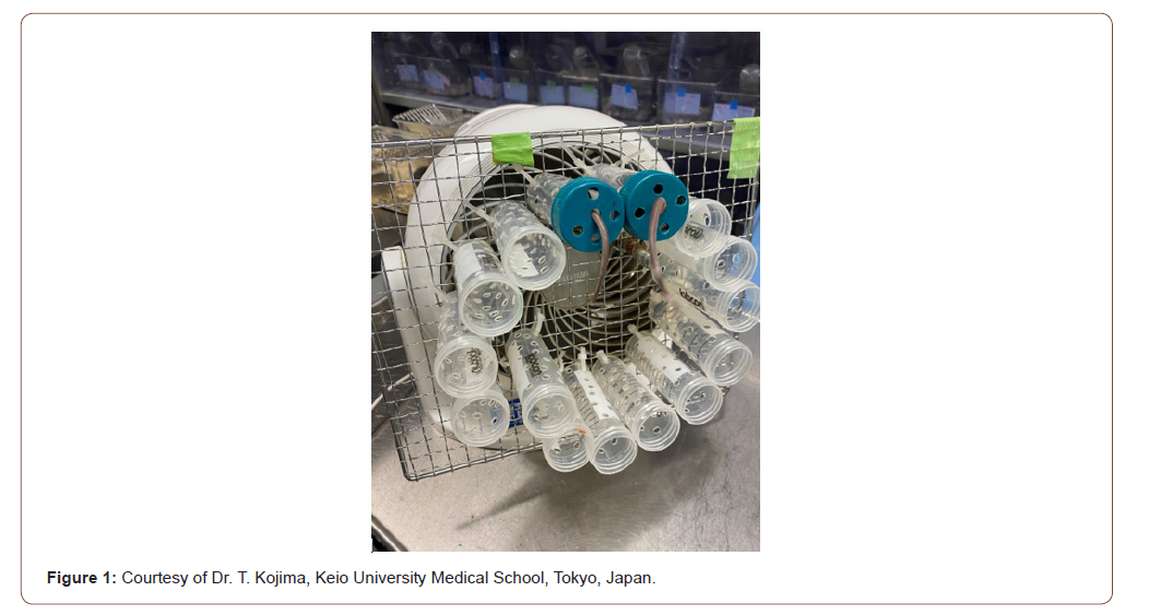
A plethora of hyaluronic acid (HA; hyaluronan; sodium hyaluronate) eye drops are available outside the United States as preferred tear substitutes for the treatment of DED. They differ in HA molecular weight and concentration [25]. HA behaves as a signal molecule in the human as well as animal body. While high molecular weight HA (HMWHA) suppresses inflammation low molecular weight HA (LMWHA) enhances inflammation [26]. A recent study on different molecular weight and concentration of HA demonstrated the suppression of the ocular immune response by HMWHA eye drops in mice under environmental stress conditions [27]. Mice develop ocular surface inflammation when exposed for five hours per day for three consecutive days to air from a fan (see Figure 1).

HMWHA has the potential to suppress the pain receptors of afferent nerves [28-30]. In a recent study on patients with very severe DED, HMWHA eye drops had been successfully applied as a substitute for autologous serum eye drops (ASED) in cases where even CsA eye drops had not resulted in acceptable relieve from symptoms [31]. The results of a multicenter randomized study on the use of HMWHA eye drops in patients suffering from severe DED will soon be published and will provide additional evidence on the potential future role of HMWHA eye drops in the treatment of DED.
The developing knowledge on the role of corneal nerves and the signaling function of HMWHA is likely to provide new insights and options also for dry eye treatment in the veterinary clinic.
Acknowledgement
None
Conflict of Interest
None
References
- Stern ME, Pflugfelder SC (2017) What We Have Learned From Animal Models of Dry Eye. Int Ophthalmol Clin 57(2): 109-118.
- Stapleton F, Alves M ,Bunya VY, Jalbert I ,Lekhanont K,et al. (2017) TFOS DEWS II Epidemiology Report. Ocul Surf 15(3): 334-365.
- Craig JP, Nichols KK, Akpek EK,Caffery B, Dua HS, et al. (2017) TFOS DEWS II Definition and Classification Report. Ocul Surf 15(3): 276-283.
- Ong ES, Felix ER, Levitt RC, Feuer WJ, Sarantopoulos CD, et al. (2018) Epidemiology of discordance between symptoms and signs of dry eye. The British journal of ophthalmology 102(5): 674-679.
- Bartlett JD, Keith MS, Sudharshan L,Snedecor SJ(2015) Associations between signs and symptoms of dry eye disease: a systematic review. Clin Ophthalmol 9: 1719-1730.
- Tsubota K, Yokoi N, Watanabe H, Dogru M, Kojima T, et al. (2020) Members of The Asia Dry Eye S,A New Perspective on Dry Eye Classification: Proposal by the Asia Dry Eye Society. Eye & contact lens, 46(1): S2-S13.
- Maggio F (2019) Ocular surface disease in dogs part 1: aetiopathogenesis and clinical signs. Companion Animal 24(5): 240-245.
- Berger SL,King VL (1998) The fluctuation of tear production in the dog. J Am Anim Hosp Assoc 34(1): 79-83.
- Beech J, Zappala RA, Smith G, Lindborg S (2003) Schirmer tear test results in normal horses and ponies: effect of age, season, environment, sex, time of day and placement of strips. Vet Ophthalmol 6(3): 251-254.
- Piccione G,Giannetto C,Fazio F, Giudice E (2008) Daily rhythm of tear production in normal horse. Vet Ophthalmol 11(1): 57-60.
- Piccione G, Giannetto C, Fazio F, Assenza A, Caola G (2009) Daily rhythm of tear production in normal dog maintained under different Light/Dark cycles. Res Vet Sci 86(3): 521-524.
- Miyasaka, K,Kazama Y,Iwashita H, Wakaiki S, Saito A (2019) A novel strip meniscometry method for measuring aqueous tear volume in dogs: Clinical correlations with the Schirmer tear and phenol red thread tests. Vet Ophthalmol 22(6): 864-871.
- Williams D L (2008) Immunopathogenesis of keratoconjunctivitis sicca in the dog. Vet Clin North Am Small Anim Pract 38(2): 251-268 vi.
- Dodi PL (2015) Immune-mediated keratoconjunctivitis sicca in dogs: current perspectives on management. Vet Med (Auckl) 6: 341-347.
- Maggio F (2019) Ocular surface disease in dogs part 2: diagnosis and treatment. Companion Animal 24(6): 319-328.
- Belmonte C (2019) Pain, Dryness, and Itch Sensations in Eye Surface Disorders Are Defined By a Balance Between Inflammation and Sensory Nerve Injury. Cornea 38(1): S11-S24.
- Shaheen BS,Bakir M, Jain S (2014) Corneal nerves in health and disease. Surv Ophthalmol 59(3): 263-285.
- Al-Aqaba MA,Dhillon VK, Mohammed I,Said DG, Dua HS (2019) Corneal nerves in health and disease. Progress in retinal and eye research 73: 100762.
- Galor A, Batawi H, Felix ER, Margolis TP,Sarantopoulos KD, et al. (2016)Incomplete response to artificial tears is associated with features of neuropathic ocular pain. The British journal of ophthalmology 100(6): 745-749.
- Muller LJ, Marfurt CF, Kruse F,Tervo TM (2003) Corneal nerves: structure, contents and function. Experimental eye research 76(5): 521-542.
- Ljubimov AV (2017) Diabetic complications in the cornea. Vision Res 139: 138-152.
- Niaz K,Maqbool F,Khan F, Hassan FI, Momtaz S (2018) Comparative occurrence of diabetes in canine, feline, and few wild animals and their association with pancreatic diseases and ketoacidosis with therapeutic approach. Vet World 11(4): 410-422.
- Fall T,Hamlin HH, Hedhammar A, Kampe O,Egenvall A (2007) Diabetes mellitus in a population of 180,000 insured dogs: incidence, survival, and breed distribution. J Vet Intern Med 21(6): 1209-1216.
- O Neill DG, Gostelow R, Orme C, Church DB, Niessen SJ, et al. (2016) Epidemiology of Diabetes Mellitus among 193,435 Cats Attending Primary-Care Veterinary Practices in England. J Vet Intern Med 30(4): 964-972.
- Müller Lierheim WGK (2020) Why Chain Length of Hyaluronan in Eye Drops Matters. Diagnostics (Basel) 10(8): E511.
- Jiang D,Liang J, Noble PW (2011)Hyaluronan as an immune regulator in human diseases. Physiological reviews 91(1): 221-264.
- Kojima T,Nagata T, Kudo H, Müller-Lierheim WGK, van Setten GB, et al. (2020) The Effects of High Molecular Weight Hyaluronic Acid Eye Drop Application in Environmental Dry Eye Stress Mice. Int. J. Mol. Sci 21(10): 3516.
- Gomis A, Pawlak M, Balazs EA, Schmidt RF, Belmonte C (2004) Effects of different molecular weight elastoviscous hyaluronan solutions on articular nociceptive afferents. Arthritis and rheumatism 50(1): 314-32
- Caires R, Luis E,Taberner F J,Fernandez-Ballester G, Ferrer-Montiel A, et al. (2015) Hyaluronan modulates TRPV1 channel opening, reducing peripheral nociceptor activity and pain. Nature communications 6: 8095.
- Ferrari LF,Khomula EV, Araldi D, Levine JD (2018) CD44 Signaling Mediates High Molecular Weight Hyaluronan-Induced Antihyperalgesia. J Neurosci 38(2): 308-321.
- Beck R, Stachs O, Koschmieder A, Mueller-Lierheim WGK, Peschel S, et al. (2019) Hyaluronic Acid as an Alternative to Autologous Human Serum Eye Drops: Initial Clinical Results with High-Molecular-Weight Hyaluronic Acid Eye Drops. Case reports in ophthalmology 10(2): 244-255.
-
Wolfgang G.K. Müller Lierheim. Dry Eye in Animals – What Can We Learn from the Human Eye?. Arch Animal Husb & Dairy Sci. 2(2): 2020. AAHDS.MS.ID.000534.
-
Cucumber Plants; Cucumis sativus; Red spider; Tetranychus urticae; Two-spotted spider.
-

This work is licensed under a Creative Commons Attribution-NonCommercial 4.0 International License.


