 Case Report
Double Orifice Mitral Valve Associated with Bicuspid Aortic Valve: A Case Report
Torres-Martel JM1*, Barrera JC2, Flores J3, Garcia-Tejada BA4, Begazo-Paredes JE5 and Garcia-Cruz E6
1Professor, Faculty of Medicine, Autonomous University of Queretaro, Mexico
2University Center for Health Sciences, University of Guadalajara, Maternal and Child Hospital Esperanza López Mateos, Jalisco Health Secretary, Mexico
3General Zone Hospital of the Mexican Institute of Social Security (IMSS), México
4Goyeneche, Hospital III, Arequipa, Peru
5Yanahuara, Hospital III, Arequipa, Peru
6Adult Congenital Heart Disease Clinic, National Institute of Cardiology of Mexico “Ignacio Chávez”, Mexico
Torres-Martel JM, Professor, Faculty of Medicine, Autonomous University of Queretaro, Mexico.
Received Date: June 07, 202; Published Date: June 22, 2023
Double orifice mitral valve (DOMV) es a extremely rare, congenital or acquired anomaly characterized by a two-channel atrioventricular valve in the left ventricle. We describe a case of an 18-year-old women with previous history of bicuspid aortic valve with an incidental finding of DOMV. Transthoracic echocardiography (TTE) revealed a bicuspid aortic valve (BAV), on para-sternal short-axis view, two separate asymmetric mitral valve orifices were visualized. Management is related to the type and severity of mitral valve dysfunction, with mitral regurgitation being the predominant finding.
Keywords:Congenital heart disease; Double orifice mitral valve; Bicuspid aortic valve
Introduction
Double orifice mitral valve (DOMV) is a extremely rare, congenital, or acquired anomaly characterized by a two-channel atrioventricular valve in the left ventricle that is typically asymptomatic allowing a normal blood flow between the two chambers of the left heart. When symptomatic, it is due to the development of mitral valve dysfunction such as stenosis or regurgitation or symptoms due to the association with other cardiac congenital anomalies such left-sided obstructive lesions, ventricular septal defects, anomalies of the tricuspid valve and atrioventricular septal defects. The first description DOMV was made by William Smith Greenfield a British pantologist in 1876. The exact incidence is unknow, but some studies have shown an incidence of 0.06-0.05% in general population and is present in 1% of the patients with congenital heart disease. The majority of cases are detected by 2-dimensional transthoracic echocardiography; however, transesophageal echocardiography also provides a more detailed analysis of both the structures and functions of the mitral valve [1-3]. We describe a case of 18-year-old women with previous history of bicuspid aortic valve with an incidental finding of DOMV.
Case Report
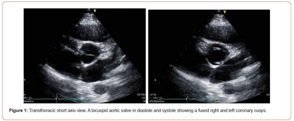
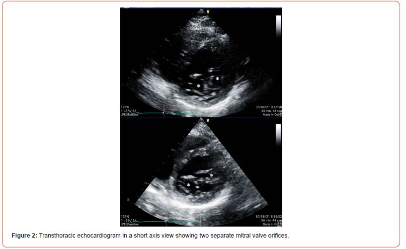
An 18-years old girl patient was referred to the outpatient adult congenital heart disease clinic, with no complaining. She has previous history of bicuspid aortic valve with no symptoms and no echocardiographic previous findings of stenosis or aortic valve regurgitation. Her physical examination was unremarkable, and her pulse rate was regular, with blood pressure of 110/70mmHg. She had normal heart auscultation with no abnormal sounds. Moreover, the electrocardiography and chest X ray test revealed no abnormalities. Transthoracic echocardiography (TTE) showed the size of 4 cardiac chambers view normal with proper right ventricle (RV) and left ventricle (LV) ejection fraction, the short axis view of the heart base revealed a bicuspid aortic valve (BAV) with two leaflets with fused cusps between the two components of the right and left coronary cusps, with no aortic stenosis (AS) (aortic mean gradient= 3.58mmHg, peak gradient= 5.81mmHg) and a mild aortic regurgitation (Figure 1), the mitral inflow was normal and there was no stenosis (mitral valve mean gradient= 1.06mmHg, peak gradient= 2.60mmHg) and a mild mitral regurgitation. On para-sternal short-axis view, two separate asymmetric mitral valve orifices were visualized without stenosis and a mild mitral regurgitation (Figure 2). No other congenital cardiac abnormality was detected on echocardiography.


Discussion
Double-orifice mitral valve is a rare congenital malformation, the exact incidence is still unknown, autopsy studies have shown a 1% of incidence of DOMV In the adult population, a retrospective study involving echocardiographic data revealed an estimated incidence of 0.06 % in the adult tertiary referral echocardiography laboratory. The embryology is incompletely understood, theories include abnormal embryonic fusion of mitral leaflets, fusion of the endocardial cushion tissue of one or the other major cushion with that of the lateral cushion, and incomplete fusion of the two AV endocardial cushions. It is characterized by a mitral valve with a single fibrous annulus and two separate valve orifices of varying sizes in association with the abnormalities of the sub valvular apparatus, especially the tensor apparatus, the characteristic feature is that all the chordae of one papillary muscle go to one ostia while all of the chordae of the other papillary muscle go to the other ostium. DOMV is always associated with anomaly of either the chorda tendinea or the papillary muscles. The associated anomalies of the sub valvular apparatus such as accessory and redundant chordal attachments, chordal ring, accessory septal attachment, parachute chordal attachments and multiple, unequal, or fused papillary muscles. It may have no hemodynamic significance or may cause clinically significant mitral stenosis or mitral regurgitation. It is associated with endocardial cushion defect, bicuspid aortic valve, coarctation of the aorta, interrupted aortic arch, ventricular septal defect and patent ductus arteriosus. The most commonly associated anomaly is partial atrioventricular septal defect. A BAV is relatively common and is character- ized by the abnormal fusion of two leaflets of the aortic valve (AV) during development, resulting in a two leaf- let valve instead of the normal tricuspid AV [4-6].
There are no specific clinical signs suggestive of DOMV, is usually an incidental finding in the diagnosis of the elderly patient and may be missed or undiagnosed in asymptomatic or even symptomatic patients. Patients with significantly dysfunctional DOMV may present with symptoms due to heart failure and can range from tachypnea, dyspnea, and wheezing, requiring initial medical therapy, the extent of symptoms depends on the grade of left atrial pressure resulting in pulmonary congestion. In patients with an associated congenital heart defect, it is usual for that particular disorder to manifest on clinical examination as there are no signs specific to DOMV. The isolated cases of DOMV do not need therapy and can be followed up only by echocardiographic examinations. However, clinic manifestation and management depend not only on the severity of mitral valve dysfunction, but also on associated malformations, which cause pulmonary hypertension due to pulmonary hyper flow from intracardiac shunt [7].
Echocardiography is the modality of choice for diagnosis, best assessed in the parasternal short axis view, color Doppler contributes important information about flow through the mitral valve and can show valve regurgitation or stenosis. Based on echocardiographic features, there are three morphological types; a) eccentric type (85%), characterized by a larger main orifice and a smaller accessory orifice located either at the posteromedial or anterolateral commissure; b) central or bridge type (15%) characterized by a central bridge of fibrous tissue connecting the two leaflets of the mitral valve. The two orifices may be of the same size or unequal; c) duplicate mitral valve type, is comprised of two mitral valve annuli and valves, each individually with its own set of leaflets, commissures, papillary muscles, and chordae [8]. The presence of two separate regurgitant jets on transthoracic parasternal short-axis view should arouse the suspicion of DOMV. In adults transesophageal echocardiography (TEE) is usually necessary for a definite diagnosis of DOMV [9].
Management is related to the type and severity of mitral valve dysfunction, with mitral regurgitation being the predominant finding. All symptomatic patients with a regurgitant valve should be considered for surgical repair with leaflet plication and possible division of the bridging tissue. If there are surgical indications for mitral stenosis, mitral valve balloon dilatation or mitral valve replacement is required [10,11].
Conclusion
Patients with both DOMV and BAV are rare. We described a young girl with BAV and DOMV which did not require either medical or surgical interventions.
Acknowledgement
We want to thank the echo lab for the images.
Conflict of Interest
No conflict of interest.
References
- Bayat F, Hasan M, Khani M, Shekarkhar S, Fatehi A, et al. (2020) Double orifice mitral valve in a patient with bicuspid aortic valve and coarctation of the aorta: A rare presentation. Clin Case Rep 8: 1021-1024.
- Khani M, Rohani A (2017) Double-orifice mitral valve associated with bicuspid aortic valve. Asian Cardiovasc Thorac Ann 25(5): 386-387.
- Samei N, Dehghan H, Pourmojib M, Mohebbi A, Hosseini S, et al. (2017) Isolated double-orifice mitral valve in a young girl. ARYA Atheroscler 13(6): 295-298.
- Lee C, Danielson G, Schaff H, Puga F, Mair D (1985) Surgical treatment of double-orifice mitral valve in atrioventricular canal defects. J THoRAc Cardiovas Surg 90: 700-705.
- Proenca G, Freitas A, Baptista S, Thomas B, Fragata J, et al. (2004) Double Orifice Mitral Valve in an Asymptomatic Adult with an Unusual Combination of Congenital Malformations: A Case Report. Rev Port Cardiol 23(2): 233-236.
- Sun F, Tan X, Sun A, Zhang X, Liang Y, et al. (2021) Rare double orifice mitral valve malformation associated with bicuspid aortic valve in Turner syndrome: diagnosed by a series of novel three-dimensional echocardiography and literature review. BMC Cardiovasc Disord 21: 377.
- Dias M, Menardi A, Almeida-Fiho O, Andrade W, Barbosa P (2019) Double-Orifice Mitral Valve: An Educational Presentation. Braz J Cardiovasc Surg 34(3): 377-379.
- Abdul R, Chowdhury YS (2021) Double Orifice Mitral Valve. In: StatPearls [Internet]. Treasure Island (FL): StatPearls Publishing.
- Erkol A, Karagöz A, Özkan A, Koca F, Yilman F, et al. (2009) Double-orifice mitral valve associated with bicuspid aortic valve: a rare case of incomplete form of Shone’s complex. Eur J Echocardiogr 10: 801-803.
- Arevalo-Guerrero E, Restrepo-Molina G, Cardona I, Canaza-Apaza R, Lòpez J (2018) Double-orifice mitral valve as an isolated finding in an adult patient. RETIC 1: 19-21.
- Li H, Wang H, Zhang W, Cheng L (2021) Echocardiographic diagnosis of congenital double orifice mitral valve malformation: A case report. J Clin Ultrasound 49(5): 509-511.
-
Torres-Martel JM*, Barrera JC, Flores J, Garcia-Tejada BA, Begazo-Paredes JE and Garcia-Cruz E. Double Orifice Mitral Valve Associated with Bicuspid Aortic Valve: A Case Report. On J Cardio Res & Rep. 7(4): 2023. OJCRR.MS.ID.000667.
-
Congenital heart disease, Double orifice mitral valve, Bicuspid aortic valve, Atrioventricular valve, Echocardiography, Mitral valve, Blood pressure, Endocardial cushion tissue, Redundant chordal attachments, Chordal ring, Accessory septal attachment, Parachute chordal attachments Unequal, Fused papillary muscles
-

This work is licensed under a Creative Commons Attribution-NonCommercial 4.0 International License.


